ABSTRACT
Primary vertebral tumors and tumor-like lesions in skeletally immature patients are uncommon yet clinically consequential causes of pain, deformity, and neurological compromise. Compared with adults, pediatric lesions arise in a spine with open physes, evolving marrow composition, and substantial remodeling capacity—features that shape tumor biology and imaging appearance. This pictorial review summarizes malignant tumors, benign tumors, and tumor-like lesions of the pediatric spine, adopting a modality-first, compartment-aware approach. Useful highlights include vertebra plana in Langerhans cell histiocytosis, posterior-element bias in lesions such as osteoid osteoma/osteoblastoma, and the role of whole-body magnetic resonance imaging patterns in chronic nonbacterial osteomyelitis/chronic recurrent multifocal osteomyelitis. We also update terminology and concepts for entities such as vertebral hemangioma (venous malformation) and aneurysmal bone cyst. Throughout, we couple modality choice with pediatric anatomy and growth considerations, as well as illustrating the discussion with real-life cases from our teaching files.
Keywords:
Spinal neoplasms; Bone neoplasms; Pediatrics; Magnetic Resonance Imaging; Tomography, X-ray computed; Osteomyelitis/physiopathology.
RESUMO
Os tumores e pseudotumores vertebrais primários em pacientes esqueleticamente imaturos são incomuns, porém clinicamente relevantes como causas de dor, deformidade e comprometimento neurológico. Em relação aos adultos, as lesões pediátricas surgem em uma coluna com fises abertas, medula óssea em evolução e grande capacidade de remodelação—características que moldam a biologia tumoral e a aparência nas imagens. Esta revisão pictorial resume tumores malignos, tumores benignos e lesões pseudotumorais da coluna pediátrica, adotando uma abordagem centrada na modalidade e atenta ao compartimento. Destaques úteis incluem a vertebra plana na histiocitose de células de Langerhans, o predomínio de acometimento dos elementos posteriores em lesões como osteoma osteoide/osteoblastoma e o papel dos padrões de ressonância magnética de corpo inteiro na osteomielite não bacteriana crônica/osteomielite multifocal recorrente crônica, além da atualização de terminologia e conceitos em malformação venosa (“hemangioma” vertebral) e cisto ósseo aneurismático. Integramos as modalidades de imagem às particularidades anatômicas e de crescimento pediátricas e ilustramos a discussão com casos do nosso arquivo de ensino.
Palavras-chave:
Neoplasias da coluna vertebral; Neoplasias ósseas; Pediatria; Imageamento por ressonância magnética; Tomografia computadorizada por raios X; Osteomielite/fisiopatologia.
INTRODUCTION
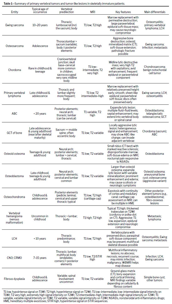
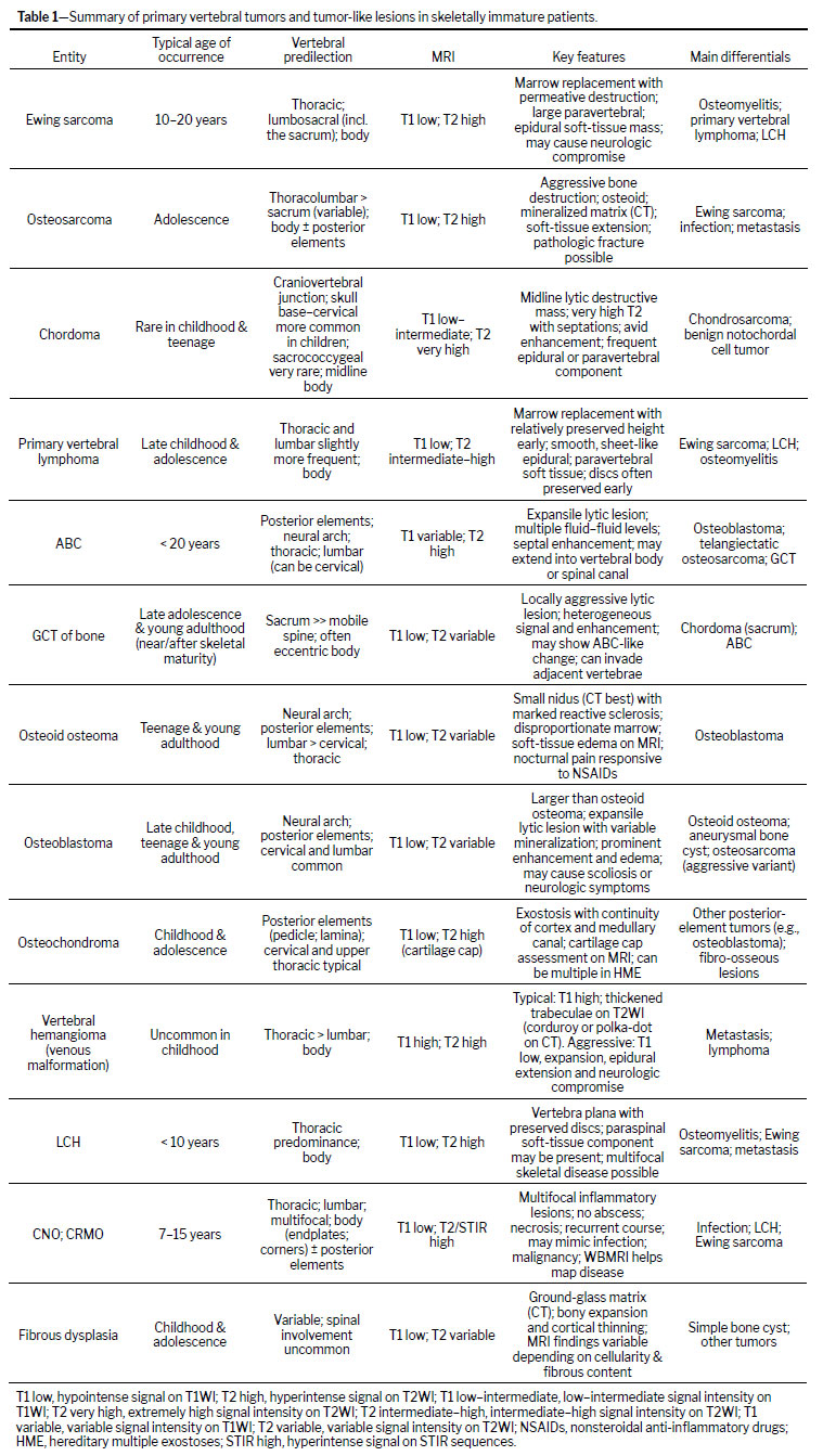
When children and adolescents present with spinal red flags—night pain, pain persisting beyond four weeks, focal neurological deficits, progressive spinal deformity, or systemic symptoms—imaging of the spine to screen for neoplastic and tumor-like etiologies is essential(1–4). The skeletally immature spine differs from the adult spine in marrow composition, vascularity, and the presence of multiple open physes and synchondroses; these features influence tumor predilection (for example, a posterior-element bias), patterns of spread, signal characteristics, and, in some entities, a capacity for height reconstitution after collapse(1,4). Initial localization (intradural vs. extradural; and vertebral body vs. neural arch) narrows the differential diagnosis and guides management(4). In this pictorial review, we integrate modality choices with pediatric anatomy and growth considerations, illustrating the text with real-life cases drawn from our teaching files, rather than from a single institutional cohort. Table 1 summarizes the entities covered in this pictorial review, including typical age/peak incidence, vertebral predilection, key imaging findings, and the main differential diagnoses.
Conventional radiography remains the first look for deformity, collapse, and matrix calcifications and is the classic modality for identifying vertebra plana in Langerhans cell histiocytosis (LCH), though early findings may be subtle
(2,3,5). Computed tomography (CT) excels at matrix characterization—distinguishing “cloud-like” osteoid matrix from rings-and-arcs chondroid matrix—and at depicting cortical permeation and tiny calcifications; thin-section targeted reconstructions improve detection of a small nidus and support safe biopsy planning
(6,7). Magnetic resonance imaging (MRI) maps marrow infiltration, as well as epidural/paravertebral spread, and is indispensable for staging Ewing sarcoma and chordoma; edema-predominant benign lesions (osteoid osteoma/osteoblastoma) may obscure a nidus, inflammatory marrow can mimic tumor, and aggressive vascular lesions can be hypointense on T1-weighted imaging (T1WI)—the correlation with CT is often decisive
(3,5,8–15). Whole-body MRI (WBMRI) is preferred for baseline mapping and follow-up in chronic nonbacterial osteomyelitis/chronic recurrent multifocal osteomyelitis (CNO/CRMO) and LCH, capturing silent vertebral lesions and lesion-distribution patterns that facilitate the differential diagnosis and therapy monitoring
(16–19).
Nuclear medicine is sensitive but nonspecific for multifocal disease; in children, WBMRI is favored when feasible
(16–18). Biopsy planning should leverage cross-sectional imaging to minimize the number of passes and avoid contaminating future resection planes, coordinated with the surgical team
(3,6,12,13,20).
Clinical and imaging presentationMalignant tumorsEwing sarcoma
At-a-glance—Demographic profile: children and adolescents; Segment: sacrum and thoracolumbar most common; Portion: vertebral body, often with a large soft-tissue mass.
Ewing sarcoma is the most common primary malignant vertebral tumor in children and adolescents, found mainly in the sacrum or thoracolumbar spine
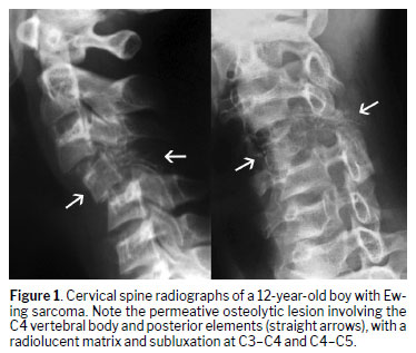
(12–14,20). Imaging typically shows permeative lysis with cortical destruction (Figure 1), often with a disproportionately large soft-tissue mass on CT or MRI
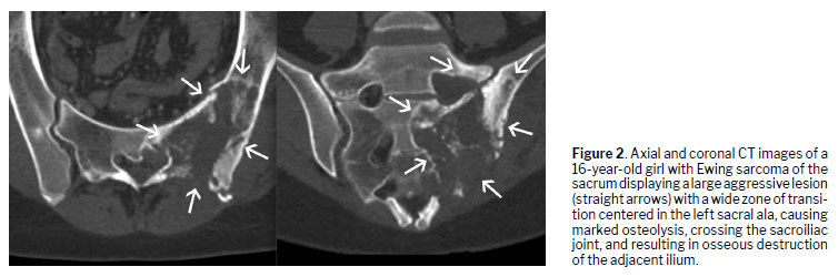
(3,5,9,21). In the sacrum, the lesions may cross the sacroiliac joint space, with contiguous involvement of the adjacent ilium
(21), as shown in Figure 2. On MRI, marrow replacement shows a low-intensity signal on T1WI, shows a high-intensity signal on fluid-sensitive sequences, and is heterogeneously enhancing; epidural extension and canal compromise are common
(11,12). Osteomyelitis and lymphoma can mimic Ewing sarcoma; the combination of aggressive osseous destruction and bulky soft-tissue disease favors Ewing sarcoma
(3,5,11,12). When feasible, wide or en bloc margins improve local control, although it is challenging to achieve safe margins in the spine and pelvis; pediatric planning must weigh stabilization against growth
(20).
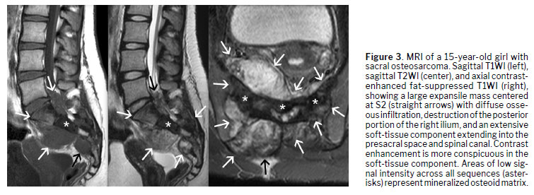
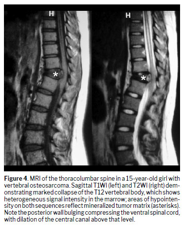
OsteosarcomaAt-a-glance—Demographic profile: adolescents; Segment: thoracolumbar > sacrum (variable); Portion: vertebral body, with or without posterior elements.
Although spinal osteosarcoma is uncommon in children, it is aggressive when it occurs
(12,20,22). Computed tomography shows a mixed lytic–sclerotic lesion with a “cloud-like” osteoid matrix or, less often (in osteoblastic forms), an ivory vertebra
(9,12). The use of MRI allows the delineation of intramedullary extent, cortical breakthrough, epidural spread, and soft-tissue masses; the mineralized component of the tumor—whether osseous or in the soft tissues—appears hypointense on T1WI and T2WI
(12), as illustrated in Figures 3 and 4. Telangiectatic osteosarcoma may show fluid–fluid levels and can mimic an aneurysmal bone cyst; solid enhancing components, an aggressive periosteal reaction, and permeative margins support osteosarcoma
(5,9,12–14,22). Surgical planning must balance decompression/stabilization with oncologic control; local recurrence is lower with wide margins when achievable
(12,20).
Chordoma
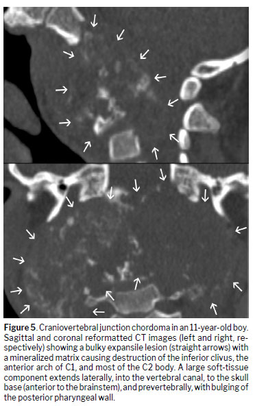
At-a-glance—Demographic profile: rare in children/adolescents, with an overall bimodal age distribution (small early-age cluster, larger adult peak); Segment: sacrum > clivus > mobile spine and, in children, sacrococcygeal disease is very rare compared with craniovertebral (skull base/cervical) involvement; Portion: midline of the vertebral body (central).
Chordoma arises from notochordal remnants. In the general population, it more often affects the sacrum than the craniovertebral junction. However, in children, sacrococcygeal chordoma is distinctly uncommon; instead, skull-base and cervical disease account for the clear majority
(23). A CT scan shows central lysis with faint calcifications
(12), as depicted in Figure 5. An MRI scan depicts a lobulated mass that shows very high-intensity signal on T2WI, with thin hypointense septa and heterogeneous enhancement, commonly with epidural/paravertebral extension
(11,12,24). The main differential diagnoses are as follows
(11,12,24): benign notochordal cell tumor, characterized by no cortical breach or soft-tissue mass; giant cell tumor (GCT), characterized by a less gelatinous appearance on T2WI, often with aneurysmal bone cyst (ABC)-like change; and chondrosarcoma, characterized by a rings-and-arcs pattern of calcification.
Primary vertebral lymphoma
At-a-glance—Demographic profile: children/adolescents; Segment: thoracic and lumbar slightly more common; Portion: vertebral body with smooth, sheet-like epidural/paravertebral soft tissue, often with early preservation of vertebral height and adjacent discs.
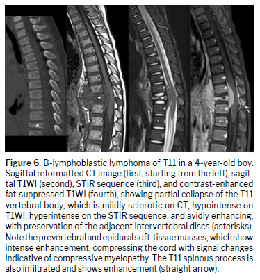
In children, primary vertebral lymphoma (primary bone lymphoma of the spine) typically presents with insidious spinal pain, sometimes with radicular symptoms or early neurologic compromise. On MRI, marrow replacement shows diffuse, homogeneous hypointensity on T1WI and hyperintensity on short-tau inversion recovery (STIR) sequences, with relatively uniform enhancement and a smooth, plaque-like epidural or paravertebral soft-tissue component that may be disproportionately large relative to the degree of cortical destruction (Figure 6); vertebral height and adjacent discs are often preserved in the early stages
(3,4,11,12,25). Restricted diffusion is common in the marrow and soft tissue, and CT may show subtle lysis or sclerosis with minimal periosteal reaction
(11,12,25). Compared with Ewing sarcoma, cortical permeation and aggressive periosteal change are typically less pronounced, and the soft-tissue envelope is more uniform
(3,4,11,12). Although fluorodeoxyglucose positron emission tomography/computed tomography helps confirm osseous unifocality, exclude nodal/visceral disease, and establish a baseline for response assessment, histopathologic confirmation remains mandatory
(11,12,25).
Benign tumorsABCAt-a-glance—Demographic profile: first two decades of life; Segment: thoracic/lumbar (can be cervical); Portion: neural arch/posterior elements.
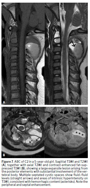
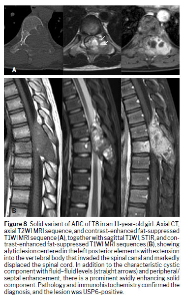
An ABC is now regarded as a true neoplasm driven by the ubiquitin-specific protease 6 (USP6) gene rearrangements; USP6 encodes a deubiquitinating enzyme whose overexpression promotes cyst formation and local bone remodeling
(26,27). The previously termed “secondary ABC,” denoting cystic-hemorrhagic change within another tumor, is best called “ABC-like changes” in contemporary World Health Organization usage; USP6 testing—by fluorescence in situ hybridization or next-generation sequencing—supports primary (USP6-positive) ABC and is typically negative in ABC-like change
(26–28). Conventional radiographs and CT usually show an expansile, septated lytic lesion with cortical thinning/remodeling and internal trabeculations; CT may also depict fluid–fluid levels and small dependent layering, which makes it valuable for preprocedural planning
(2,12,26,27). An MRI scan demonstrates fluid–fluid levels, internal septa, and peripheral/septal enhancement, with scant solid tissue in typical cases (Figure 7); when substantial solid enhancing components are present, consider ABC-like changes over another tumor or the rare solid variant of ABC, which remains uncommon but has been reported in children and adolescents—including in the spine
(12,27,28), as shown in Figure 8.
GCT of boneAt-a-glance—Demographic profile: skeletally mature/near-mature individuals (late teens/young adults); Segment: sacrum far more common than the mobile spine; Portion: vertebral body (often eccentric) with possible posterior extension.
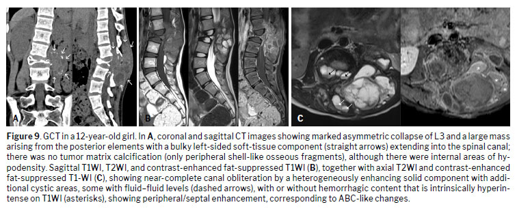
A GCT is locally aggressive
(11,12). In the mobile spine, aggressive cases are typically osteolytic with cortical destruction, are vertebral body-centered, and, in many cases, show epidural extension with pathologic fracture/collapse; cystic areas are common, and fluid–fluid levels occur in a subset of cases
(29), as illustrated in Figure 9. Invasion of an adjacent vertebra has been reported and should be actively sought on sagittal and axial imaging
(30). In the sacrum, GCTs are often eccentric and may extend across the sacroiliac joint into the ilium, a useful discriminator from midline sacral chordoma
(31). Although mobile-spine and sacral lesions may both display ABC-like change and avid enhancement, a midline sacral chordoma—with very high signal intensity on T2WI and a midline epicenter—remains the key differential
(11,12,24,31).
Osteoid osteoma
At-a-glance—Demographic profile: teens, predominantly males; Segment: lumbar > cervical/thoracic; Portion: neural arch/posterior elements.
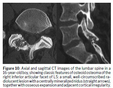
Osteoid osteoma classically presents with nocturnal pain relieved by nonsteroidal anti-inflammatory drugs and is a leading cause of painful scoliosis when spinal, often with convexity away from the lesion and associated facet synovitis
(2). A CT scan is decisive for identifying the < 1.5–2.0 cm nidus—frequently with central mineralization—within reactive cortical/sclerotic bone (Figure 10); thin, targeted reconstructions maximize conspicuity and guide ablation
(5–7). The use of MRI can be pitfall-prone in the spine: extensive marrow and soft-tissue edema, joint effusion, and enhancement can overwhelm or obscure a tiny nidus; recognition of a focal enhancing dot within edema is helpful, but correlative CT is often mandatory
(10,14,32). Although bone scintigraphy typically shows focal hyperemia and uptake (often a “double-density” sign), WBMRI is increasingly favored in children when the differential diagnosis includes multifocal processes to limit ionizing radiation
(16–19,32).
Osteoblastoma
At-a-glance—Demographic profile: older children/teens, young adults; Segment: cervical and lumbar predilection; Portion: neural arch/posterior elements.
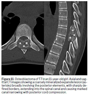
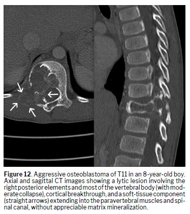
Osteoblastoma spans a broad spectrum of presentations on imaging, from lesions that appear giant osteoid osteoma-like (well-circumscribed, dominant nidus-like core with surrounding sclerosis, as in Figure 11) to frankly aggressive tumors with cortical breakthrough, epidural/paravertebral extension, and prominent soft-tissue reaction
(11,12), as depicted in Figure 12. Approximately 30–40% arise in the spine, usually from the neural arch with potential vertebral body extension, and canal/foraminal compromise often drives presentation
(5,11,12). An osteoblastoma is heterogeneous on fluid-sensitive MRI sequences with avid, variable enhancement; marrow/soft-tissue edema can mask the core lesion, so CT correlation is essential to demonstrate internal osteoid matrix and any coarse calcifications
(11,12,14,33), as illustrated in Figure 11. There can be ABC-like change, which may complicate differentiation from primary ABC and telangiectatic osteosarcoma; attention to solid enhancing tumor, matrix, and periosteal response helps avoid misclassification
(11,12,33).
Osteochondroma
At-a-glance—Demographic profile: children/adolescents, with a higher risk in hereditary multiple exostoses; Segment: cervical and upper thoracic typical; Portion: neural arch/posterior elements (pedicle, lamina, spinous process).
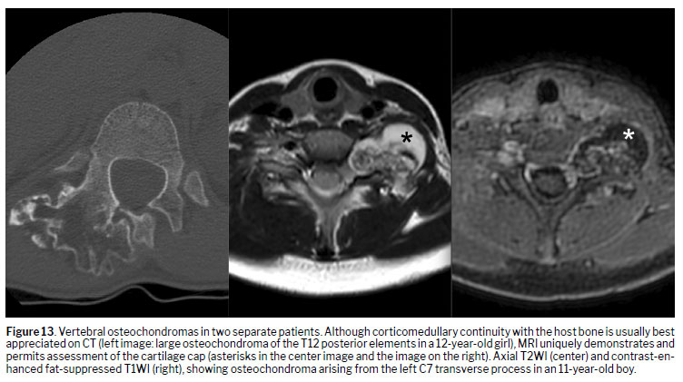
Spinal osteochondromas show cortical and medullary continuity with the parent bone on CT and a T2-bright cartilage cap on MRI (Figure 13); lesions arise from the pedicle, lamina, or spinous process and can encroach upon the canal or foramina, producing radiculopathy or myelopathy
(2,10). Bursitis and reactive edema around the cap can mimic aggression, although a lack of destructive change and confirmation of continuity favor a benign diagnosis
(2,10). In children and adolescents, cartilage caps can be physiologically thicker during active growth; a threshold of > 3 cm (≈ 30 mm) is more commonly used as a threshold for malignant transformation, given that values up to ≈ 2–3 cm can be observed in skeletally immature patients
(34,35). In contrast, in skeletally mature patients, a threshold of > 2 cm is typically used to raise suspicion.
Vertebral hemangiomaAt-a-glance—Demographic profile: uncommon in children; Segment: thoracic > lumbar; Portion: vertebral body.
Vertebral hemangiomas—biologically venous malformations per the International Society for the Study of Vascular Anomalies—remain widely labeled “hemangiomas” in daily practice
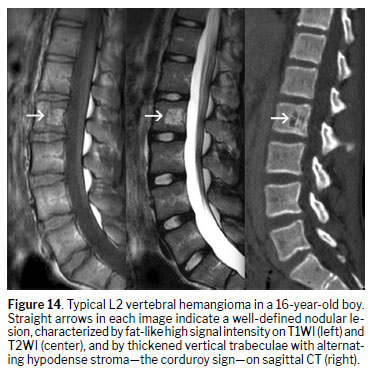
(36). They consist of vascular channels interlaced with thickened vertical trabeculae, producing the “polka-dot” sign on axial CT and vertical striations on sagittal/coronal images
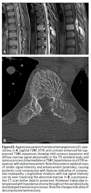
(5,10), as illustrated in Figure 14. Aggressive variants can be hypointense on T1WI, hyperintense on T2WI, and avidly enhancing, with cortical erosion and epidural/paravertebral extension (Figure 15a); preservation of vertical trabeculae on CT (Figure 15b) favors a venous malformation over malignancy
(13,14,37,38).
Tumor-like lesionsLCHAt-a-glance—Demographic profile: young children (peak at early school age); Segment: thoracic; Portion: vertebral body (vertebra plana), discs preserved.
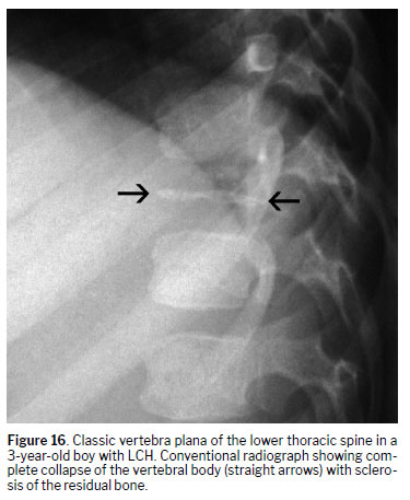
The leading cause of vertebra plana in children is LCH, typically with preserved intervertebral discs and a relatively small paravertebral/epidural component compared with that seen in malignant collapse
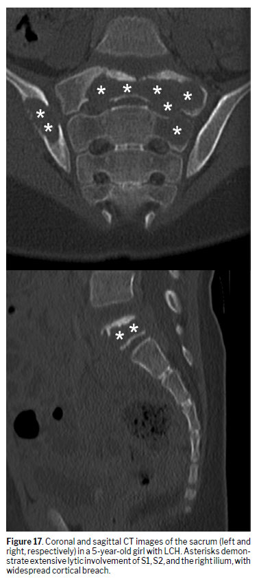
(3,41). Conventional radiographs may show an evolving anterior wedge that progresses to complete collapse (vertebra plana), as shown in Figure 16, whereas CT confirms lysis, defining cortical integrity and any small epidural component when MRI is not immediately available
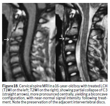
(5,11,13,41), as illustrated in Figure 17. An MRI scan shows a hypointense signal on T1WI and a hyperintense fluid-sensitive signal within the vertebral body, with variable enhancement and with the posterior elements often spared (Figure 18); over time, partial height reconstitution is common and supports conservative management when neurologically safe
(5,11,13,41). Because LCH can be multifocal, WBMRI helps detect additional skeletal sites and tailor follow-up in a radiation-free manner
(16).
CNO/CRMOAt-a-glance—Demographic profile: school age to adolescence; Segment: predominantly thoracic, often multifocal; Portion: vertebral body (endplates/corners) with posterior-element involvement common.
For baseline mapping and follow-up of CNO/CRMO, WBMRI is preferred; classic extra-spinal foci in the metaphyses of the long bones often clinch the diagnosis when interpreted alongside the spinal pattern—particularly when the presentation is spine-predominant or spine-only
(16–19). In children, clavicular disease is typically isolated to the clavicle—most often the medial third with lateral progression—while sparing the sternoclavicular joint and sternum
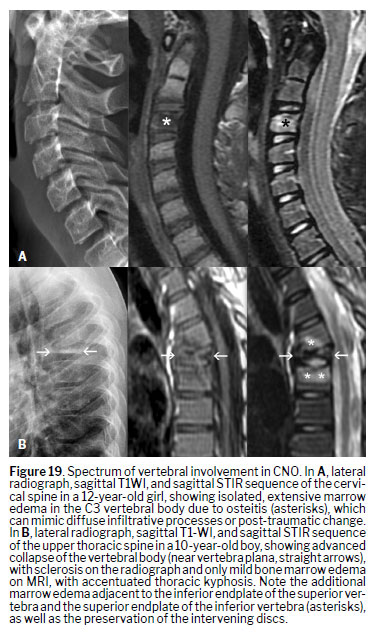
(16–19). An MRI of the spine typically shows marrow edema that is hypointense on T1WI and hyperintense on STIR sequences, together with endplate/corner changes and mild paraspinal reaction with preserved discs (Figure 19); abscess is atypical
(17,18). Complications include pathologic fractures/vertebra plana; unlike LCH, a collapsed vertebra in CNO/CRMO generally does not re-expand, which guides counseling and follow-up
(39). Management escalates from nonsteroidal anti-inflammatory drugs to disease-modifying antirheumatic drugs/biologics, with bisphosphonates effective in vertebral disease; most lesions partially resolve, although there can be new-level recurrence
(18,40).
Fibrous dysplasia
At-a-glance—Demographic profile: children/adolescents (monostotic or polyostotic, consider McCune–Albright features); Segment: variable (spinal involvement is uncommon and usually polyostotic); Portion: body or posterior elements.
Fibrous dysplasia is a fibro-osseous lesion driven by post-zygotic GNAS mutations (encoding the stimulatory G-protein alpha subunit, Gsα), which cause constitutive Gs signaling and replacement of normal bone by fibrous tissue and disorganized woven bone
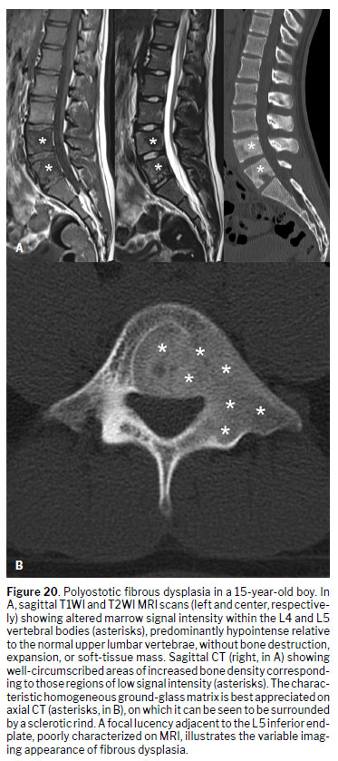
(42). Conventional radiography and CT show a ground-glass matrix with smooth expansion and a sclerotic rim; MRI findings are variable and nonspecific, with low-to-intermediate signal strength on T1WI and intermediate-to-high signal strength on T2WI (Figure 20), depending on trabeculae and cystic/hemorrhagic change
(5,10). In the spine, monostotic disease is rare; most vertebral cases are polyostotic, and management ranges from observation to stabilization for deformity/instability
(42).
CONCLUSIONPrimary vertebral tumors and tumor-like lesions in skeletally immature patients are heterogeneous and frequently imaging-driven diagnoses. A structured, modality-wise approach—grounded in pediatric anatomy and growth, precise compartmental localization, and judicious use of radiography, CT, MRI, and WBMRI—enables timely, accurate differentiation among malignant, benign, and tumor-like entities
(1–4). Recognizing pediatric-specific signatures—including vertebra plana with potential height reconstitution in LCH versus lack of re-expansion in CNO/CRMO; posterior-element bias and the broad yet condensed spectrum of osteoblastoma; WBMRI patterns in CNO/CRMO—and applying updated terminology and concepts refine the interpretation and management
(16–19,23,24,36–38).
REFERENCES1. Schatteman S, Jaremko J, Jans L, et al. Update on pediatric spine imaging. Semin Musculoskelet Radiol. 2023;27:566-79.
https://doi.org/10.1055/s-0043-17713332. Joaquim AF, Ghizoni E, Valadares MGC, et al. Spinal tumors in children. Rev Assoc Med Bras (1992). 2017;63:459-65.
https://doi.org/10.1590/1806-9282.63.05.4593. Menashe SJ, Iyer RS. Pediatric spinal neoplasia: a practical imaging overview of intramedullary, intradural, and osseous tumors. Curr Probl Diagn Radiol. 2013;42:249-65.
https://doi.org/10.1067/j.cpradiol.2013.05.0034. Kumar N, Tan WLB, Wei W, et al. An overview of the tumors affecting the spine—inside to out. Neurooncol Pract. 2020;7(Suppl 1):i10-i17.
https://doi.org/10.1093/nop/npaa0495. Riahi H, Mechri M, Barsaoui M, et al. Imaging of benign tumors of the osseous spine. J Belg Soc Radiol. 2018;102:13.
https://doi.org/10.5334/jbsr.13806. Abdel Razek AAK, Castillo M. Imaging appearance of primary bony tumors and pseudo-tumors of the spine. J Neuroradiol. 2010;37:37-50.
https://doi.org/10.1016/j.neurad.2009.08.0067. Garcia DAL, Aivazoglou LU, Garcia LAL, et al. Diagnostic imaging of primary bone tumors of the spine. Curr Radiol Rep. 2017;5:22.
https://doi.org/10.1007/s40134-017-0220-18. Ropper AE, Cahill KS, Hanna JW, et al. Primary vertebral tumors: Part II—locally aggressive and malignant tumors. Neurosurgery. 2012;70:211-9.
https://doi.org/10.1227/NEU.0b013e31822d5f179. Ropper AE, Cahill KS, Hanna JW, et al. Primary vertebral tumors: Part I—benign tumors. Neurosurgery. 2011;69:1171-80.
https://doi.org/10.1227/NEU.0b013e31822b810710. Ariyaratne S, Jenko N, Iyengar KP, et al. Primary osseous malignancies of the spine. Diagnostics (Basel). 2023;13:1801.
https://doi.org/10.3390/diagnostics1310180111. Patnaik S, Jyotsnarani Y, Uppin SG, et al. Imaging features of primary tumors of the spine: a pictorial essay. Indian J Radiol Imaging. 2016;26:279-89.
https://doi.org/10.4103/0971-3026.18441312. Mechri M, Riahi H, Sboui I, et al. Imaging of malignant primitive tumors of the spine. J Belg Soc Radiol. 2018;102:56.
https://doi.org/10.5334/jbsr.141013. Nguyen TT, Thelen JC, Bhatt AA. Bone up on spinal osseous lesions: a case review series. Insights Imaging. 2020;11:80.
https://doi.org/10.1186/s13244-020-00883-614. Reijnierse M. Axial skeleton bone marrow changes in inflammatory rheumatologic disorders. Semin Musculoskelet Radiol. 2023;27:91–102.
https://doi.org/10.1055/s-0043-176149615. Cederberg KB, Iyer RS, Chaturvedi A, et al. Imaging of pediatric bone tumors: a COG Diagnostic Imaging Committee/SPR Oncology Committee white paper. Pediatr Blood Cancer. 2023;70 Suppl 4:e30000.
https://doi.org/10.1002/pbc.3000016. Andronikou S, Mendes da Costa T, Hussien M, et al. Radiological diagnosis of chronic recurrent multifocal osteomyelitis using whole-body MRI-based lesion distribution patterns. Clin Radiol. 2019;74:737.e3–737.e15.
https://doi.org/10.1016/j.crad.2019.02.02117. Byrdy-Daca M, Duczkowski M, Sudoł-Szopińska I, et al. Spinal involvement in patients with chronic non-bacterial osteomyelitis (CNO): distinctive imaging features. J Clin Med. 2023;12:7419.
https://doi.org/10.3390/jcm1223741918. Lanckoroński M, Gietka P, Mańczak M, et al. Whole-body MRI at initial presentation of chronic recurrent multifocal osteomyelitis, juvenile idiopathic arthritis, their overlapping syndrome, and non-specific arthropathy. J Clin Med. 2024;13:998.
https://doi.org/10.3390/jcm1304099819. Viana SL, Ribeiro MCM, Machado BB. Imaging of chronic recurrent multifocal osteomyelitis and autoinflammatory bone disorders. In: Joint Imaging in Childhood and Adolescence. 2nd ed. Cham: Springer; 2019. p. 155-182.
https://doi.org/10.1007/978-3-030-11342-1_620. Helenius IJ, Krieg AH. Primary malignant bone tumours of spine and pelvis in children. J Child Orthop. 2021;15:337–45.
https://doi.org/10.1302/1863-2548.15.21008521. Murphey MD, Senchak LT, Mambalam PK, et al. From the archives of the AFIP: Ewing sarcoma family of tumors—radiologic-pathologic correlation. Radiographics. 2013;33:803-31.
https://doi.org/10.1148/rg.33313500522. Crombé A, Simonetti M, Longhi A, et al. Imaging of osteosarcoma: presenting findings, metastatic patterns, and features related to prognosis. J Clin Med. 2024;13:5710.
https://doi.org/10.3390/jcm1319571023. Sebro R, DeLaney T, Hornicek F, et al. Differences in sex distribution, anatomic location and MR imaging appearance of pediatric compared to adult chordomas. BMC Med Imaging. 2016;16:53.
https://doi.org/10.1186/s12880-016-0149-524. Lee SH, Kwok KY, Wong SM, et al. Chordoma at the skull base, spine, and sacrum: a pictorial essay. J Clin Imaging Sci. 2022;12:44.
https://doi.org/10.25259/JCIS_62_202225. Tannenbaum MF, Noda S, Cohen S, et al. Imaging musculoskeletal manifestations of pediatric leukemia and lymphoma. AJR Am J Roentgenol. 2020;214:455–64.
https://doi.org/10.2214/ajr.19.2183326. WHO Classification of Tumours Editorial Board. Soft Tissue and Bone Tumours. 5th ed. Vol. 3. Lyon: IARC; 2020.
27. Restrepo R, Zahrah D, Pelaez L, et al. Update on aneurysmal bone cyst: pathophysiology, histology, imaging and treatment. Pediatr Radiol. 2022;52:1601–14.
https://doi.org/10.1007/s00247-022-05396-628. Nasri E, Reith JD. Aneurysmal bone cyst: a review. J Pathol Transl Med. 2023;57:81–7.
https://doi.org/10.4132/jptm.2023.02.2329. Yuan B, Zhang L, Yang SM, et al. Imaging features of aggressive giant cell tumors of the mobile spine: retrospective analysis of 101 patients from single center. Global Spine J. 2022;12:1449–61.
https://doi.org/10.1177/219256822098228030. Jiang G, Sun LL, Ye YJ, et al. Giant cell tumors of the mobile spine with invasion of adjacent vertebrae: an unusual imaging finding. BMC Musculoskelet Disord. 2021;22:726.
https://doi.org/10.1186/s12891-021-04610-031. Vanheule E, Huysse W, Herregods N, et al. Sacral tumours on MRI: a pictorial essay. J Belg Soc Radiol. 2019;103:62.
https://doi.org/10.5334/jbsr.1887.32. Carneiro BC, Da Cruz IAN, Ormond Filho AG, et al. Osteoid osteoma: the great mimicker. Insights Imaging. 2021;12:32.
https://doi.org/10.1186/s13244-021-00978-833. Liu J, Han S, Li J, et al. Spinal osteoblastoma: a retrospective study of 35 patients’ imaging findings with an emphasis on MRI. Insights Imaging. 2020;11:122.
https://doi.org/10.1186/s13244-020-00934-y34. Rueda-de-Eusebio A, Gomez-Pena S, Moreno-Casado MJ, et al. Hereditary multiple exostoses: an educational review. Insights Imaging. 2025;16:46.
https://doi.org/10.1186/s13244-025-01899-635. Tepelenis K, Papathanakos G, Kitsouli A, et al. Osteochondromas: an updated review of epidemiology, pathogenesis, clinical presentation, radiological features and treatment options. In Vivo. 2021;35:681-91.
https://doi.org/10.21873/invivo.1230836. Goldenberg DC, Vikkula M, Penington A, et al. Updated classification of vascular anomalies: a living document from the International Society for the Study of Vascular Anomalies Classification Group. J Vasc Anom (Phila). 2025;6:e113.
https://doi.org/10.1097/jova.000000000000011337. Strauss SB, Steinklein JM, Phillips CD, et al. Intraosseous venous malformations of the head and neck. AJNR Am J Neuroradiol. 2022;43:1090-8.
https://doi.org/10.3174/ajnr.A757538. Kato K, Teferi N, Challa M, et al. Vertebral hemangiomas: a review on diagnosis and management. J Orthop Surg Res. 2024;19:310.
https://doi.org/10.1186/s13018-024-04799-539. Sá Silva J, Bettencourt S, Madureira I, et al. Chronic nonbacterial osteomyelitis in neuroradiology – behavior and evolution of vertebral and mandibular lesions on imaging. Pediatr Radiol. 2024;54:2046-59.
https://doi.org/10.1007/s00247-024-06079-040. Hug NF, Purger DA, Li D, et al. Neurosurgical management of vertebral lesions in pediatric chronic recurrent multifocal osteomyelitis: patient series. J Neurosurg Case Lessons. 2023;5:CASE22179.
https://doi.org/10.3171/case2217941. Khung S, Budzik JF, Amzallag-Bellenger E, et al. Skeletal involvement in Langerhans cell histiocytosis. Insights Imaging. 2013;4:569–79.
https://doi.org/10.1007/s13244-013-0271-742. Kushchayeva YS, Kushchayev SV, Glushko TY, et al. Fibrous dysplasia for radiologists: beyond ground glass bone matrix. Insights Imaging. 2018;9:1035–56.
https://doi.org/10.1007/s13244-018-0666-61. Tesla Diagnóstico por Imagem, Brasília, DF, Brazil
2. Hospital da Criança de Brasília José Alencar, Brasília, DF, Brazil
3. Dasa/Exame Medicina Diagnóstica, Brasília, DF, Brazil
4. Instituto Dasa de Ensino e Pesquisa, São Paulo, SP, Brazil
5. School of Medicine, University of Brasília, Brasília, DF, Brazil
6. Gastren Clínica Médica e Radiológica, Iúna, ES, Brazil
7. Clínica Radiológica Med Imagem, Cachoeiro de Itapemirim, ES, Brazil
8. Clínica Radiológica Radial, Cachoeiro de Itapemirim, ES, Brazil
9. Santa Casa de Misericórdia, Cachoeiro de Itapemirim, ES, Brazil
10. Unimed Sul Capixaba, Cachoeiro de Itapemirim, ES, Brazil
a.
https://orcid.org/0000-0002-3580-9147b.
https://orcid.org/0000-0001-6660-2766c.
https://orcid.org/0000-0002-2571-7775Correspondence:Sergio Lopes Viana
MD. SMHN, Via W1 Norte, salas 7-8, Mezanino, 70710-146, Brasília, DF, Brazil.
Email:
sergiolopesviana@gmail.comEditor in charge: Dr. Valdair Francisco Muglia.
Data availability: Not applicable.
Received in
September 20 2025.
Accepted em
February 24 2026.
Publish in
September 25 2026.

 |
|






 PDF English
 Print
Print
 Send this article by email
 How to cite this article
 Submit a comment
 Mendeley
 Pocket
 Share on Facebook
 Share on Twitter