ABSTRACT
Fasciculations are a key manifestation of lower motor neuron dysfunction in amyotrophic lateral sclerosis (ALS). Although electromyography is considered the gold-standard method for the detection of fasciculations, its sensitivity for that purpose depends on the muscle sampled and the examination time. Muscle ultrasound has emerged as a dynamic, noninvasive technique with high sensitivity for detecting spontaneous muscle activity. This updated systematic review compares ultrasound and electromyography in terms of their performance for the detection of muscle fasciculations in ALS, with stratification by muscle group and observation time. A comprehensive search of the literature identified studies evaluating both techniques. Data were synthesized descriptively and, when possible, quantitatively. Ultrasound demonstrated higher or comparable detection rates across most muscle groups, particularly in muscles that are large or deep, such as the biceps brachii, tibialis anterior, and vastus lateralis. Longer ultrasound observation times, especially 60 s, were associated with substantially higher detection rates without a loss of specificity. Although electromyography is still essential for electrophysiological characterization, ultrasound offers broader muscle coverage, excellent reproducibility, and a noninvasive approach. These findings support muscle ultrasound as a valuable complementary tool for the detection and assessment of fasciculations in patients with motor neuron disease.
Keywords:
Ultrasonography; Electromyography; Fasciculation; Amyotrophic lateral sclerosis; Motor neuron disease.
RESUMO
As fasciculações representam uma manifestação central da disfunção do neurônio motor inferior na esclerose lateral amiotrófica (ELA). A eletroneuromiografia é considerada o método de referência para sua detecção, porém sua sensibilidade depende da extensão da amostragem muscular e do tempo de exame. A ultrassonografia muscular surgiu como uma técnica dinâmica e não invasiva, com elevada sensibilidade para identificar atividade muscular espontânea. Esta revisão sistemática atualizada compara o desempenho da ultrassonografia e da eletroneuromiografia na detecção de fasciculações em pacientes com ELA, com estratificação por grupos musculares e tempo de observação. A literatura disponível foi analisada de forma descritiva e, quando possível, quantitativa. De modo geral, a ultrassonografia apresentou taxas de detecção superiores ou semelhantes às da eletroneuromiografia na maioria dos músculos, especialmente em músculos grandes ou profundos, como bíceps braquial, tibial anterior e vasto lateral. Protocolos com maior tempo de observação, particularmente 60 s, aumentaram significativamente a taxa de detecção pela ultrassonografia sem comprometer a especificidade. Embora a eletroneuromiografia permaneça fundamental para a caracterização eletrofisiológica, a ultrassonografia oferece maior cobertura muscular, excelente reprodutibilidade e caráter não invasivo, configurando-se como método complementar valioso na avaliação das doenças do neurônio motor.
Palavras-chave:
Ultrassonografia; Eletromiografia; Fasciculação; Esclerose lateral amiotrófica; Doenças do neurônio motor.
INTRODUCTION
Fasciculations are brief, spontaneous twitches of muscle fibers innervated by a single motor unit(1). They may occur in a wide spectrum of conditions, including benign conditions—such as benign fasciculation syndrome, physiological variants, metabolic disturbances, and drug-induced states—as well as in pathological conditions requiring accurate diagnosis, including amyotrophic lateral sclerosis (ALS), other motor neuron diseases, spinal cord lesions, and neuromyotonia. Fasciculations are not a typical feature of primary myopathies, given that most originate from dysfunction of the anterior horn cell or distal motor nerve terminal(2). Fasciculations can be identified clinically, electrophysiologically (by electromyography), or visually (by ultrasonography). Each method offers complementary insights into lower motor neuron (LMN) dysfunction(3).
Fasciculations arise from spontaneous depolarization of unstable LMN or terminal axons, generating involuntary discharges that activate a single motor unit. Although magnetic resonance imaging techniques, including advanced motor unit assessment and diffusion-weighted imaging, have also been explored for detecting neuromuscular abnormalities associated with fasciculations, they still play a complementary role and are not always accessible in routine clinical practice. The discharges that provoke fasciculations may result from membrane hyperexcitability, collateral sprouting, ion-channel dysfunction, or impaired axonal integrity—mechanisms commonly associated with denervation and reinnervation processes(2). In ALS, fasciculations reflect ongoing motor-unit instability and increased axonal excitability, often preceding clinically detectable weakness and therefore representing one of the earliest indicators of LMN degeneration(3).
Clinically, fasciculations carry substantial diagnostic relevance in ALS. Their presence in multiple body regions, especially when associated with weakness, atrophy, and upper motor neuron signs, increases the likelihood of ALS and contributes to meeting the Awaji and revised El Escorial criteria(4–6). Fasciculations in specific muscle groups—such as the tongue, paravertebral, and distal limb muscles—are particularly suggestive of motor neuron disease. Early and accurate detection of fasciculations can therefore shorten the time to diagnosis, guide disease monitoring, and facilitate the differentiation from benign fasciculation syndromes and other neuromuscular disorders.(2,3,7).
Electromyography is still the gold-standard test for evaluating LMN impairment in ALS(8). However, it has inherent limitations—its sensitivity depends on the number of muscles examined and the duration of screening for each muscle(9). In addition, electromyography primarily samples small areas of muscle and cannot assess atrophy or structural integrity(10). Ultrasound, in contrast, is a dynamic, noninvasive, and highly motion-sensitive technique capable of detecting displacements as small as 5 µm, with frame rates exceeding 80 fps(11). It allows real-time visualization of superficial and deep muscles, enabling analysis of muscle atrophy and architecture, as well as fasciculations(11,12).
A prior systematic review by our group demonstrated that ultrasound shows greater overall accuracy than electromyography for detecting fasciculations in ALS(13). However, that analysis did not stratify results by muscle group or duration of the observation. Since then, additional studies have explored muscle-specific detection rates, including protocols with 60-s observations(14,15). Given the anatomical and physiological variability in fasciculation frequency across different muscles, understanding how electromyography and ultrasound perform within specific muscle groups may guide the standardization of imaging protocols and optimize diagnostic yield. Therefore, the aim of this updated systematic review and meta-analysis was to compare electromyography and ultrasound in terms of the rate of detection of fasciculations across individual muscles, integrating all available evidence up to December 24, 2025.
METHODS
Study design
This was a systematic review of diagnostic performance studies comparing ultrasound and electromyography for the detection of muscle fasciculations in ALS. This review represents an updated synthesis expanding on our previously published systematic review(12), incorporating newly available evidence and extended observation protocols.
The review was conducted in accordance with the Preferred Reporting Items for Systematic Reviews and Meta-Analyses guidelines(16), registered on the International Prospective Register of Systematic Reviews platform (Registration no. CRD42017078388), and approved by the local institutional research ethics committee. Because the study involved no human or animal subjects, the institutional review board waived the requirement for formal institutional review.
Eligibility criteria
We included studies that directly compared ultrasound and electromyography as means of detecting fasciculations in patients with ALS, reporting absolute numbers of muscles or patients with and without fasciculations for each method. All study designs (prospective, cross-sectional, and case-control) were eligible if muscle-specific data were available. No restrictions were applied regarding language or publication status.
Study populations
Studies enrolling adults diagnosed with ALS or its variants, according to the Awaji or revised El Escorial criteria, were included. Studies limiting their samples to healthy volunteers or patients with other neuromuscular conditions were excluded from quantitative synthesis but could be discussed qualitatively for methodological comparison.
Index and comparator tests
Ultrasound
On ultrasound, fasciculations were defined as spontaneous, irregular, brief contractions of muscle fibers observed in real time on B-mode imaging. Observation times of 10, 30, or 60 s per muscle were accepted.
Electromyography
On electromyography, fasciculations were defined as spontaneous discharges of motor unit potentials in relaxed muscle.
Reference standard
Because all participants had a confirmed diagnosis of ALS, the disease served as the clinical context but not as the diagnostic reference standard. The objective was to compare electromyography and ultrasound in terms of their performance in the detection of fasciculations, not their diagnostic accuracy for ALS itself.
Search strategy
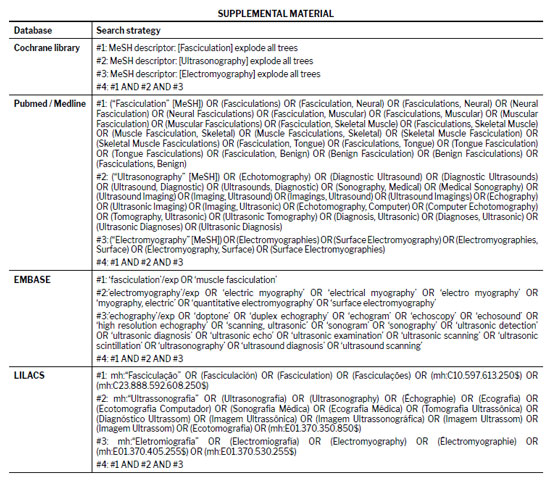
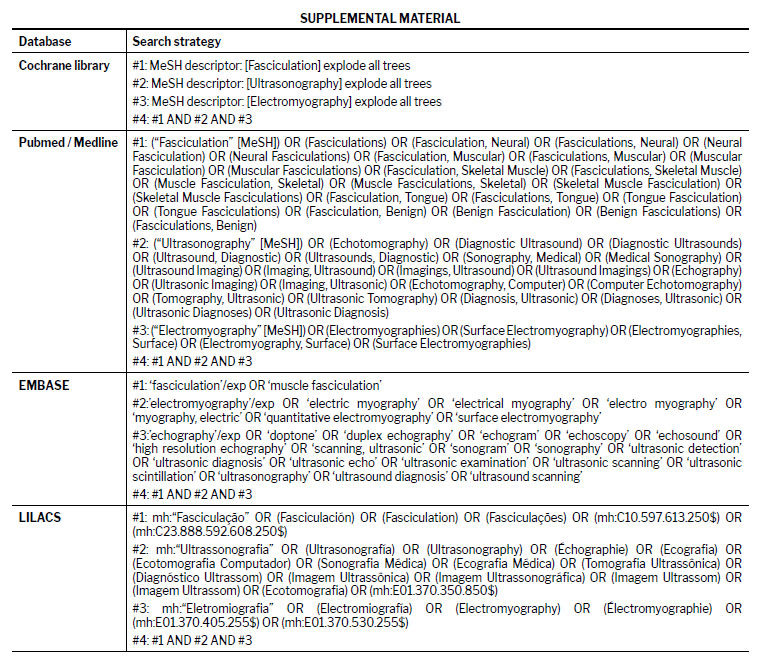
A comprehensive search was conducted across the MEDLINE (via PubMed), EMBASE, Cochrane Library, and Latin-American and Caribbean Health Sciences Literature databases, from their inception to December 24, 2025. The search combined controlled vocabulary and free-text terms for “fasciculation”, “ultrasonography”, and “electromyography” (Supplemental material). Manual searches of the bibliographies of all included articles and relevant reviews were conducted to identify additional studies.
Study selection and data extraction
Two reviewers, working independently, screened all titles and abstracts, retrieved full-text articles, and extracted data using a standardized form. Discrepancies were resolved by consensus with a third reviewer. The selection process was carried out on the Rayyan platform (https://rayyan.qcri.org), as previously described(17). The following data were extracted: publication year, authors, and country; study design and sample size; muscles examined and observation time per muscle; and the numbers of positive and negative fasciculation detections for each method (ultrasound and electromyography).
Quality assessment
Although all included studies evaluated patients with clinically confirmed ALS, the main objective of this update was not to determine diagnostic accuracy for the disease itself, but rather to compare the technical performance of ultrasound and electromyography in detecting muscle fasciculations. Therefore, no clinical gold standard was applied for diagnostic validation, and the comparison focused solely on the relative difference in detection performance between the two techniques. In this context, the Quality Assessment of Diagnostic Accuracy Studies 2 tool—designed for assessing bias in diagnostic accuracy studies—was deemed inappropriate for the scope of this review. Instead, we employed only the Research Triangle Institute item bank, because it is more suitable for observational and comparative performance studies, allowing a more precise evaluation of methodological clarity, population description, and analytical consistency of the included works(18,19).
Statistical analysis
For each study and muscle group, the fasciculation detection rate was calculated as the proportion of muscles showing visible or recordable fasciculation events relative to the total number of muscles examined. Comparisons between ultrasound and electromyography were expressed as absolute numbers and percentages. When at least two studies evaluated the same muscle, pooled detection rates for each method were calculated by using RevMan 5.3 (Cochrane Collaboration, Oxford, UK) and Meta-DiSc 1.4 (Hospital Ramón y Cajal, Madrid, Spain). If only one study provided data for a given muscle, the results are presented descriptively. Comparisons were stratified by muscle group (e.g., tongue, biceps brachii, and tibialis anterior) and the duration of observation (10, 30, and 60 s). Risk ratios with 95% confidence intervals were used as the effect measure. For homogeneous subgroups (e.g., analyses restricted to a single observation time or muscle), pooled estimates were calculated with the Mantel–Haenszel fixed-effects model. When heterogeneity across studies or muscles was expected (e.g., when pooling all muscles within the same observation time), the Mantel–Haenszel random-effects model was applied. Between-study heterogeneity was assessed by calculating the I2 statistic, with values above 40% being considered indicative of moderate-to-high heterogeneity.
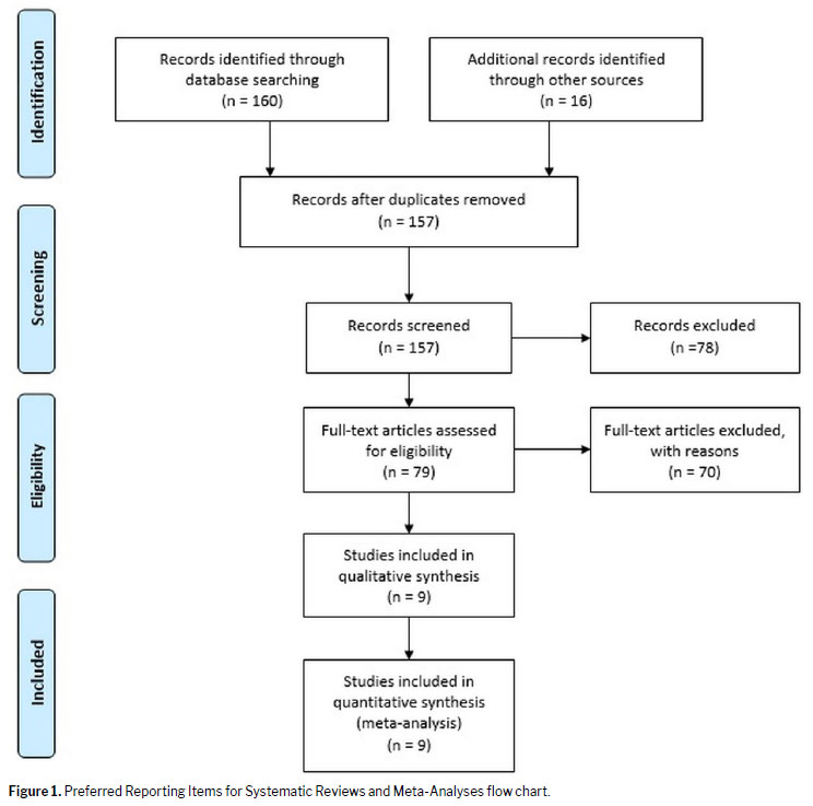
The initial search retrieved 176 records related to the topic in question (Figure 1). After titles, abstracts, and full texts had been screened, nine studies met all of the inclusion criteria and were included in the final analysis. Three studies, despite having compared electromyography and ultrasound, were excluded: a study conducted by Regensburger et al.
(20) was excluded because ultrasound and electromyography were performed simultaneously by the same operator, thus precluding adequate blinding between methods and introducing potential observer bias; Botter et al.
(21) was excluded because it was a technical study conducted exclusively in healthy volunteers rather than ALS patients; and a study conducted by Bibbings et al.
(22), developed by the Sheffield group, was excluded because it compared ultrasound and electromyography using an automated video-analysis system instead of a human observer, thus allowing assessment of temporal effects and methodological innovations not previously addressed. Compared with our previous systematic review
(13), this update included two additional eligible studies—both extending the ultrasound observation time to 60 s
(14,15).
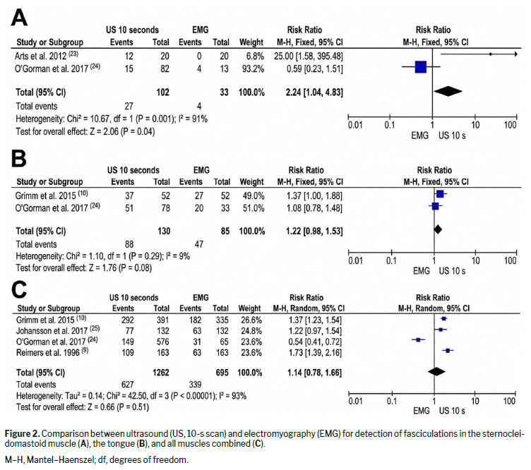
Evaluation with electromyography and 10-s ultrasound, by muscleTwo studies—Arts et al.
(23) and O’Gorman et al.
(24)—evaluated sternocleidomastoid muscles (N = 135, collectively) and reported exact numbers of the muscles testing positive for fasciculations. In those two studies (collectively), fasciculations were detected in four (12.1%) of 33 muscles evaluated by electromyography and in 27 (26.4%) of 102 muscles evaluated for 10 s on ultrasound (Figure 2A). The tongue was also evaluated in two studies—Grimm et al.
(10) and O’Gorman et al.
(24)—collectively comprising 215 muscles, also evaluated. In those two studies, fasciculations were found in 47 (55.4%) of 85 muscles evaluated by electromyography and in 88 (67.7%) of 130 muscles evaluated by 10-s ultrasound (Figure 2B). When all muscles were evaluated together—in Grimm et al.
(10), Johansson et al.
(25), O’Gorman et al.
(24), and Reimers et al.
(9); N = 1,957)—fasciculations were identified in 339 (48.8%) of 695 muscles evaluated by electromyography and in 627 (49.7%) of 1,262 muscles evaluated by 10-s ultrasound, showing comparable detection rates between the two modalities (Figure 2C).
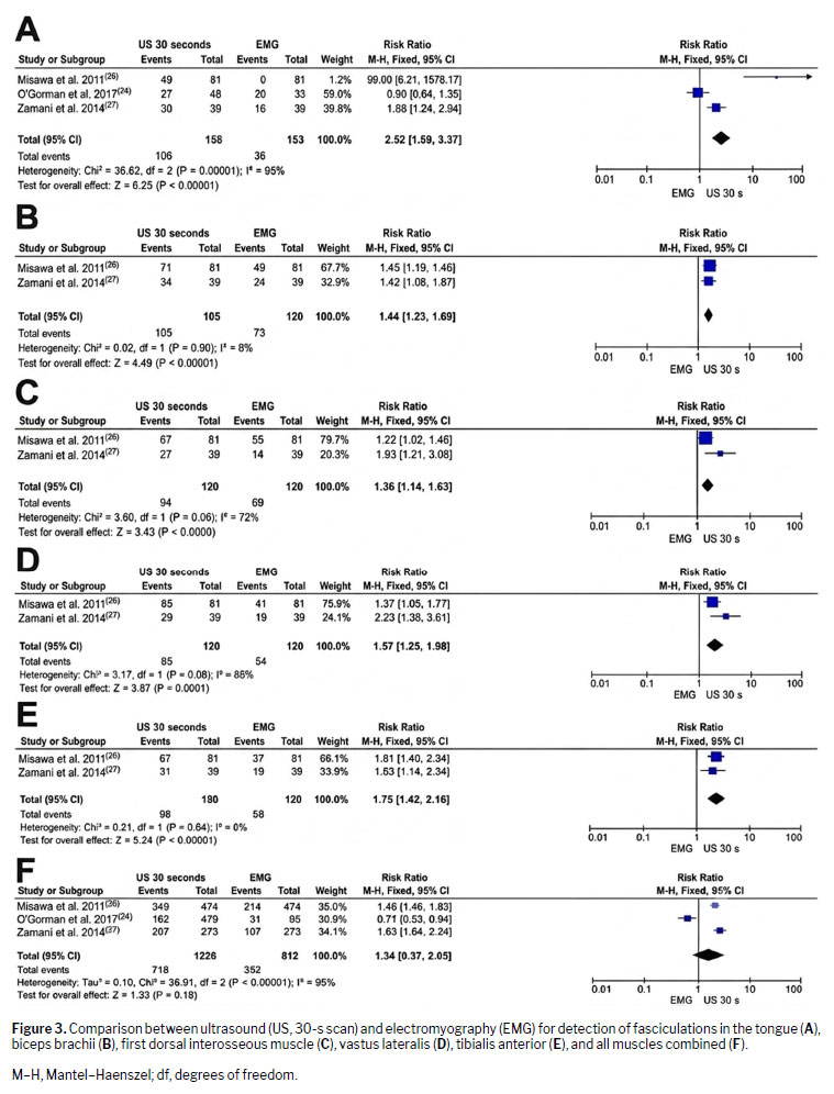
Evaluation with electromyography and 30-s ultrasound, by muscleThree studies—Misawa et al.
(26), Zamani et al.
(27), and O’Gorman et al.
(24)— provided data on muscle-specific comparisons between electromyography and 30-s ultrasound. For the tongue, fasciculations were detected in 36 (11.2%) of 153 muscles evaluated by electromyography and in 106 (63.1%) of 168 muscles evaluated by 30-s ultrasound. Misawa et al.
(26) reported that, in 81 patients, electromyography identified no fasciculations, because of continuous voluntary contraction or respiratory activity (Figure 3A). For the biceps brachii, fasciculations were detected in 73 (60.8%) of 120 muscles evaluated by electromyography and in 105 (87.5%) of 120 muscles evaluated by 30-s ultrasound (Figure 3B). For the first dorsal interosseous, 69 (57.5%) of 120 muscles were positive for fasciculations on electromyography and 94 (78.3%) of 120 muscles were positive for fasciculations on 30-s ultrasound (Figure 3C). For the vastus lateralis, positivity for fasciculation was seen in 54 (45.0%) of 120 muscles on electromyography and in 85 (70.8%) of 120 muscles on 30-s ultrasound (Figure 3D). For the tibialis anterior, 56 (46.7%) of 120 muscles were positive on electromyography and 98 (81.7%) of 120 muscles were positive on 30-s ultrasound (Figure 3E). In the studies that considered all muscles together—Misawa et al.
(26), Zamani et al.
(27), and O’Gorman et al.
(24); N = 2,038—fasciculations were observed in 352 (43.3%) of 812 muscles by electromyography and in 718 (58.6%) of 1,226 muscles by 30-s ultrasound, showing a consistent increase in detection with ultrasound as the observation time doubled (Figure 3F).
Evaluation with electromyography and 60-s ultrasound, by muscleThe most recent studies—Bokuda et al.
(14) and Rajula et al.
(15)—extended the observation time to 60 s per muscle, allowing a detailed evaluation of the temporal influence on detection rates. Both studies included patients with confirmed ALS and compared electromyography and ultrasound under blinded conditions.
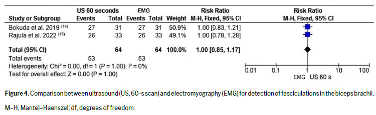
Biceps brachiiThe biceps brachii was the only muscle assessed by both studies
(14,15), totaling 64 muscles (31 and 33, respectively).
Bokuda et al.
(14): fasciculations detected by 60-s ultrasound and by electromyography in 27 (87.1%) of the 31 muscles evaluated.
Rajula et al.
(15): fasciculations detected by 60-s ultrasound and by electromyography in 26 (78.8%) of the 33 muscles evaluated.
The combined results indicate that the detection rate for the two methods is high and equivalent when a full 60-s scan is applied in ultrasound (Figure 4).
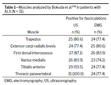
Other musclesIn Bokuda et al.
(14), seven muscles were examined: the trapezius, biceps brachii, extensor carpi radialis brevis, first dorsal interosseous, vastus medialis, tibialis anterior, and thoracic paravertebral muscles. Ultrasound demonstrated higher detection rates than did electromyography in most muscles, particularly in those that are larger or deeper (Table 1).
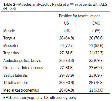
In Rajula et al.
(15), nine muscles were examined by electromyography and by 60-s ultrasound: the tongue, masseter, trapezius, biceps brachii, abductor pollicis brevis, first dorsal interosseous, vastus lateralis, tibialis anterior, and medial gastrocnemius muscles. In that study, 60-s ultrasound showed consistently higher detection rates than did electromyography in all of the muscles evaluated (Table 2). The differences in favor of ultrasound were greatest in the vastus lateralis, tibialis anterior, and gastrocnemius muscles, supporting the hypothesis that muscles that are larger or deeper require longer ultrasound scan durations for optimal detection of fasciculations.
Overall detection performanceIn the two studies that employed electromyography and 60-s ultrasound, a total of 415 muscles were examined: 217 in Bokuda et al.
(14); and 198 in Rajula et al.
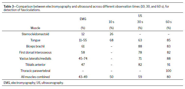
(15). Among those 415 muscles, fasciculations were detected by ultrasound in 332 (80.0%) and by electromyography in 270 (65.0%), translating to an absolute difference of 15 percentage points in favor of ultrasound. These results confirm that increasing ultrasound observation time improves the detection of fasciculations, particularly in muscles that are large or deep, without a significant loss of specificity in comparison with electromyography (Table 3).
DISCUSSIONThis systematic review confirms that the diagnostic performance of ultrasound is superior to that of electromyography for detecting fasciculations in ALS, across all observation times and muscle groups. Previous studies have demonstrated the overall accuracy of ultrasound
(28,29), and the present update underscores those findings through an expanded analysis including 10-, 30-, and 60-s protocols, with stratification by muscle.
It is important to note that the sensitivity of electromyography also increases with longer examination times. Prolonged, careful sampling improves the likelihood of detecting fasciculations, particularly in muscles with low firing frequency, and that could partially reduce the performance gap between electromyography and ultrasonography in optimized protocols.
(30)At 10 s and 30 s, detection rates were comparable in general, yet ultrasound consistently identified a greater number of fasciculations within each muscle.
(31,32) The difference was particularly evident in muscles that are larger or deeper—such as the biceps brachii, vastus lateralis, and tibialis anterior—in which the sensitivity of electromyography tends to decline because of limited needle sampling area and muscle depth.
(33) In contrast, muscles such as the tongue and first dorsal interosseous, which are more superficial and easily relaxed, showed smaller differences between techniques.
Two complementary studies also evaluated 30-s observation protocols using distinct methodological approaches. Botter et al.
(21) conducted a technical validation study in healthy volunteers, demonstrating a strong correlation between surface electromyography signals and fasciculations visually detected on ultrasound. Bibbings et al.
(22) applied automated video processing through a motion-detection algorithm (“foreground detection”), comparing electromyographic findings with those of 30-s ultrasound sequences in ALS patients. Although neither study was included in the main clinical meta-analysis, both underscore the diagnostic accuracy of ultrasound and highlight its technological evolution for the identification of fasciculations.
In studies extending the ultrasound observation time to 60 s, detection rates increased substantially. Bokuda et al.
(14) and Rajula et al.
(15) demonstrated that longer dynamic evaluation improves the identification of fasciculations, particularly in muscles that are large or deep, such as the vastus lateralis, tibialis anterior, and paravertebral muscles. For proximal muscles such as the biceps brachii, which was assessed in both studies, the authors found the performance of ultrasound to be equivalent or superior to that of electromyography, even under blinded conditions and with longer scan durations. These results confirm that extending the scan duration enhances the likelihood of visualizing spontaneous fasciculations that occur intermittently or at low frequency. When all observation times were pooled, ultrasound maintained higher detection rates than electromyography across all muscles, which confirms the robustness and consistency of this finding.
The higher sensitivity of ultrasound in comparison with electromyography can be explained by its ability to monitor a broader muscle volume and detect contractions arising from multiple motor units, whereas intramuscular electromyography registers activity from a limited number of fibers surrounding the needle tip.
(34) In addition, ultrasound has the advantages of being noninvasive, painless, and widely available, while allowing simultaneous assessment of muscle atrophy and architecture.
(14) Furthermore, it has been reported that interobserver agreement for identifying fasciculations on ultrasound is excellent, underscoring the reproducibility of the method.
The morphological characteristics of fasciculations may also provide diagnostic clues. Benign fasciculations tend to be stable, regular, and of lower amplitude, reflecting preserved motor-unit integrity. In ALS, fasciculations are typically unstable, variable in morphology, and irregular in frequency, often accompanied by fibrillation potentials and positive sharp waves on electromyography.
(35-37) Although ultrasound reliably displays the mechanical expression of fasciculations, it cannot differentiate between stable and unstable discharges, because that distinction depends on the characterization of motor-unit potential morphology and electrophysiological aspects.
(26,29) Therefore, although ultrasound is better at detecting fasciculations, electromyography is still essential for distinguishing benign fasciculation syndrome from pathological fasciculations associated with ALS.
(35)Electromyography continues to be the only technique capable of distinguishing between stable and unstable fasciculations through motor-unit potential analysis—a major limitation of ultrasound that can impede disease staging and prognosis. Therefore, the two techniques should be regarded as complementary rather than competitive: electromyography provides detailed electrophysiological characterization, whereas ultrasound offers broader visualization and a dynamic context.
Descriptive synthesis (unpooled analysis)Across the two studies employing 60-s ultrasound protocols
(14,15), the mean detection rates varied by muscle group, ranging from 80% to 95% for 60-s ultrasound and from 65% to 80% for electromyography. The sensitivity gain with ultrasound was particularly pronounced in the vastus lateralis, tibialis anterior, and paravertebral muscles, confirming that a longer observation time improves sensitivity, especially in deep or slow-contracting muscles.
Study limitationsOur study has some limitations, most of which relate to the quality and heterogeneity of the primary studies included. The studies varied in muscle selection, sample size, and observation protocols, which restricted the feasibility of pooled meta-analyses for certain muscle groups. Many investigations were conducted in single-center settings with relatively small ALS cohorts, and in several cases the operators were aware of the clinical diagnosis—factors that might have introduced selection or observer bias. In addition, because the reference standard relied on technical rather than clinical comparisons, the results reflect relative detection performance rather than absolute diagnostic accuracy. Reporting of interobserver agreement was inconsistent, and the absence of standardized protocols for image acquisition and interpretation continues to be a barrier to broader clinical applicability. Despite these study-level constraints, the present review adhered strictly to the principles established in the Cochrane Handbook for Diagnostic Test Accuracy Reviews, which ensured a high standard of methodological rigor. Independent dual data extraction and structured quality appraisal were employed to minimize bias and enhance transparency. This robust, systematic approach supports the reliability of our conclusions and provides the most comprehensive synthesis to date on detection of fasciculations by ultrasound and electromyography in ALS.
Clinical implications and future perspectivesAlthough the aforementioned limitations must be considered, our findings underscore the clinical utility of muscle ultrasound as a complementary diagnostic tool and, in some cases, a tool for earlier diagnosis in ALS. Ultrasound provides a noninvasive approach to screening for fasciculation and is particularly valuable in muscles that are large or deep, in which electromyography has limited sensitivity. Integrating real-time ultrasound into neurophysiological assessment may enhance diagnostic accuracy, reduce the number of electromyography insertions, and improve the patient experience.
Future multicenter studies with standardized muscle selection, observation times, and interpretation criteria are needed in order to establish consensus protocols. Emerging technologies—such as automated motion detection and artificial intelligence-based analysis—may further improve reproducibility and allow objective quantification of fasciculation frequency and morphology over time. Such advances could enable ultrasound not only to complement electromyography but also to serve as a longitudinal biomarker of disease progression and treatment response in ALS.
CONCLUSIONIn several muscle groups and across observation protocols, ultrasound demonstrated higher rates of detection of fasciculations in ALS than did electromyography. That advantage was more evident in muscles that are large or deep—such as the vastus lateralis, tibialis anterior, and paravertebral muscles—particularly when longer observation times were applied, suggesting that extended scanning increases sensitivity without compromising specificity.
By enabling real-time visualization of superficial and deep musculature, ultrasound allows broader sampling of motor units and facilitates the identification of spontaneous muscle activity that may be missed by intramuscular electromyography. However, electromyography continues to be essential for characterizing motor-unit potential morphology and for distinguishing between stable and unstable fasciculations.
Taken together, our findings support the role of ultrasound as a complementary, noninvasive, reproducible tool that can enhance the assessment of LMN dysfunction, improve patient comfort, and potentially reduce the need for extensive needle sampling. The combined use of ultrasound and electromyography may therefore provide a more comprehensive evaluation strategy. Multicenter studies with standardized acquisition protocols and the incorporation of automated image-analysis techniques are warranted to further define the role of ultrasound as a quantitative biomarker for monitoring disease progression and treatment response in ALS.
REFERENCES1. van Baalen A, Stephani U. Fibration, fibrillation, and fasciculation: say what you see. Clin Neurophysiol. 2007 Jun;118(6):1418-20.
2. Carvalho M, Swash M. Fasciculation discharge frequency in amyotrophic lateral sclerosis and related disorders. Clin Neurophysiol. 2016 May;127(5):2257-62.
3. Joyce NC, Carter GT. Electrodiagnosis in persons with amyotrophic lateral sclerosis. PM R. 2013 May;5(5 Suppl):S89-95.
4. Brooks BR. El Escorial World Federation of Neurology criteria for the diagnosis of amyotrophic lateral sclerosis. J Neurol Sci. 1994 Jul;124 Suppl:96-107.
5. Brooks BR, Miller RG, Swash M, et al. El Escorial revisited: revised criteria for the diagnosis of amyotrophic lateral sclerosis. Amyotroph Lateral Scler Other Motor Neuron Disord. 2000 Dec;1(5):293-9.
6. de Carvalho M, Dengler R, Eisen A, et al. Electrodiagnostic criteria for diagnosis of ALS. Clin Neurophysiol. 2008 Mar;119(3):497-503.
7. Sonoo M, Kuwabara S, Shimizu T, et al. Utility of trapezius EMG for diagnosis of amyotrophic lateral sclerosis. Muscle Nerve. 2009 Jan;39(1):63-70.
8. Takamatsu N, Nodera H, Mori A, et al. Which muscle shows fasciculations by ultrasound in patients with ALS? J Med Invest. 2016;63(1-2):49-53.
9. Reimers CD, Ziemann U, Scheel A, et al. Fasciculations: clinical, electromyographic, and ultrasonographic assessment. J Neurol. 1996 Aug;243(8):579-84.
10. Grimm A, Prell T, Décard BF, et al. Muscle ultrasonography as an additional diagnostic tool for the diagnosis of amyotrophic lateral sclerosis. Clin Neurophysiol. 2015 Apr;126(4):820-7.
11. Walker FO, Donofrio PD, Harpold GJ, et al. Sonographic imaging of muscle contraction and fasciculations: a correlation with electromyography. Muscle Nerve. 1990 Jan;13(1):33-9.
12. Cartwright MS. Neuromuscular ultrasound for the evaluation of amyotrophic lateral sclerosis [Msc thesis]. Winston-Salem, NC: Wake Forest University; 2012.
13. Duarte ML, Iared W, Oliveira ASB, et al. Ultrasound versus electromyography for the detection of fasciculation in amyotrophic lateral sclerosis: systematic review and meta-analysis. Radiol Bras. 2020 Mar-Apr;53(2):116-121.
14. Bokuda K, Shimizu T, Kimura H, et al. Relationship between EMG-detected and ultrasound-detected fasciculations in amyotrophic lateral sclerosis: A prospective cohort study. Clin Neurophysiol. 2020 Jan;131(1):259-264.
15. Rajula RR, Saini J, Unnikrishnan G, et al. Muscle ultrasonography in detecting fasciculations: A noninvasive diagnostic tool for amyotrophic lateral sclerosis. J Clin Ultrasound. 2022 Feb;50(2):286-291.
16. Cochrane Diagnostic Test Accuracy Working Group. Cochrane Handbook for Systematic Reviews of Diagnostic Test Accuracy Version 5.1. The Cochrane Collaboration; 2013.
17. Ouzzani M, Hammady H, Fedorowicz Z, et al. Rayyan-a web and mobile app for systematic reviews. Syst Rev. 2016 Dec 5;5(1):210.
18. Margulis AV, Pladevall M, Riera-Guardia N, et al. Quality assessment of observational studies in a drug-safety systematic review, comparison of two tools: the Newcastle-Ottawa Scale and the RTI item bank. Clin Epidemiol. 2014 Oct 10;6:359-68.
19. Viswanathan M, Berkman ND. Development of the RTI item bank on risk of bias and precision of observational studies. J Clin Epidemiol. 2012 Feb;65(2):163-78.
20. Regensburger M, Tenner F, Möbius C, et al. Detection radius of EMG for fasciculations: Empiric study combining ultrasonography and electromyography. Clin Neurophysiol. 2018;129(2):487-93.
21. Botter A, Carbonaro M, Vieira TM, et al. Identification of muscle fasciculations from surface EMG: comparison with ultrasound-based detection. Annu Int Conf IEEE Eng Med Biol Soc. 2019 Jul;2019:5117-5120.
22. Bibbings K, Harding PJ, Loram ID, et al. Foreground Detection Analysis of Ultrasound Image Sequences Identifies Markers of Motor Neurone Disease across Diagnostically Relevant Skeletal Muscles. Ultrasound Med Biol. 2019 May;45(5):1164-1175.
23. Arts IM, Overeem S, Pillen S, et al. Muscle ultrasonography: a diagnostic tool for amyotrophic lateral sclerosis. Clin Neurophysiol. 2012 Aug;123(8):1662-7.
24. O’Gorman CM, Weikamp JG, Baria M, et al. Detecting fasciculations in cranial nerve innervated muscles with ultrasound in amyotrophic lateral sclerosis. Muscle Nerve. 2017 Dec;56(6):1072-1076.
25. Johansson MT, Ellegaard HR, Tankisi H, et al. Fasciculations in nerve and muscle disorders – A prospective study of muscle ultrasound compared to electromyography. Clin Neurophysiol. 2017 Nov;128(11):2250-2257.
26. Misawa S, Noto Y, Shibuya K, et al. Ultrasonographic detection of fasciculations markedly increases diagnostic sensitivity of ALS. Neurology. 2011 Oct 18;77(16):1532-7.
27. Zamani B, Rohani M, Emami K, et al. The accuracy of direct ultrasound study of the muscles in diagnosis of ALS comparing to electromyography in patients referred to Rasoul Akram hospital during 2012-2013. Cerebrovasc Dis. 2014;37(Suppl 2):55. [Persian]
28. Noto YI, Shibuya K, Shahrizaila N, et al. Detection of fasciculations in amyotrophic lateral sclerosis: The optimal ultrasound scan time. Muscle Nerve. 2017 Dec;56(6):1068-1071.
29. Pillen S, Van Alfen N. Muscle ultrasound from diagnostic tool to outcome measure-Quantification is the challenge. Muscle Nerve. 2015 Sep;52(3):319-20.
30. Mills KR. Detecting fasciculations in amyotrophic lateral sclerosis: duration of observation required. J Neurol Neurosurg Psychiatry. 2011 May;82(5):549-51.
31. Harding PJ, Loram ID, Combes N, et al. Ultrasound-Based Detection of Fasciculations in Healthy and Diseased Muscles. IEEE Trans Biomed Eng. 2016 Mar;63(3):512-8.
32. Simon NG. Dynamic muscle ultrasound–Another extension of the clinical examination. Clin Neurophysiol. 2015;126(8):1466-7.
33. Kleine BU, Boekestein WA, Arts IM, et al. Fasciculations and their F-response revisited: high-density surface EMG in ALS and benign fasciculations. Clin Neurophysiol. 2012 Feb;123(2):399-405.
34. Wenzel S, Herrendorf G, Scheel A, et al. Surface EMG and myosonography in the detection of fasciculations: a comparative study. J Neuroimaging. 1998 Jul;8(3):148-54.
35. Katzberg HD, Bril V, Breiner A. Ultrasound in Neuromuscular Disorders. J Clin Neurophysiol. 2016 Apr;33(2):80-5.
36. Oliveira AS, Pereira RD. Amyotrophic lateral sclerosis (ALS): three letters that change the people’s life. For ever. Arq Neuropsiquiatr. 2009 Sep;67(3A):750-82.
37. Buainain R, Moura LS, Oliveira A. Fasciculação. Rev Neurocienc. 2000;8:31-4.
1. Department of Radiology, Universidade de Ribeirão Preto – Campus Guarujá, Guarujá, SP, Brazil
2. Department of Radiology, Diagnósticos da América S/A, São Paulo, SP, Brazil
3. Instituto de Ensino e Pesquisa DASA (IEPD), São Paulo, SP, Brazil
4. Department of Ultrasonography, Diagnósticos da América S/A, São Paulo, SP, Brazil
5. Department of Neurology, Universidade Federal de São Paulo/Escola Paulista de Medicina (UNIFESP/EPM), São Paulo, SP, Brazil
6. Department of Internal Medicine, Universidade de Ribeirão Preto – Campus Guarujá, Guarujá, SP, Brazil
7. Department of Human Movement Sciences and Postgraduate Program in Evidence-Based Health, Universidade Federal de São Paulo (UNIFESP), São Paulo, SP, Brazil
a.
https://orcid.org/0000-0002-7874-9332b.
https://orcid.org/0000-0002-6426-5636c.
https://orcid.org/0000-0002-6986-4937d.
https://orcid.org/0000-0001-7897-1198e.
https://orcid.org/0000-0003-0329-4588Correspondence:Márcio Luís Duarte
Radiologist. Universidade de Ribeirão Preto (UNAERP) – Campus Guarujá, Av. D. Pedro I, 3.300, Enseada, Guarujá, SP, Brazil, 11440-003.
Email:
marcioluisduarte@gmail.com Telephone: +55 (13) 3398-1000
Editor in charge: Dr. Valdair Francisco Muglia
Received in
January 27 2026.
Accepted em
June 1 2026.
Publish in
September 22 2026.

 |
|







 PDF English
 Print
Print
 Send this article by email
 How to cite this article
 Submit a comment
 Mendeley
 Pocket
 Share on Facebook
 Share on Twitter