ABSTRACT
Takotsubo cardiomyopathy is an important differential diagnosis for acute chest pain. Imaging tests, such as ventriculography, echocardiography, computed tomography of the heart, and cardiac magnetic resonance, are valuable tools for diagnostic confirmation in this context. This study reviews the literature and exemplifies the spectrum of typical and atypical cardiac magnetic resonance findings in this disease, on the basis of the experience of our facility. Recognition of these characteristics underscores the roles that radiologists and cardiologists play in the care of patients with acute chest pain, enabling an accurate diagnosis and appropriate treatment.
Keywords:
Chest pain; Takotsubo cardiomyopathy; Cardiac magnetic resonance.
RESUMO
A cardiomiopatia de takotsubo é um diagnóstico diferencial importante de dor torácica aguda. Exames de imagem, seja por ventriculografia, ecocardiograma, tomografia computadorizada do coração ou ressonância magnética cardíaca, são ferramentas valiosas para a confirmação diagnóstica nesse contexto. O presente estudo revisa a literatura e exemplifica o espectro dos achados típicos e atípicos dessa doença observados na ressonância magnética cardíaca, com base na experiência do nosso serviço. O reconhecimento dessas características reforça o papel do médico radiologista/cardiologista na linha de cuidado de pacientes com dor torácica aguda, possibilitando o diagnóstico e tratamento adequado.
Palavras-chave:
Dor torácica; Cardiomiopatia de takotsubo; Ressonância magnética cardíaca.
INTRODUCTION
Takotsubo cardiomyopathy, also known as broken-heart syndrome, stress-induced cardiomyopathy, and apical ballooning syndrome, was first described by Sano et al., in 1990(1). Although its exact incidence is unknown, it has been observed in 1–2% of patients with suspected acute coronary syndrome, mainly affecting postmenopausal women(1).
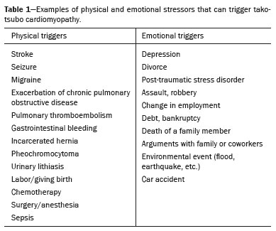
The clinical presentation of takotsubo cardiomyopathy resembles that of acute coronary syndrome, including precordial pain, dyspnea, electrocardiographic changes, and elevated troponin. One peculiar characteristic is that up to two thirds of patients can identify a physically or emotionally stressful event (Table 1) that occurred prior to the onset of symptoms(2,3).

Although the pathophysiology of takotsubo cardiomyopathy is not completely understood, it is believed to be related to excessive catecholamine discharge, secondary to the stressful event, associated with differences in the density and distribution of adrenergic receptors in the myocardium. Reduced estrogen levels also appear to be involved
(1,3).
Over the years, various diagnostic criteria have been proposed to help identify takotsubo cardiomyopathy in a patient complaining of acute chest pain. Here, we highlight those proposed by the Mayo Clinic
(4), which include the following:
• Transient hypokinesia, akinesia, or dyskinesia of the middle segments of the left ventricle (LV), with or without apical involvement, with segmental changes in contractility extending beyond a coronary territory, with or without an identified stressor stimulus
• Absence of obstructive coronary artery disease or angiographic evidence of acute plaque rupture
• New electrocardiographic changes (ST-segment elevation, T-wave inversion, or both) or slight elevation of troponin
• No pheochromocytoma or myocarditis.
From the time of diagnosis of takotsubo cardiomyopathy, cardiac magnetic resonance (CMR) has a well-established role in the evaluation of patients with the disease, allowing a detailed anatomical analysis, identification of segmental changes in myocardial contractility, and quantification of ventricular function, as well as the detection of edema and myocardial necrosis/fibrosis. It also allows the detection of complications, such as pericardial effusion, pleural effusion, dynamic obstruction of the LV outflow tract, acute pulmonary edema, intracavitary thrombus, and systemic thromboembolism. Finally, it is useful to exclude other pathologies with clinical presentations similar to that of acute coronary syndrome, such as myocarditis
(1).
Evaluation with CMR is also important for patient follow-up; it is recommended that it be performed three to six months after the acute event. In most cases, the prognosis is good, with normalization of segmental changes in contractility, recovery of the ventricular ejection fraction, and resolution of edema in the follow-up examination
(1).
TYPICAL FINDINGSClassically, takotsubo cardiomyopathy is characterized by segmental changes in contractility—dyskinesia, hypokinesia, or akinesia of the mid-apical segments of the LV, accompanied by hyperkinesia of the basal segments—this combination resulting in the typical morphology
(1,5,6), as illustrated in Figures 1 to 4 and described as similar to a jar used in Japan to capture octopuses, known as a takotsubo.
In addition to contractile changes, CMR allows the detection of myocardial edema, present in at least 80% of patients with takotsubo cardiomyopathy, traditionally on T2-weighted fast spin-echo sequences with triple inversion recovery (Figure 5) and, more recently, on T1 and T2 maps (Figure 6). In most cases, this edema is not accompanied by changes in the image on delayed enhancement sequences (Figure 7), unlike what happens in other conditions, such as acute myocardial infarction and myocarditis
(1,5,6).
ATYPICAL FINDINGSThe terms "atypical takotsubo cardiomyopathy" and "variant of takotsubo cardiomyopathy" are used when dyskinesia, hypokinesia, or akinesia affects nontraditional segments, sparing the cardiac apex and involving basal or mid-ventricular segments (Figures 8 to 11), which occurs in up to 40% of cases
(4,5).
In addition to unusual segmental changes in contractility, a small proportion of patients may also present with delayed myocardial enhancement (Figure 12), which manifests as small, scattered foci with a clearly non-ischemic pattern, often identified only by quantitative analysis with the aid of software and not persisting in follow-up examinations performed a few weeks after the acute event. Although the pathophysiology of that finding is not yet fully understood, it is believed to be related to changes in proteins (collagen type 1) in the myocardium during the acute phase of the disease, and not to myocardial necrosis or fibrosis
(1).
The clinical significance of the variants of takotsubo cardiomyopathy is still an open question. Some clinical differences, such as the involvement of women of a slightly younger age (mean of 62 years) and the association with neurological diseases, have been described. Depression of the ST-segment, lower B-type natriuretic peptide values on admission, and less pronounced changes in the left ventricular ejection fraction are also characteristics that stand out in these cases
(7–9).
CONCLUSIONTakotsubo cardiomyopathy is a diagnosis that should be considered in the context of chest pain in an emergency care setting. The use of CMR allows noninvasive diagnosis, providing information additional to that obtained by echocardiography and enabling the detection of any complications
(10,11).
REFERENCES1. Plácido R, Lopes BC, Almeida AG, et al. The role of cardiovascular magnetic resonance in takotsubo syndrome. J Cardiovasc Magn Reson. 2016;18:68.
2. Boyd B, Solh T. Takotsubo cardiomyopathy: review of broken heart syndrome. JAAPA. 2020;33:24–9.
3. Ghadri JR, Wittstein IS, Prasad A, et al. International expert consensus document on takotsubo syndrome (Part I): clinical characteristics, diagnostic criteria, and pathophysiology. Eur Heart J. 2018;39: 2032–46.
4. Madhavan M, Prasad A. Proposed Mayo Clinic criteria for the diagnosis of Tako-Tsubo cardiomyopathy and long-term prognosis. Herz. 2010;35:240–3.
5. Gunasekara MY, Mezincescu AM, Dawson DK. An update on cardiac magnetic resonance imaging in takotsubo cardiomyopathy. Curr Cardiovasc Imaging Rep. 2020:13:17.
6. Eitel I, von Knobelsdorff-Brenkenhoff F, Bernhardt P, et al. Clinical characteristics and cardiovascular magnetic resonance findings in stress (takotsubo) cardiomyopathy. JAMA. 2011;306:277–86.
7. Ghadri JR, Cammann VL, Napp LC, et al. Differences in the clinical profile and outcomes of typical and atypical takotsubo syndrome: data from the International Takotsubo Registry. JAMA Cardiol. 2016;1:335–40.
8. Yalta K, Yetkin E, Taylan G. Atypical variants of takotsubo cardiomyopathy: mechanistic and clinical implications. J Geriatr Cardiol. 2020;17:447–8.
9. Murthy A, Arora J, Singh A, et al. Takotsubo cardiomyopathy: typical and atypical variants, a two-year retrospective cohort study. Cardiol Res. 2014;5:139–44.
10. Mileva N, Paolisso P, Gallinoro E, et al. Diagnostic and prognostic role of cardiac magnetic resonance in MINOCA: systemic review and meta-analysis. JACC Cardiovasc Imaging. 2023;16:376–89.
11. Daneshrad JA, Ordovas K, Sierra-Galan LM, et al. Role of cardiac magnetic resonance imaging in the evaluation of MINOCA. J Clin Med. 2023;12:2017.
1. Department of Imaging, Hospital Israelita Albert Einstein, São Paulo, SP, Brazil
a.
https://orcid.org/0000-0001-6812-506X b.
https://orcid.org/0000-0003-0337-9469 c.
https://orcid.org/0000-0002-1941-7899 d.
https://orcid.org/0000-0002-5909-5126 e.
https://orcid.org/0000-0002-0233-0041Correspondence: Dra. Camila Vilas Boas Machado
Hospital Israelita Albert Einstein, Departamento de Imagem
Avenida Albert Einstein, 627, Vila Leonor
São Paulo, SP, Brazil, 05652-900
Email:
danieleconcli@yahoo.com.br
Received in
February 14 2025.
Accepted em
April 28 2025.
Publish in
August 5 2025.

 |
|

