Radiologia Brasileira - Publicação Científica Oficial do Colégio Brasileiro de Radiologia
AMB - Associação Médica Brasileira CNA - Comissão Nacional de Acreditação
 Vol. 43 nº 2 - Mar. / Apr. of 2010
|
CASE REPORT
|
|
Spontaneous pneumomediastinum following vocal effort: a case report |
|
|
Autho(rs): Flávia Paiva Lobo Lopes, Edson Marchiori, Gláucia Zanetti, Talita Fonseca Medeiros da Silva, Laura Brasil Herranz, Maria Isabel de Brito Almeida |
|
|
Keywords: Spontaneous pneumomediastinum, Mediastinal emphysema, Computed tomography, Vocal effort, Chest |
|
|
Abstract:
Flávia Paiva Lobo LopesI; Edson MarchioriII; Gláucia ZanettiIII; Talita Fonseca Medeiros da SilvaIV; Laura Brasil HerranzIV; Maria Isabel de Brito AlmeidaIV IPhD, Researcher for Laboratory of Cells and Molecules Labeling, Department of Radiology, Universidade Federal do Rio de Janeiro (UFRJ), Rio de Janeiro, RJ, Brazil
INTRODUCTION Pneumomediastinum is characterized by the presence of air in the mediastinum and may cause chest pain irradiating towards the neck, dyspnea, subcutaneous emphysema and crepitus associated with heart sounds on auscultation. Association with pneumothorax is frequently observed(1,2). By definition, in spontaneous pneumomediastinum there is no evidence of traumatism, iatrogenesis or previous pneumopathies. Considering the uncommon nature of this condition, it eventually may not be diagnosed, with harmful consequences for the patient since the disease may progress with potentially fatal complications(1,3–5). The present article reports the case of spontaneous pneumomediastinum following increased vocal effort during a soccer game in a 14-year-old, male teenager, emphasizing the radiological findings of the disease.
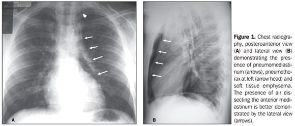
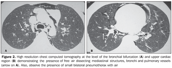
CASE REPORT A male, previously healthy 14-year-old patient, with no history of traumatism, drugs usage, asthma or other respiratory disease, presented with retrosternal chest pain following increased vocal effort during a soccer game. The pain irradiated towards the neck, improving in the knee-chest position, and was associated with hoarseness and dyspnea. At clinical examination, the patient was lucid, oriented, acyanotic, anicteric, with axillary temperature at 36.8°C, arterial pressure, 130 × 80 mmHg, heart rate 72 bpm, respiratory rate 21 irpm, 94% O2 saturation. Cardiovascular system: regular cardiac rhythm with normal heart sounds, normophonetic heart sounds with no pathological jugular turgescence. Respiratory system with no abnormality. Abdomen: flacid, tympanic, peristaltic and nonpainful on palpation. Crepitus on palpation at the anterior chest wall. Electrocardiogram demonstrated sinus rhythm and diffuse, nonspecific alteration of the ventricular repolarization. Chest radiography (Figure 1) demonstrated pneumomediastinum associated with bilateral soft tissue emphysema, larger at left, and small bilateral pneumothorax. Such findings were confirmed at chest computed tomography (CT) (Figure 2). Imaging studies did not demonstrate parenchymal lesions. Esophageal series was performed to evaluate a possible esophageal rupture, demonstrating a normal aspect.
Once the hypothesis of pneumomediastinum secondary to esophageal rupture had been ruled out, the diagnosis of spontaneous mediastinum was reached by exclusion. The patient was referred to the intensive care unit for follow-up, with radiography after 12 and 24 hours. Echocardiogram was inconclusive because of the poor visualization of the cardiac structures as a function of the presence of pneumomediastinum and subcutaneous emphysema. Worsening of the picture was not observed at clinical evaluation within the following 24 hours. Chest CT was performed after 48 hours, demonstrating resorption of more than 50% of the pneumomediastinum, with the patient being discharged 72 hours after admission.
DISCUSSION Spontaneous pneumomediastinum or mediastinal emphysema is a self-limited, benign condition that affects young patients with ages ranging from 17 and 25 years. The incidence rate is extremely low, with the condition being observed in approximately 1/30,000 hospital admissions. The clinical picture may range from asymptomatic to severe or even fatal in some cases. Retrosternal pain predominates among the described symptoms(2,6–9). Main causes of spontaneous pneumomediastinum are the following: intense physical exercises, labor, pulmonary barotrauma, very deep diving, intense paroxysmal cough, emesis, asthma, narcotic inhalation, bronchial asthma and thin biotype. Narcotics use has been reported by some authors as the main cause of spontaneous pneumomediastinum(10). In the present case, the patient did not present any of the previously mentioned factors, except for the vocal effort. Oura et al.(11) have reported two cases of spontaneous pneumomediastinum secondary to vocal effort, where the patients presented with chest and cervical discomfort, with radiological confirmation and favorable clinical progression after Five-day conservative therapy, corroborating the data reported in the present study. Chest radiography still remains as the gold standard in the diagnosis of spontaneous pneumomediastinum. The sensitivity of the posteroanterior and lateral views in spontaneous pneumomediastinum is almost 100%. It is know that the absence of the lateral view may lead to misdiagnosis in approximately half of patients(1,11–13). Radiological findings of spontaneous pneumomediastinum include linear images of gas in the mediastinum, generally extending towards the cervical region, air bubbles or extensive air collections outlining mediastinal blood vessels, gross caliber airways, esophagus or heart. The presence of interstitial emphysema is an aid in the diagnosis of pneumomediastinum. A relevant sign of pneumomediastinum at radiography is the presence of air dissecting both inferiorly and laterally the thymus. The thymus outlining by air is a specific sign of pneumomediastinum and may constitute the primary sign for diagnosis assurance(13). Levin(14) has described the continuous diaphragm sign in pneumomediastinum. Usually, the central portion of the diaphragm is not visible at the images because of its contact with the heart, considering the similar radiological density of such structures. Gas interposition between the diaphragm and the heart is observed in the continuous diaphragm sign, allowing the visualization of the entire diaphragm from one side to the other. In case of clinical suspicion with normal or inconclusive chest radiography, CT may be performed because it allows the anatomical localization of the air on cross sectional sections. Once specific causes of pneumomediastinum are excluded, the patient with spontaneous pneumomediastinum must remain under observation. Resolution occurs in most of cases. In the present case, esophagography was also performed to rule out esophageal rupture, considering that the pain onset was coincidental with food ingestion. Finally, despite the fact that spontaneous mediastinum is a rare, self-limited condition with a benign prognosis, it should be considered in the differential diagnosis of sudden chest pain. For such a purpose, the mentioned radiological parameters must be present on chest radiographs. Additionally both posteroanterior and lateral views are essential in the differential diagnosis of pneumomediastinum, pneumopericardium and pneumothorax. In dubious cases, CT constitutes an extremely valuable diagnostic tool.
REFERENCES 1. Ba-Ssalamah A, Schima W, Umek W, et al. Spontaneous pneumomediastinum. Eur Radiol. 1999; 9:724–7. [ ] 2. Dekel B, Paret G, Szeinberg A, et al. Spontaneous pneumomediastinum in children: clinical and natural history. Eur J Pediatr. 1996;155:695–7. [ ] 3. Chapdelaine J, Beaunoyer M, Daigneault P, et al. Spontaneous pneumomediastinum: are we overinvestigating? J Pediatr Surg. 2004;39:681–4. [ ] 4. James M, Miguel M, Fancher T. Spontaneous pneumomediastinum. J Hosp Med. 2007;2:283–4. [ ] 5. Langwieler TE, Steffani KD, Bogoevski DP, et al. Spontaneous pneumomediastinum. Ann Thorac Surg. 2004;78:711–3. [ ] 6. Gerazounis M, Athanassiadi K, Kalantzi N, et al. Spontaneous pneumomediastinum: a rare benign entity. J Thorac Cardiovasc Surg. 2003;126:774–6. [ ] 7. Yellin A, Gapany-Gapanavicius M, Lieberman Y. Spontaneous pneumomediastinum: is it a rare cause of chest pain? Thorax. 1983;38:383–5. [ ] 8. Nounla J, Tröbs R, Bennek J, et al. Idiopathic spontaneous pneumomediastinum: an uncommon emergency in children. J Pediatric Surg. 2004;39:E23–4. [ ] 9. Shen G, Chai Y. Spontaneous pneumomediastinum in adolescents. Chin Med J (Engl). 2007;120:2329–30. [ ] 10. López Penza P, Odriozola M, Ruso L. Neumomediastino espontáneo asociado al consumo de drogas inhalantes. Rev Med Urug. 2007;23:378–82. [ ] 11. Oura H, Aikawa H, Ishiki M. Simultaneous spontaneous pneumomediastinum caused by vocal exercise: report of 2 cases. Kyobu Geka. 2004;57:1149–52. [ ] 12. Kumar VM, Grant CA, Hughes MW, et al. Role of routine chest radiography after percutaneous dilatational tracheostomy. Br J Anaesth. 2008;100:663–6. [ ] 13. Bejvan SM, Godwin JD. Pneumomediastinum: old signs and new signs. AJR Am J Roentgenol. 1996;166:1041–8. [ ] 14. Levin B. The continuous diafragm sign. A newly-recognized sign of pneumomediastinum. Clin Radiol. 1973;24:337–8. [ ] Received June 4, 2008. * Study developed at Universidade Federal do Rio de Janeiro (UFRJ), Rio de Janeiro, RJ, Brazil. |
|
Av. Paulista, 37 - 7° andar - Conj. 71 - CEP 01311-902 - São Paulo - SP - Brazil - Phone: (11) 3372-4544 - Fax: (11) 3372-4554


