Radiologia Brasileira - Publicação Científica Oficial do Colégio Brasileiro de Radiologia
AMB - Associação Médica Brasileira CNA - Comissão Nacional de Acreditação
 Vol. 40 nº 1 - Jan. /Feb. of 2007
|
ORIGINAL ARTICLE
|
|
Comparison between computed tomography and mediastinoscopy in the assessment of mediastinal nodal involvement in non-small cell bronchial carcinoma |
|
|
Autho(rs): Décio Valente Renck, Daniel Brito de Araújo, Nilton Haertel Gomes, Rodrigo Mendonça |
|
|
Keywords: Lung cancer, Computed tomography, Mediastinoscopy |
|
|
Abstract:
IMaster Degree in Sciences of Health by Universidade Católica de Pelotas, Radiologist at School-Hospital of Fundação de Apoio ao Universitário da Universidade Federal de Pelotas and at Service of Radiology of Santa Casa de Misericórdia de Pelotas
INTRODUCTION Since the second decade of the twenty century, the incidenceof lung cancer has been alarmingly increasing in all over theworld, the surgical resection being the most effective treatmentfor non-small cell lung carcinoma. The clinical stage of thetumor as well as functional condition of the patient, determinethe feasibility and applicability of surgical resection as aprimary method of treatment. The patients who are candidates tosurgery are those with tumors in stages I or II, and a smallnumber with tumors in stage III. Only in exceptional situations,patients with tumors in stages IIIB or IV may be considered aspotentially surgical. In 1959, Carlens(1) described themediastinoscopy procedure. This technique has been rapidlydiffused, representing a significant progress in the lung cancermanagement, since, for the first time, it provided an accuratepresurgical evaluation of the existence and extent of mediastinalmetastatic disease. The mediastinoscopy has established apractical way to determine the "N" of the TNM classification, andstill remains as the golden-standard in the diagnosis ofmediastinal involvement. Early in the nineties, the development of the thoracoscopy hastriggered a series of procedures for evaluation of primaryneoplastic lesions and mediastinal nodes. Besides beingsatisfactory for evaluation of mediastinal nodes, this techniqueallows the performance of maneuvers for surgical dissection inthe suspect of contact, compression or invasion of hilar ormediastinal structures by the tumor. Computed tomography (CT) and magnetic resonance imaging (MRI) have shown 60% accuracy in relation to the nature of intrathoracic lesions. Therefore, depending on the involvement degree, the patient should not be deprived of a surgical treatment only with basis on such imaging findings. Imaging studies, however, might guide a more limited surgical exploration to prevent that patients with unresectable diseases are submitted to more extensive surgeries.
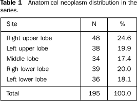
MATERIALS AND METHODS In the period between April/2001 and December/2002, all the patients diagnosed with lung cancer in our service were prospectively studied. Of the initial 412 patients, 195 were included in the present study, with basis on the following inclusion criteria: chest CT performed previously to invasive procedures for staging and/or definite surgical treatment; confirmed histological diagnosis of bronchial carcinoma; performance of mediastinoscopy, and absence of previous oncologic therapy. Of these 195 patients, 124 (63.6%) were men, and 71 (36.4%) were women. Ages ranged between 36 and 96 years (mean = 64.2, median = 67 years). The histological types of tumors identified in the present series were the following: 112 (57.4%) patients with squamous carcinoma, 59 (30.3%) with adenocarcinoma and subtypes, and 24 (12.3%) patients with undifferentiated large cell carcinoma; cases of small cell bronchial carcinoma were excluded. The most frequent site of lesions was the right lung in 121 (62.0%) cases, and 74 (38.0%) cases in the left lung, the anatomical distribution being shown on Table 1.
CT scans were performed in a Siemens Somatom AR.SP equipment,with the patient in dorsal decubitus, during maximum inspiration.For the mediastinum evaluation, there was a soft tissue filter;and during pulmonary parenchyma evaluation, a bone filter wasemployed. Scan time was 1.7 second. Wide, medium and narrowfields of view were utilized. Slices thickness for pulmonary andmediastinal tissues were, respectively, 10 mm and 5 mm or 10 mm,with helical technique and 1.5 pitch. Intravenous contrast agentHypaque 50% (Sanofi Winthrop Farmacêutica; Brazil) wasemployed; except for patients with contraindications. Mediastinal nodes classification followed the AmericanThoracic Society, 1983 criteria. The definition of thelocalization of lymph nodes visualized on CT and mediastinoscopywas based on this classification. The mediastinoscope utilized was a Storz 10970 model, 17 cm inlength (Karl Storz Endoscopy; Germany) coupled with a 250 Whalogen light source. The patients underwent cervicalmediastinoscopy as recommended by Carlens(1)under general anesthesia. The patients with lesion in the left upper lobe and negativeresults for neoplasm in mediastinal node freezing biopsies weresubmitted to paraesternal mediastinotomy — with theChamberlain(2) technique. The main objective ofthis procedure was the histological study of the region in theaortopulmonary window (region 5). The ganglions were separated according to the nodal region, aspreviously described. The frozen biopsied specimens of nodalregions were included in paraffin blocks allowing histology. The findings of chest CT and mediastinoscopy were comparedwith the anatomopathological results of nodal biopsy withmediastinoscopy as the golden standard. The normality criterion as to the mediastinal nodes size wasbased on the study developed by Glazer etal.(3), where chest CT studies definedganglions with > 1.0 cm in diameter in their shortest axis asmetastatic. All the tomographic evaluations were considered as "positive"or "negative", according to criteria as to the presence ofneoplasm in mediastinal nodes and compared with the goldenstandard provided by anatomopathological studies of these samenodes biopsied by means of mediastinoscopy. So, the measurements of diagnostic performance (sensitivity,specificity, positive and negative predictive values with theirrespective confidence intervals) were calculates for chestCT.
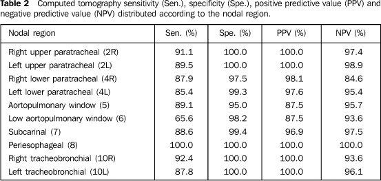
RESULTS Results concerning mediastinal nodal involvement were the following: 66 (33.9%) patients with N1, 90 (46.1%) patients with N2, and 39 (20%) patients with N3. Sensitivity, specificity, positive and negative predictive values for detection of nodal metastasis by CT were calculated (Table 2).
Nodal regions presenting lower sensitivity values were: lower aortopulmonary window, 5 (65.6%), left peribronchial, 10L (87.8%), and upper aortopulmonary window, 5 (89.1%). Best results were found in the following regions: left upper paratracheal 2L (89.5%), right peribronchial 10R (92.4%) and right upper paratracheal 2R (91.1%) (Table 2). Staples et al.(4) have found similar resultsin their study, also evaluating CT accuracy as per regions. Theregion of the aortopulmonary window is problematic because of thevascular confluence interfering in the evaluation. This situationis particularly significant in cases of tumor at left, wherefrequently the upper portion of the pulmonary artery might beconfused with a ganglion. Also, the transverse pericardial sinusmight be erroneously confused with a lymph node. The regionsalong the main bronchi, right tracheobronchial, and leftperibronchial present vascular confluence which, for a correcttomographic evaluation, must be correctly contrast enhanced, andis investigates with thin slices, to avoid that CT results areaffected. The upper right paratracheal region is surrounded bylarge vessel involved in fat. The misinterpretation of the nodedensity might confuse the findings in this site.
DISCUSSION Determining the mediastinal nodes involvement is essential fordefinition of the therapy and prognosis. Although there is agreat number of studies on the chest CT appropriateness fordetermining mediastinal involvement in patients with non-smallcell bronchial carcinoma, there is not a precise rule to beapplied in these cases, and this investigation remainscontroversial. One of the greatest problems in staging tumors with chest CTis the poor standardization of nodes in certain anatomicalregions. Many studies suggest the normality criterion formediastinal lymph nodes is 1.0 cm in diameter in its longestaxis; with high sensitivity (79% to 91%)(4–11).Although this diameter has high sensitivity, its specificity islow. Considering up to 0.5 cm in diameter in its longest axis,sensitivity achieves 95%, but specificity may decrease to 69%,with an increase in the number of false-positive results,reinforcing the necessity of mediastinoscopy. Even so, 5%–10% ofmediastinal nodal metastases are not detected by chestCT(4). Glazer et al.(12) havedetermined that the node diameter in the shortest axis is moreaccurate than the node diameter in the longest axis, since thelatest is highly dependent on the spatial orientation of lymphnodes. Generally, the greater the size of the node to beconsidered as involved, the greater the loss of sensitivity ofthe method. Our results related to mediastinal involvement, reinforce thecurrent consensus that all the patients with abnormal mediastinalfindings on chest CT should undergo an invasive investigation oflymph nodes. Two review series(3,13) on thismatter show that one third of patients with enlarged mediastinalnodes detected on CT, do not present tumor dissemination.Additionally, a recent metanalysis(14) hasdemonstrated that 29% of chest CTs presenting enlarged nodescorresponded to false-positive results. Therefore, enlargedmediastinal nodes detected on tomographic studies are an absoluteindication for mediastinoscopy. Cases where CT does not demonstrate mediastinal involvementremain controversial. Many studies(4,6,13,15)have reported high negative predictive values in thedetermination of mediastinal metastases, suggesting the anegative chest CT could avoid a mediastinoscopy and that thesepatients should directly undergo thoracotomy, considering that asmall number of unnecessary thoracotomies would be balanced by agreat number of avoided mediastinoscopies. In contrast, Pearson(16) recommends thatmediastinoscopy must be performed in every T2 and T3 tumors andin T1 tumors with diagnosis of adenocarcinoma and large cellcarcinoma, even with negative findings on chest CT. It is our understanding that the main objective of thepresurgical staging is to avoid an unnecessary thoracotomy.Considering this procedure inherent morbimortality, a highsensitivity is essential in the presurgical investigation. Interms of CT, this does not seem to be the reality yet. Additionally, the identification of patients with stage IIIAdisease before pulmonary resection has gained significance. Inthe past, patients diagnosed with stage N2 by mediastinoscopy,many times used to be submitted to non-surgical therapeuticmodalities. Although the surgery was technically feasible,several series(17–26) have shown a very poorsurvival in patients for whom pulmonary resection was themodality of choice. However, new studies suggest that neoadjuvantchemotherapy improves the resectability and results, downgradingthe pathological stage IIIA, and changing the mediastinoscopyobjective. Instead of just diagnosing the unresectablemediastinal disease, presently, the mediastinoscopy defines thepatients with minimal N2 disease who might be included inprotocols with chemotherapy as a neoadjuvant treatment, with orwithout radiotherapy, and, later, surgical resection. In summary, based on our findings, and supported by thecurrent literature, we believe that it is not possible yet toutilize chest CT as a mean to avoid unnecessarymediastinoscopies, however, chest CT remains as an important toolfor mediastinal mapping, selecting and guiding the surgicalintervention which will yield the definite anatomopathologicaldiagnosis.
REFERENCES 1. Carlens E. Mediastinoscopy: a method for inspection and tissue biopsy in the superior mediastinum. Dis Chest 1959;36:343–352. [ ] 2. McNeill TM, Chamberlain JM. Diagnostic anterior mediastinotomy. Ann Thorac Surg 1966;2: 532–539. [ ] 3. Glazer GM, Orringer MB, Gross BH, Quint LE. The mediastinum in non-small cell lung cancer: CT-surgical correlation. AJR Am J Roentgenol 1984;142:1101–1105. [ ] 4. Staples CA, Müller NL, Miller RR, Evans KG, Nelems B. Mediastinal nodes in bronchogenic carcinoma: comparison between CT and mediastinoscopy. Radiology 1988;167:367–372. [ ] 5. Baron RL, Levitt RG, Sagel SS, White MJ, Roper CL, Marbarger JP. Computed tomography in the preoperative evaluation of bronchogenic carcinoma. Radiology 1982;145:727–732. [ ] 6. Daly BDT Jr, Faling LJ, Bite PACG, et al. Mediastinal lymph node evaluation by computed tomography in lung cancer. An analysis of 345 patients grouped by TNM staging, tumor size, and tumor location. J Thorac Cardiovasc Surg 1987; 94:664–672. [ ] 7. Görich J, Beyer-Enke SA, Flentje M, Zuna I, Vogt-Moykopf I, Van Kaick G. Evaluation of recurrent bronchogenic carcinoma by computed tomography. Clin Imaging 1990;14:131–137. [ ] 8. Kaplan DK. Mediastinal lymph node metastases in lung cancer: is size a valid criterion? Thorax 1992;47:332–333. [ ] 9. Lewis JW Jr, Pearlberg JL, Beute GH, et al. Can computed tomography of the chest stage lung cancer? Yes and no. Ann Thorac Surg 1990;49: 591–596. [ ] 10. Martini N, Heelan R, Westcott J, et al. Comparative merits of conventional, computed tomographic, and magnetic resonance imaging in assessing mediastinal involvement in surgically confirmed lung carcinoma. J Thorac Cardiovasc Surg 1985;90:639–648. [ ] 11. Patterson GA, Ginsberg RJ, Poon PY, et al. A prospective evaluation of magnetic resonance imaging, computed tomography, and mediastinoscopy in the preoperative assessment of mediastinal node status in bronchogenic carcinoma. J Thorac Cardiovasc Surg 1987;94:679–684. [ ] 12. Glazer GM, Gross BH, Quint LE, Francis IR, Bookstein FL, Orringer MB. Normal mediastinal lymph nodes: number and size according to American Thoracic Society mapping. AJR Am J Roentgenol 1985;144:261–265. [ ] 13. Bollen ECM, Goei R, van't Hof-Grootenboer BE, Versteege CWM, Engelshove HA, Lamers RJ. Interobserver variability and accuracy of computed tomographic assessment of nodal status in lung cancer. Ann Thorac Surg 1994;58:158–162. [ ] 14. Suzuki K, Nagai K, Yoshida J, Nishimura M, Takahashi K, Nishiwaki Y. Clinical predictors of N2 disease in the setting of a negative computed tomographic scan in patients with lung cancer. J Thorac Cardiovasc Surg 1999;117:593–598. [ ] 15. Glazer GM, Orringer MB, Chenevert TL, et al. Mediastinal lymph nodes: relaxation time/pathologic correlation and implications in staging of lung cancer with MR imaging. Radiology 1988; 168:429–431. [ ] 16. Pearson FG. Staging of the mediastinum. Role of mediastinoscopy and computed tomography. Chest 1993;103(4 Suppl):346S–348S. [ ] 17. Choi NC, Carey RW, Daly W, et al. Potential impact on survival of improved tumor downstaging and resection rate by preoperative twice-daily radiation and concurrent chemotherapy in stage IIIA non-small-cell lung cancer. J Clin Oncol 1997;15:712–722. [ ] 18. Dillman RO, Herndon J, Seagren SL, Eaton WL Jr, Green MR. Improved survival in stage III non-small-cell lung cancer: seven-year follow-up of cancer and leukemia group B (CALGB) 8433 trial. J Natl Cancer Inst 1996;88:1210–1215. [ ] 19. Feld R, Rubinstein L, Thomas PA. Adjuvant chemotherapy with cyclophosphamide, doxorubicin, and cisplatin in patients with completely resected stage I non-small-cell lung cancer. The Lung Cancer Study Group. J Natl Cancer Inst 1993;85: 299–306. [ ] 20. Figlin RA, Piantodosi S. A phase 3 randomized trial of immediate combination chemotherapy vs delayed combination chemotherapy in patients with completely resected stage II and III non-small cell carcinoma of the lung. Chest 1994; 106(6 Suppl):310S–312S. [ ] 21. Krasna MJ, Reed CE, Nugent WC, et al. Lung cancer staging and treatment in multidisciplinary trials: cancer and leukemia group B cooperative group approach. Thoracic surgeons of CALGB. Ann Thorac Surg 1999;68:201–207. [ ] 22. Le Chevalier T, Arriagada R, Quoix E, et al. Radiotherapy alone versus combined chemotherapy and radiotherapy in nonresectable non-small-cell lung cancer: first analysis of a randomized trial in 353 patients. J Natl Cancer Inst 1991;83:417–423. [ ] 23. Mandell L, Hilaris B, Sullivan M, et al. The treatment of single brain metastasis from non-oat cell lung carcinoma. Surgery and radiation versus radiation therapy alone. Cancer 1986;58:641–649. [ ] 24. Ratto GB, Zino P, Mirabelli S, et al. A randomized trial of adoptive immunotherapy with tumor-infiltrating lymphocytes and interleukin-2 versus standard therapy in the postoperative treatment of resected nonsmall cell lung carcinoma. Cancer 1996;78:244–251. [ ] 25. Rosenthal SA, Curran WJ Jr, Herbert SH, et al. Clinical stage II non-small cell lung cancer treated with radiation therapy alone. The significance of clinically staged ipsilateral hilar adenopathy (N1 disease). Cancer 1992;70:2410–2417. [ ] 26. Vansteenkiste JF, De Leyn PR, Deneffe GJ, et al. Survival and prognostic factors in resected N2 non-small cell lung cancer: a study of 140 cases. Leuven Lung Cancer Group. Ann Thorac Surg 1997;63:1441–1450. [ ]
Received November 16, 2005.
* Study developed at Santa Casa de Misericórdia de Pelotas, Pelotas, RS, Brazil. |
|
Av. Paulista, 37 - 7° andar - Conj. 71 - CEP 01311-902 - São Paulo - SP - Brazil - Phone: (11) 3372-4544 - Fax: (11) 3372-4554


