Radiologia Brasileira - Publicação Científica Oficial do Colégio Brasileiro de Radiologia
AMB - Associação Médica Brasileira CNA - Comissão Nacional de Acreditação
 Vol. 41 nº 3 - May / June of 2008
|
ORIGINAL ARTICLE
|
|
Measurement of fetal nasal bone length in the period between 11 and 15 gestational weeks in a Brazilian population: a preliminary study |
|
|
Autho(rs): Paulo Sérgio Cossi, Edward Araujo Júnior, Luiz Cláudio de Silva Bussamra, Hélio Antonio Guimarães Filho, Luciano Marcondes Machado Nardozza, Antonio Fernandes Moron |
|
|
Keywords: Fetal biometry, Nasal bone, Normality range, Ultrasound |
|
|
Abstract:
IMaster, Department of Obstetrics - Universidade Federal de São Paulo/Escola Paulista de Medicina (Unifesp/EPM), São Paulo, SP, Brazil
INTRODUCTION The typical low-set nose in Down syndrome patients was first reported by Langdon Down in 1866(1). Absence of ossification of the nasal bone was observed in eight cases (25.8%) in a post-mortem radiological study with 31 fetuses with trisomy 21 aborted between the 12th and 24th gestational weeks(2). In a pioneering study, Cicero et al.(3) have evidenced that the nasal bone was absent in 43 of 59 (73%) fetuses with trisomy 21, and in three of 603 (0.5%) chromosomically normal fetuses between the 11th and 14th gestational weeks. In a recent study, the addition of the nasal bone evaluation to the measurement of nuchal translucency (NT) and maternal biochemical serum screening maintained the rate of detection of trisomy 21 at 90%, however reduced the false positive rates from 5% to 2.5%(4). It is important to note that the visualization and/or measurement of the nasal bone length between the 11th and 14th gestational weeks is influenced by ethnic and racial factors(5-7). Therefore, the ethnic factor should be taken into consideration in the utilization of the nasal bone length in the screening for trisomy 21 in the first trimester of gestation(7). There are several studies determining reference values for fetal nasal bone length between the 11th-14th gestational weeks in different populations(8,9), as well as in different ethnic groups(5). However, no study has been found in the literature concerning the determination of reference values for the nasal bone length in the first trimester of gestation in the Brazilian population, with only one study reporting such evaluation in the second trimester of gestation(10). Considering the pronounced ethnic mixing, it is necessary to determine a nomogram of the nasal bone length between the 11th and 14th gestational weeks for the Brazilian population, aiming at the utilization of these parameters in the screening for trisomy 21 in the first trimester of gestation. So, the present preliminary study was aimed at determining reference values for the fetal nasal bone length between the 11th and 15th gestational weeks in a non-selected sample of the Brazilian population.
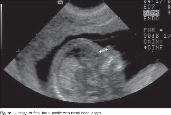
MATERIALS AND METHODS A cross-sectional study was developed in the period between January 2004 and March 2005, with the objective of evaluating the fetal nasal bone length between the 11th and 15th gestational weeks in the Department of Obstetrics at Universidade Federal de São Paulo/Escola Paulista de Medicina (Unifesp/EPM). The study sample included 171 pregnant women who had been submitted to first trimester ultrasonographic screening for chromosomopathies, whose results had suggested low risk for fetal malformations (1:300 or less). Inclusion criteria in the present study were: women with single gestation pregnancy, gestational age based on the last menses period, and confirmed by first trimester ultrasound ranging between 11 and 14 weeks and six days of gestation, absence of malformations detected during the pre- or postnatal ultrasonographic follow-up. Exclusion criteria were: abortion or fetal death during the pre-natal follow-up. The present study was approved by the Committee for Ethics in Research of Unifesp/EPM, and the patients accepted to voluntarily participate in the study, signing a term of free and informed consent. For measuring the nasal bone length, a sagittal section of the fetal profile was obtained with the ultrasound transducer at an angle between 45° and 135° to the facial plane. The image was magnified so that the fetal head and upper thorax were present on 75% of the screen. The nasal bone and nasofrontal synostosis, which appear as an anechoic area on the glabellar region, were identified, besides other two linear, parallel and echogenic images corresponding to the skin interface right above the nasal bone. The caliper position was adjusted in such a way that each movement corresponded to a 0.1 mm-displacement. Once the appropriate plane was identified, the first measurement of the nasal bone was performed (Figure 1). A second image was acquired, with a new measurement of the nasal bone. The final nasal bone length corresponded to the arithmetic mean between the two measurements.
All the ultrasound examinations were performed by three sonographists with experience in the fetal morphological evaluation at the first trimester of gestation, and accredited by the Fetal Medicine Foundation. All the examinations were performed in a Toshiba Powervision 6000 (Toshiba; Tokyo, Japan) equipment coupled with a convex transducer (3.5-5.0 MHz), and the transabdominal approach was adopted for all the measurements. The application SPSS 12.0 for Windows (Chicago, IL, USA) was utilized for statistical analysis. Polynomial regression was utilized for identifying the curves which best fit to the mean and standard deviation as a function of the gestational age. Residual analysis plots were utilized to validate the models. The 5% and 95% percentiles for the nasal bone length in the gestational interval evaluated resulted from the formula: mean ± 1.645 standard deviation. The Spearman correlation coefficient with 95% confidence interval (CI 95%) was utilized for evaluating the correlation between the nasal bone length and fetal anthropometric parameters — biparietal diameter (BPD), cranial circumference (CC), abdominal circumference (AC), femur length (FL), humerus length (HL), and crown-rump length (CRL). The significance level of 5% was utilized for all statistical analyses, that is to say, descriptive levels presenting p < 0.05 were considered as statistically significant.
RESULTS All of the 171 selected pregnant women met both the inclusion and exclusion criteria, and were included in the final statistical analysis. The fetal nasal bone was visualized and measured in 100% of the fetuses. The pregnant women ages ranged from 17 to 42 years (mean, 30 years; standard deviation, 6 years). Table 1 shows the means and respective standard deviations for fetal nasal bone length at the 11th-14th gestational weeks, demonstrating that the mean nasal bone length increased with the progression of the gestational age. On Figure 2, the behavior of the nasal bone length can be observed as a function of the gestational age.
In the period of evaluation (11 to 14 weeks and 6 days of gestation), an increase of 0.404 ± 0.026 mm (p < 0.001) was observed along the weeks. Table 2 demonstrates that the nasal bone length was statistically correlated with all the fetal anthropometric parameters (BPD, CC, AC, FL, HL, and CRL) (p < 0.001). CRL and BPD parameters demonstrated respectively the highest and lowest correlation with the nasal bone length.
The reference measurements in the 2.5%, 5%, 50%, 95% and 97.5% percentiles are shown on Table 3. Figure 3 shows the nasal bone lengths and the 5%, 50% and 95% percentiles adjustments for the gestational ages. According to Figure 3, a linear increase was observed in the nasal bone length as a function of the gestational age, with the linear adjustment resulting in an explanation coefficient (R²) = 59.4% (p < 0.001).
DISCUSSION Several studies have demonstrated the association between absent fetal nasal bone at 11-13 weeks and 6 days and trisomy 21(3,11,12), as well as other chromosomic abnormalities(13). It is important to consider the influence of the maternal ethnic origin on the incidence of fetal nasal bone absence. Prefumo et al.(6), in a prospective study have evaluated 3992 fetuses and observed that the prevalence of absent nasal bones was 5.8% in mothers of African origin, 3.4% in those of Asian origin, and only 2.6% in those of Caucasian origin. They suggest that corrections for maternal ethnicity are required for utilizing the fetal nasal bone length in the screening for trisomy 21 at the first trimester of gestation. In the present study, the fetal nasal bone could be visualized and measured in 100% of cases. The greatest majority of studies report high rates of first trimester evaluation of the fetal nasal bone. In the pioneering study developed by Cicero et al.(3) the fetal profile could be visualized in 701 cases (100% success). In a recent study involving 21074 fetuses at 11-13 weeks and 6 days, the fetal profile could not be visualized in only 243 cases (98.8% success)(4). The high rate of success of the present study in the visualization of the fetal profile is related to the relatively small number of pregnant women evaluated. In the present study, the mean nasal bone length as a function of the gestational age ranged between 1.69 mm and 2.94 mm. In a nomogram determined for the Korean population, Moon et al.(9) have found a mean variation between 1.5 mm and 2.1 mm in the nasal bone length at 11-14 weeks. In a multicentric study developed by Orlandi et al.(14) the fetal nasal bone length ranged from 2.48 mm to 3.12 mm respectively for a CRL of 45 mm and 84 mm. As regards percentiles, the nasal bone length ranged from 1.0 mm (2.5% percentile) to 3.7 mm (97.5% percentile) at 11-14 weeks of gestation. In the study developed by Sonek et al.(5) with 3537 pregnant women of different racial groups, the nasal bone length ranged from 1.3 mm (2.5% percentile) to 5.7 mm (97.5% percentile) in the same gestational period. Based on these results, one may conclude that ethnic and racial factors are extremely relevant factors in the evaluation of the fetal nasal bone, so a correction for ethnicity should be considered if the nasal bone length is included in the first-trimester screening for trisomy 21. Additionally, a linear increase was observed in the fetal nasal bone length as a function of the gestational age, similarly to the results reported by Moon et al.(9) and Chen et al.(8) respectively in the Korean and Chinese populations. In the study developed by Sonek et al.(5), these authors obtained a second order polynomial regression (R² = 0.77) as the best correlation between the fetal nasal bone length and the gestational age; however, the nasal bone length was evaluated between the 11th and 40th weeks, and not between the 11th and 14th weeks. The nasal bone length was strongly correlated with all of the anthropometric parameters, particularly CRL. The nomograms of the nasal bone length at 11 and 14 weeks of gestation in studies published to the present also demonstrate this strong correlation(8,9). This finding demonstrates that the nasal bone length is a parameter for evaluation of the fetal growth.
CONCLUSIONS Despite the preliminary character of the present study, one may consider its significant role as a pioneer in the evaluation of the nasal bone at 11-15 weeks of gestation in the Brazilian population. The preliminary results of the present study demonstrated that the nasal bone lengths were quite different from other ethnic groups, while the pronounced ethnical diversity of the Brazilian population is widely known. Therefore, additional multicentric studies with larger casuistics are necessary to include the nasal bone length as a parameter in the screening for trisomy 21.
REFERENCES 1. Down LJ. Observations on an ethnic classification of idiots. Clinical Lectures and Reports. London Hospital. 1866;3:259-62. [ ] 2. Keeling JW, Hansen BF, Kjaer I. Pattern of malformations in the axial skeleton in human trisomy 21 fetuses. Am J Med Genet. 1997;68:466-71. [ ] 3. Cicero S, Curcio P, Papageorghiou A, et al. Absence of nasal bone in fetuses with trisomy 21 at 11-14 weeks of gestation: an observational study. Lancet. 2001;358:1665-7. [ ] 4. Cicero S, Avgidou K, Rembouskos G, et al. Nasal bone in first-trimester screening for trisomy 21. Am J Obstet Gynecol. 2006;195:109-14. [ ] 5. Sonek JD, McKenna D, Webb D, et al. Nasal bone length throughout gestation: normal ranges based on 3537 fetal ultrasound measurements. Ultrasound Obstet Gynecol. 2003;21:152-5. [ ] 6.Prefumo F, Sairam S, Bhide A, et al. Maternal ethnic origin and fetal nasal bones at 11-14 weeks of gestation. BJOG. 2004;11:109-12. [ ] 7. Collado F, Bombard A, Li V, et al. Ethnic variation of fetal nasal bone length between 11-14 weeks' gestation. Prenat Diagn. 2005;25:690-2. [ ] 8. Chen M, Lee CP, Tang R, et al. First-trimester examination of fetal nasal bone in the Chinese population. Prenat Diagn. 2006;26;703-6. [ ] 9. Moon MH, Cho JY, Lee YM, et al. Nasal bone length at 11-14 weeks of pregnancy in the Korean population. Prenat Diagn. 2006;26:524-7. [ ] 10. Bunduki V, Ruano R, Miguelez J, et al. Fetal nasal bone length: reference range and clinical application in ultrasound screening for trisomy 21. Ultrasound Obstet Gynecol. 2003;21:156-60. [ ] 11. Otaño L, Aiello H, Igarzábal L, et al. Association between first trimester absence of fetal nasal bone on ultrasound and Down syndrome. Prenat Diagn. 2002;22:930-2. [ ] 12. Zoppi MA, Ibba RM, Axiana C, et al. Absence of fetal nasal bone and aneuploidies at first-trimester nuchal translucency screening in unselected pregnancies. Prenat Diagn. 2003;23:496-500. [ ] 13. Cicero S, Rembouskos G, Vandecruys H, et al. Likelihood ratio for trisomy 21 in fetuses with absent nasal bone at the 11-14-week scan. Ultrasound Obstet Gynecol. 2004;23:218-23. [ ] 14. Orlandi F, Bilardo CM, Campogrande M, et al. Measurement of nasal bone length at 11-14 weeks of pregnancy and its potential role in Down syndrome risk assessment. Ultrasound Obstet Gynecol. 2003;22:36-9. [ ] Received June 14, 2007. Accepted after revision July 24, 2007. * Study developed in the Department of Obstetrics - Universidade Federal de São Paulo/Escola Paulista de Medicina (Unifesp/EPM), São Paulo, SP, Brazil. |
|

Av. Paulista, 37 - 7° andar - Conj. 71 - CEP 01311-902 - São Paulo - SP - Brazil - Phone: (11) 3372-4544 - Fax: (11) 3372-4554