Radiologia Brasileira - Publicação Científica Oficial do Colégio Brasileiro de Radiologia
AMB - Associação Médica Brasileira CNA - Comissão Nacional de Acreditação
 Vol. 41 nº 2 - Mar. / Apr. of 2008
|
ORIGINAL ARTICLE
|
|
Morphometric study of the fetal heart: a parameter for echocardiographic analysis |
|
|
Autho(rs): Jennecy Sales Cavalcanti, Suzana Marques Duarte |
|
|
Keywords: Fetal echocardiography, Fetal circulation, Cardiac malformation, Heart |
|
|
Abstract:
IAssociate Professor of Clinical and Radiological Anatomy, Department of Anatomy, Universidade Federal de Pernambuco (UFPE), Recife, PE, Brazil
INTRODUCTION With the development of fetal echocardiography and new surgical techniques for correction of prenatally diagnosed cardiac malformations, a sound knowledge about the normal anatomy and development of the fetal heart has become absolutely necessary(1,2). Malformations may affect any region of the heart, including the atrioventricular ostia and cardiac valves, generally requiring immediate surgical correction, considering that these malformations may compromise the cardiac function, and even lead to fetal or neonatal death(3–5). The advances of ultrasonography and its application in the investigation of the fetal development have allowed the detection of malformations, especially in the cardiovascular system, between the 16th and 20th gestational weeks, besides a more accurate analysis of fetal heart structures, including the morphology and dynamics of atrioventricular valves and great vessels(1,6,7). The possibility of early identification of the presence of cardiac malformations still during the intrauterine life, by means of echocardiography, represents a relevant development both in the practice of pediatric cardiology and obstetrics. Considering that echocardiography is essential for the early diagnosis of fetal cardiac malformations, it is absolutely necessary to know the morphometric features of the fetal heart to achieve an accurate diagnosis of possible malformations as well as other conditions that may cause physiological alterations in the fetal heart progressing to cardiac failure and fetal death, if not opportunely corrected(5,8–10).
MATERIALS AND METHODS The sample of the present study included twenty 10%–formol–fixed human hearts from both male and female fetuses at gestational ages between 28 and 36 weeks, owned by the Department of Anatomy – Centro de Ciências Biológicas (Center of Biological Sciences) of Universidade Federal de Pernambuco. The approximate gestational age (GA) was based on the Balthazar–Dervieux equation (GA = fetus length × 5.6 ÷ 7). The hearts were macroscopically normal, without any type of malformation. Initially, the hearts were weighted and subsequently dissected. Atrial cavities were removed by incision along the coronary sulcus. The anteroposterior and transverse diameters of the atrioventricular valves were measured with a paquimeter with a 1/10 mm accuracy. Then, the areas of the respective valves were calculated. With the aid of the same measurement instrument, the major anteroposterior and transverse diameters of the right and left ventricular cavities were measured. Also, the thicknesses of the anterior and posterior ventricular walls, as well as the interventricular septum were measured. The data resulting from these measurements were statistically analyzed.
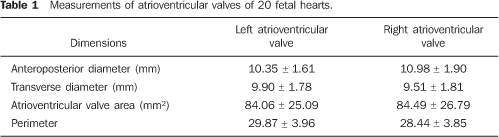
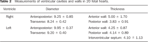
RESULTS Data regarding anteroposterior and traverse diameters, as well as areas of right and left atrioventricular valves are shown on Table 1. Table 2 demonstrates anteroposterior and transverse diameters of right and left ventricular cavities as well as thicknesses of ventricular walls and interventricular septum.
DISCUSSION Congenital cardiopathy is defined as any structural alteration affecting a fetal or neonatal heart. A significant prevalence is reported in the population — 8 to 12 cases/1000 live neonates —, with death occurring in the first month of life in 20%–30% of cases(9,11,12). Therefore, the early diagnosis of congenital cardiopathies is important, considering the sudden clinical manifestation, including the possibility of death(13). Up to some years ago, a single aspect of the fetal cardiovascular physiology could be routinely monitored: the fetal cardiac frequency(1). Early in the eighties, studies were published in the literature describing the fetal echocardiography technique, and from that time on, the analysis of the intracardiac anatomy, cardiac function and detection of fetal malformations have become feasible(1,14). An early prenatal diagnosis of severe congenital cardiopathies improves the perinatal prognosis, allowing a more appropriate planning of a perinatal strategy, considering the high probability of a sudden neonate death. Several echocardiographic studies have been performed to establish the cardiac cavities dimensions and biparietal diameters in normal fetuses(15,16). The values found present a slight variation, depending on the method, the cut (four–chamber or transverse), and the phase of cardiac cycle were the measurements were taken (systole or diastole)(1). Coincidentally, these studies report that the cardiac cavities present a three–fold or four–fold increase in size from the 17th week to the end of the gestational period(17), demonstrating a linear pattern of growth in relation to the gestational age and biparietal diameter(15–19). However, the mitral and tricuspid flow velocities do not present a significant variation along the gestation, and the maximum tricuspid peak–velocity is slightly higher than the maximum mitral peak–velocity(1). On the other hand, the mitral and tricuspid valves diameters are similar to each other, differently from the adult heart, where the tricuspid valve diameter is approximately 1.5–fold larger than the mitral valve diameter(20). Morphometric data of fetal atrioventricular valves could not be found in the reviewed literature for comparison with those found in the present study. It can be assumed that alterations in the normal dimensions of atrioventricular valves may result in disorders detectable by combined pulsatile Doppler, 2D imaging and color flow mapping(1). Blood flow velocity, direction and volume can be evaluated to aid in the determination of the presence of valvular regurgitation or stenosis(1). So, it can be presumed that the data found in the present study can be utilized as parameters for fetal echocardiographic findings. The present study, that is aimed at establishing the morphometry of the mitral and tricuspid valvular apparatuses as well as the ventricular cavities diameters and thicknesses of fetal heart walls; on the other hand, determining if a fetal heart can reproduce the same configuration observed in vivo is not feasible. Notwithstanding, other experimental studies have demonstrated that, when a heart stops beating, it becomes hyper–relaxed and, after the inevitable effect of the cardiac rigor mortis, it contracts itself, presenting with an architecture similar to the cardiac contraction at end–systole(21–23).
CONCLUSION The role of morphometric data of fetal atrioventricular valves is highly significant in the echocardiographic diagnosis of possible congenital cardiac malformations.
REFERENCES 1. Tatani BS. Ecocardiografia fetal. Apresentando o método. Arq Bras Cardiol. 1997;69:77–88. [ ] 2. Driggers RW, Spevak PJ, Crino JP, et al. Fetal anatomic and functional echocardiography: a 5–year review. J Ultrasound Med. 2003;22:45–51. [ ] 3. Allan LD, Tynan MJ, Campbell S, et al. Echocardiographic and anatomical correlates in the fetus. Br Heart J. 1980;44;444–51. [ ] 4. Lang D, Oberhoffer R, Cook A, et al. Pathologic spectrum of malformations of the tricuspid valve in prenatal and neonatal life. J Am Coll Cardiol. 1991;17:1161–7. [ ] 5. Hornberger LK, Sahn DJ, Kleinman CS, et al. Tricuspid valve disease with significant tricuspid insufficiency in the fetus: diagnosis and outcome. J Am Coll Cardiol. 1991;17:167–73. [ ] 6. Kleinman CS, Hobbins JC, Jaffe CC, et al. Echocardiographic studies of the human fetus: prenatal diagnosis of congenital heart disease and cardiac dysrhythmias. Pediatrics. 1980;65:1059–67. [ ] 7. Copel JA, Pilu G, Green J, et al. Fetal echocardiographic screening for congenital heart disease: the importance of the four–chamber view. Am J Obstet Gynecol. 1987;157:648–55. [ ] 8. Mattos SS. Fisiologia da circulação fetal e diagnóstico das alterações funcionais do coração do feto. Arq Bras Cardiol. 1997;69:205–7. [ ] 9. Carvalho CA. Ecocardiografia fetal. Novas fronteiras. Arq Bras Cardiol. 1997;69:203–4. [ ] 10. Zielinsky P. Malformações cardíacas fetais. Diagnóstico e conduta. Arq Bras Cardiol. 1997;69: 209–18. [ ] 11. Campbell M. Incidence of cardiac malformations at birth and later, and neonatal mortality. Br Heart J. 1973;35:189–200. [ ] 12. Viñals FV, Giuliano AB. Cardiopatias congênitas. Incidência antenatal. Rev Chil Obstet Ginecol. 2002;67:203–6. [ ] 13. Hagemann LL, Zielinsky P. Rastreamento populacional de anormalidades cardíacas fetais por ecocardiografia pré–natal em gestações de baixo risco no município de Porto Alegre. Arq Bras Cardiol. 2004;82:313–9. [ ] 14. Travancas PR. Cardiologia fetal: metodologia diagnóstica e manuseio das principais anomalias cardíacas fetais. Rev SOCERJ. 2000;13:23–30. [ ] 15. DeVore GR, Siassi B, Platt LD. Fetal echocardiography. IV. M–mode assessment of ventricular size and contractility during the second and third trimesters of pregnancy in the normal fetus. Am J Obstet Gynecol. 1984;150:981. [ ] 16. Cartier MS, Davidoff A, Warneke LA, et al. The normal diameter of the fetal aorta and pulmonary artery: echocardiographic evaluation in utero. AJR Am J Roentgenol. 1987;149:1003–7. [ ] 17. Wladimiroff JW, McGhie J. Ultrasonic assessment of cardiovascular geometry and function in the human fetus. Br J Obstet Gynaecol. 1981;88: 870–5. [ ] 18. St John Sutton MG, Gewitz MH, Shah B, et al. Quantitative assessment of growth and function of the cardiac chambers in the normal human fetus: a prospective longitudinal echocardiographic study. Circulation. 1984;69:645–54. [ ] 19. Allan LD, Joseph MC, Boyd EG, et al. M–mode echocardiography in the developing human fetus. Br Heart J. 1982;47:573–83. [ ] 20. Cavalcanti JS. Aparelhos valvares mitral e tricúspide. Estudo comparativo de seus componentes anatômicos. An Fac Med CCS/UFPE. 1996;41: 105–8. [ ] 21. Curti HJV, Ferreira MCF, Ferreira AS, et al. Aparelho valvar mitral: um enfoque anátomo–ecocardiográfico. Arq Bras Cardiol. 1989;53:85–92. [ ] 22. Maron BJ, Henry WL, Roberts WC, et al. Comparison of echocardiographic and necropsy measurements of ventricular wall thicknesses in patients with and without disproportionate septal thickening. Circulation. 1977;55:341–6. [ ] 23. Sonnenblick EH, Ross J Jr, Covell JW, et al. The ultrastructure of the heart in systole and diastole. Chantes in sarcomere length. Circ Res 1967;21: 423–31. [ ] Received July 10, 2006. Accepted after revision June 26, 2007. * Study developed in the Department of Anatomy – Center of Biological Sciences, Universidade Federal de Pernambuco (UFPE), Recife, PE, Brazil |
|

Av. Paulista, 37 - 7° andar - Conj. 71 - CEP 01311-902 - São Paulo - SP - Brazil - Phone: (11) 3372-4544 - Fax: (11) 3372-4554