Radiologia Brasileira - Publicação Científica Oficial do Colégio Brasileiro de Radiologia
AMB - Associação Médica Brasileira CNA - Comissão Nacional de Acreditação
 Vol. 41 nº 1 - Jan. /Feb. of 2008
|
ORIGINAL ARTICLE
|
|
Plane-parallel ionization chamber for X-radiation of conventional radiography and mammography |
|
|
Autho(rs): Alessandro Martins da Costa, Linda V. E. Caldas |
|
|
Keywords: Conventional radiography, Mammography, Quality control, Ionization chamber |
|
|
Abstract:
IPhD in Nuclear Technology, Doctor Professor, Department of Physics and Mathematics at Faculdade de Filosofia, Ciências e Letras de Ribeirão Preto da Universidade de São Paulo (FFCLRP-USP), Ribeirão Preto, SP, Brazil
INTRODUCTION A program for radiodiagnosis quality guarantee may lead to the production of high-quality radiographic images and minimal radiation exposure to the patient(1–4). Radiodiagnosis quality guarantee includes, among other aspects, evaluation of imaging quality, film rejection analyses, evaluation of the dose to patients, and evaluation of physical parameters of the different X-ray system components. Several tests for quality control are required for guaranteeing the correct X-ray equipment operation. The tests for quality control of conventional and mammographic X-ray equipment must include a representative estimation of the skin entrance dose practiced in the radiological clinic and the corresponding values must be compared with the diagnostic reference levels in radiology established by the Brazilian standards(3). It should be emphasized that the diagnostic reference levels in radiology are still to be regulated, but must be considered as a practical mechanism to promote a better local quality control. An accurate estimation of the skin entrance dose requires a precise measurement of the incident air kerma (5) at the entrance skin plane, and also of the beam half-value layer (HVL)(6). The incident air kerma is converted into skin entrance dose by the application of an appropriate backscatter factor(7,8). Also, the management of the following parameters is particularly important: tube voltage (kVp); air kerma reproducibility and linearity with the tube current product-exposure time (mAs). These parameters characteristics may vary over the time; therefore tests should be performed at regular intervals. Consequently, it is necessary to understand how an image is influenced by these parameters, and how their characteristics could be measured with appropriate tools. Incident air kerma, HVL, reproducibility and linearity of air kerma rate as a function of the mAs are generally determined by the utilization of calibrated ionization chambers(6). The knowledge about ionization chamber limitations is essential for its correct utilization(9). The specifications for an ionization chamber design must be carefully understood by the user and correctly taken into consideration before any measurement is accepted as valid(10). In Brazil there has been an increasing interest in designing and constructing low-cost ionization chambers, demonstrating the feasibility of constructing radiation detectors with materials available in the domestic market. Initially, ionization chambers were developed for X-radiation(11) and beta-radiation(12), respectively by Instituto de Radioproteção e Dosimetria, Rio de Janeiro, RJ, and Escola de Engenharia da Universidade Federal de Minas Gerais, Belo Horizonte, MG. Plane-parallel ionization chambers for low-energy X-radiation and beta-radiation(13), high-energy electrons(14–16), computed tomography(17,18), radioprotection(19), extrapolation chambers for X-radiation and beta-radiation of dermatologic and ophthalmologic applications utilized in brachytherapy(20,21) have been constructed by Instituto de Pesquisas Energéticas e Nucleares. The present study was aimed at developing a double-faced plane-parallel ionization chamber for determining air kerma and air kerma rate in X-radiation fields utilized in conventional diagnosis and mammography. One face of this ionization chamber is appropriate for measurements in conventional radiography (face A); the other (face G) is appropriate for measurements in mammography. The ionization chamber developed in this study was calibrated for standard X-ray beams and submitted to tests according to the international recommendations(22–24). Operational characteristics of linearity, angular and energy dependence were tested according to the procedures applied for other ionization chambers(13,14,17,19–21,25,26).
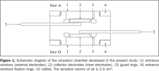
MATERIALS AND METHODS A double-faced plane-parallel ionization chamber for X-radiation beams utilized in conventional radiography and mammography was designed and constructed. The chamber is comprised of aluminized polyester entrance windows (1.7 mg/cm²), aluminium (face A) and graphite (face G) inner electrodes and guard rings (face A) and sensitive volume of air 2.5 cm³. Figure 1 shows a schematic diagram of this chamber.
Ionization currents were measured with electrodes whose relevant characteristics ser shown on Table 1.
Tests were performed with the ionization chamber coupled with a PTW Unidos electrometer. The polarization voltage was +400 V. The efficiency of the ions collection is > 99% at +400 V for both faces of the chamber(27–29). The leakage current is < 2 fA and the accuracy is > ±0.05% for both faces of the chamber. The following irradiation systems were utilized: Rigaku Denki, Geigerflex model X-ray equipment with constant potential, with a Philips tube model PW 2184/00, beryllium 1 mm window and tungsten target, operating up to 60 kV; Medicor Mövek Röntgengyara, model Neo-Diagnomax X-ray single-phase equipment, full wave rectification, with tungsten target, operating up to 125 kV in radiographic mode, and up to 100 kV in fluoroscopic mode. The X-ray systems characteristics are shown on Tables 2 and 3.
Table 4 shows the characteristics of the reference standard systems utilized in the ionization chamber calibration.
Considering that the ionization chambers utilized in the present study are unsealed, all the measurements were corrected for reference temperature and pressure conditions (20 °C and 101.3 kPa). The measurement uncertainties evaluation and expression were performed according to the "Guidelines for measurement uncertainty expression"(30).
RESULTS The linear relation between the ionization current and the air kerma rate was determined by sequential irradiation of the both faces of the chamber in the RXM35 quality of mammography Xray (Table 2) with variable tube current. The chamber was positioned at a 100 cm distance from the source, taking the entrance windows surface center as a reference. The air kerma rates were determined according to the standard system for mammography X-rays (Table 4). The data obtained are shown on Figure 2. The straight lines represent the results from linear adjustments of these data. The uncertainty obtained for the angular coefficient, i.e., the uncertainty obtained for the response linearity was 0.86% for face A, and 0.92% for face G. Also, the RXM35 quality of mammography X-ray was utilized for evaluating the chamber angular dependence. The chamber was irradiated in the air at 59.5 mGy/min air kerma rate, 100 cm focus-chamber distance, taking the entrance windows surface as reference. The response was measured with the incidence angle ranging between 0° and ±5°, where 0° corresponds to a frontal irradiation. The results obtained are shown on Table 5. The responses were normalized for 0° corresponding to the average of five successive measurements. It may be observed that the chamber is in compliance with the IEC standard requirements(23): response variation up to ±3,0% because of incidence angle variation of ±5°. Maximum response variation was 0.8%.
X-ray qualities utilized in the chamber calibration are shown on Tables 2 and 3. The chamber was irradiated in the air, and positioned at the calibration distance taking the entrance window surface as reference. The calibration coefficients were obtained with the standard systems for each energy range (Table 4). Calibration coefficients obtained for the face A of the chamber in the qualities of conventional diagnostic X-rays, and for the face G in the qualities of mammography X-radiation are presented as a function of the half-value layer on Figures 3A and 3B. On these Figures, the right vertical scales represent correction factors normalized for reference qualities for each case. The response energy dependence was 0.8% for face A in the qualities of conventional diagnostic X-rays, and 2.4% for the face G of the chamber in the qualities of mammography X-radiation. It may be observed that the chamber is in compliance with the IEC standard requirements(23): response variation up to ±5% with the radiation quality.
DISCUSSION For a consistent quality of the images produced by manual techniques, the air kerma rate linearity as a function of the mAs for a determined kVp must be < 20%(6). It is important that the measured linearity is a characteristic of the X-ray equipment and is not affected by the lack of response linearity or inaccuracy of the ionization chamber utilized for the measurement. The standard uncertainty for any mAs value can be reduced, by increasing the number of measurements, provided all the measurements uncertainties are random and normally distributed. The accuracy is > ±0,05% for both faces of the chamber developed in the present study, limiting the number of exposures required for the measurement of the air kerma rate linearity as a function of the mAs and is not significant for the measurement uncertainty. The uncertainty obtained from the chamber response linearity is a systematic uncertainty and cannot be minimized by the measurements repetition. This uncertainty is small (< 1%) as compared with the criterion for an X-ray equipment performance (< 20%). So, the chamber response linearity does not affect the determination of the air kerma rate linearity as a function of the mAs of an X-ray equipment. The air kerma rate for a determined kVp and mAs should not vary more than 10% across four consecutive exposures(6), i.e. the air kerma rate must be < 10%. The accuracy of the chamber developed in the present study is much higher than the one acceptable for the air kerma rate of an X-ray equipment, and has no adverse effect on the reproducibility measurement. Plane-parallel ionization chambers are designed to be utilized with the entrance windows facing the radiation source and perpendicular to beam axis. The angular dependence of the chamber developed in the present study was evaluated, considering the possibility of a little variation in the radiation incidence angle due the chamber positioning. It may be observed that the response variation caused by this little variation in the incidence angle does not result in a significant measurement uncertainty. An ionization chamber response energy dependence is one of the main sources of uncertainty in the determination of an X-ray beam air kerma rate and HVL for a certain kVp. The ionization chamber utilized should present a minimal response energy dependence for each energy interval corresponding to the X-ray equipment. The response energy dependence is minimal both for the face A of the ionization chamber in the qualities of conventional diagnostic X-ray and for the face G in the qualities of mammography X-rays. The face A can be utilized for measuring the air kerma and the air kerma rate, and for determining the half-value layer in the qualities of conventional diagnostic X-rays, and face G in the qualities of mammography X-rays.
CONCLUSIONS The performance tests demonstrated that the ionization chamber developed in the present study can be routinely utilized for determining the air kerma rate in a program for controlling the quality of X-ray equipment. The advantage of this chamber is that it can be utilized both for measurements in conventional radiography and mammography, provided the appropriate face is selected. Typically a specific type of ionization chamber is utilized for measurements in conventional radiography, and another type of chamber in mammography. The construction of this chamber corroborates the feasibility of producing radiation detectors with materials available in the domestic market. Acknowledgements Partial financial support for the development of the present study was granted by Conselho Nacional de Desenvolvimento Científico e Tecnológico (CNPq) and Fundação de Amparo à Pesquisa do Estado de São Paulo (Fapesp).
REFERENCES 1. World Health Organization. Quality assurance in diagnostic radiology. Geneva: WHO; 1982. [ ] 2. Food and Agriculture Organization of the United Nations, International Atomic Energy Agency, International Labour Organisation, Nuclear Energy Agency of the Organisation for Economic Co-operation and Development, Pan American Health Organization, World Health Organization. International Basic Safety Standards for Protection Against Ionizing Radiation and for the Safety of Radiation Sources, Safety Series No. 115. Vienna: IAEA; 1996. [ ] 3. Brasil. Ministério da Saúde. Secretaria de Vigilância Sanitária. Diretrizes de proteção radiológica em radiodiagnóstico médico e odontológico. Portaria nº 453, 1 de junho de 1998. Diário Oficial da União, Brasília, 2 de junho de 1998. [ ] 4. International Atomic Energy Agency, Pan American Health Organization, World Health Organization. Radiological protection for medical exposure to ionizing radiation. Safety Standards Series No. RS-G-1.5. Vienna: IAEA; 2002. [ ] 5. International Commission on Radiation Units and Measurements. ICRU Report 74: Patient dosimetry for X rays used in medical imaging. J ICRU. 2005;5(2). [ ] 6. Brasil. Ministério da Saúde. Agência Nacional de Vigilância Sanitária. Radiodiagnóstico médico: segurança e desempenho de equipamentos. Brasília: Anvisa; 2005. [ ] 7. Petoussi-Henss N, Zankl M, Drexler G, et al. Calculation of backscatter factors for diagnostic radiology using Monte Carlo methods. Phys Med Biol. 1998;43:2237–50. [ ] 8. European Commission. European protocol on dosimetry in mammography. Report EUR 16263 EN. Luxembourg: EC; 1996. [ ] 9. Wagner LK, Cerra F, Conway B, et al. Energy and rate dependence of diagnostic x-ray exposure meters. Med Phys. 1988;15:749–53. [ ] 10. DeWerd LA, Wagner LK. Characteristics of radiation detectors for diagnostic radiology. Appl Radiat Isot. 1999;50:125–36. [ ] 11. Austerlitz C, Nette P, Cordilha A. Construção, calibração e teste de uma câmara de ionização para medidas de exposição X e gama entre 40 e 1250 keV. Rev Fís Aplic Instr. 1986;1:320–8. [ ] 12. Silva I. Projeto e construção de uma câmara de extrapolação para dosimetria beta. (Dissertação de Mestrado). Belo Horizonte: Universidade Federal de Minas Gerais; 1985. [ ] 13. Albuquerque MPP, Caldas LVE. New ionization chambers for beta and X-radiation. Nucl Instrum Meth A. 1989;280:310–3. [ ] 14. Souza CN, Caldas LVE, Sibata CH, et al. Two new parallel-plate ionization chambers for electron beam dosimetry. Radiat Meas. 1996;26:65–74. [ ] 15. Bulla RT, Caldas LVE. Determinação de dose absorvida em feixes de elétrons utilizando câmaras de ionização de placas paralelas. Radiol Bras. 2004;37:171–7. [ ] 16. Bulla RT, Caldas LVE. Comparação entre fatores de calibração em termos de dose absorvida no ar para uma câmara de ionização de placas paralelas. Radiol Bras. 2006;39:203–7. [ ] 17. Maia AF, Caldas LVE. A new extended-length parallel-plate ionization chamber. Phys Med Biol. 2005;50:3837–47. [ ] 18. Maia AF, Caldas LVE. Calibração das câmaras de ionização para feixes de tomografia computadorizada no Brasil: a realidade atual. Radiol Bras. 2006;39:209–13. [ ] 19. Vivolo V. Desenvolvimento de um sistema de referência para determinação do equivalente de dose pessoal e da constância de feixes de radiação-X. (Tese de Doutorado). São Paulo: Universidade de São Paulo; 2006. [ ] 20. Dias SK, Caldas LVE. Development of an extrapolation chamber for the calibration of beta-ray applicators. IEEE T Nucl Sci. 1998;45:1666–9. [ ] 21. Oliveira ML, Caldas LVE. A special mini-extrapolation chamber for calibration of 90Sr+90Y sources. Phys Med Biol. 2005;50:2929–36. [ ] 22. International Atomic Energy Agency. Calibration of dosemeters used in radiotherapy, Technical Reports Series No. 374. Vienna: IAEA; 1994. [ ] 23. International Electrotechnical Commission. IEC 61674: Medical electrical equipment dosimeters with ionization chambers and/or semi-conductor detectors as used in X-ray diagnostic imaging. Geneva: IEC; 1997. [ ] 24. International Electrotechnical Commission. IEC 61267: Medical diagnostic X-ray equipment: radiation conditions for use in the determination of characteristics. Geneva: IEC; 2005. [ ] 25. Caldas LVE. A sequential Tandem system of ionisation chambers for effective energy determination of X-radiation fields. Radiat Prot Dosim. 1991;36:47–50. [ ] 26. Caldas LVE, Albuquerque MPP. Angular dependence of parallel-plate ionisation chambers. Radiat Prot Dosim. 1991;37:55–7. [ ] 27. Geleijns J, Broerse JJ, Zweers D. General ion recombination for ionization chambers used under irradiation conditions relevant for diagnostic radiology. Med Phys. 1995;22:17–22. [ ] 28. Das IJ, Akber SF. Ion recombination and polarity effect of ionization chambers in kilovoltage x-ray exposure measurements. Med Phys 1998; 25:1751–7. [ ] 29. International Atomic Energy Agency. Absorbed dose determination in external beam radiotherapy: an international code of practice for dosimetry based on standards of absorbed dose to water. Technical Reports Series No. 398. Vienna: IAEA; 2002. [ ] 30. Associação Brasileira de Normas Técnicas. Instituto Nacional de Metrologia, Normatização e Qualidade Industrial. Guia para a expressão da incerteza de medição. 3ª ed. Rio de Janeiro: ABNT, Inmetro; 2003. [ ]
Received May 11, 2007. Accepted after revision June 19, 2007.
* Study developed at Instituto de Pesquisas Energéticas e Nucleares – Comissão Nacional de Energia Nuclear (IPEN-CNEN), São Paulo, SP, Brazil. |
|
Av. Paulista, 37 - 7° andar - Conj. 71 - CEP 01311-902 - São Paulo - SP - Brazil - Phone: (11) 3372-4544 - Fax: (11) 3372-4554


