Radiologia Brasileira - Publicação Científica Oficial do Colégio Brasileiro de Radiologia
AMB - Associação Médica Brasileira CNA - Comissão Nacional de Acreditação
 Vol. 42 nº 2 - Mar. / Apr. of 2009
|
REVIEW ARTICLE
|
|
Use of images for human identification in forensic dentistry |
|
|
Autho(rs): Suzana Papile Maciel Carvalho, Ricardo Henrique Alves da Silva, César Lopes-Júnior, Arsenio Sales Peres |
|
|
Keywords: Radiology, Forensic dentistry, Human identification |
|
|
Abstract:
IMaster in Collective Health, Faculdade de Odontologia de Bauru da Universidade de São Paulo (FOB-USP), Pediatric Dentistry, Bauru, SP, Brazil
INTRODUCTION Identification corresponds to a set of different procedures aimed at individualizing a person or object(1). For both legal and humanitarian reasons, personal identification is highly relevant in forensic medicine, and this process is frequently initiated even before the cause of the death is determined. Based on human identification, individuals can preserve their rights as well as being charged with civil or criminal penalties. Post-mortem identification represents one of the great branches of study and research in forensic dentistry and medicine, considering that both sciences deal with a same material - the human body at different stages: ripped, lacerated, carbonized, macerated, putrefied, skeletonized - always with a single objective, that is to say, to establish the human identity(2). The identification process is characterized by the utilization of appropriate techniques and means to discover an identity, and can be developed either by skilled technicians (judiciary or police authorities) or by professionals with differentiated and specific knowledge in the area of biology (forensic medicine or dentistry), with a practically unlimited array of appropriate techniques and means to achieve human identification(2). The present study is aimed at describing radiological methods of human identification utilized in forensic dentistry as an aid to the judicial authority.
MATERIALS AND METHODS A bibliographic survey covering the last ten years was performed in the following scientific search sites: PubMed (a service of the U.S. National Library of Medicine), available at www.ncbi.nlm.nih.gov/pubmed/, and Bireme (Regional Medicine Library, a specialized database of the Pan-American Health Organization), specifically at the databases Medline, Lilacs, SciELO and Cochrane, available at www.bireme.br, utilizing the following terms as descriptors in Portuguese: radiologia, odontologia legal, identificação humana; and in English: radiology, forensic dentistry and human identification. For the systematic literature review, the following inclusion criteria were considered: 1) the articles should have been published in the period between 1997 and 2007; 2) the subject of the article should relate to the study object; 3) a clear objective and fidelity to the study developed; 4) the article should be based on previous literature; 5) conclusion in accordance with the study findings. The articles selection was based on compatibility with the study structure and methodology. Data reported in previous studies were recovered considering the production recorded on the above mentioned databases. A total of 45 articles were found on the above mentioned databases and those included in the present literature review were selected according to the already mentioned inclusion criteria; and after methodological analysis 19 articles were utilized.
HUMAN IDENTIFICATION AND DENTISTRY The activity of the dental surgeon in the forensic environment is assured by the pertinent federal regulation, the Law No. 5.081, of August 24, 1966, regulating the practice of dentistry in Brazil(3). The forensic dentist field of activity is not restricted just to the examination of dental remains, extending to several areas such as anthropology, genetics, biochemistry, forensic ballistic, thanatology and forensic traumatology, radiology, computing and images mixing, and is regulated by a pertinent federal law(4). Forensic dentistry is present in processes of post-mortem human identification from the early procedures (general identification) comprising estimation of age and sex, determination of ethnic group, skin color and other personal characteristics such as height and diagnosis of skin spots or fluids present or originating from the oral cavity, or even in the definition of time and cause of the death, to the irrefutable possibility of individual identification(2). The contribution of forensic dentistry in this field can be measured on innumerable scientific reports(5-9) and quantified including by persons unaware of the dentistry terminology and forensic sciences, like in the case of the victims of the TAM airplane crash occurred in São Paulo, SP, Brazil, in 1996, where the media highlighted the relevance of identification procedures(2). In the meantime, the two major airplane crashes in Brazil can be mentioned, the first one occurred in September 2006, involving a GOL airlines' airplane, causing the death of 154 people, and the second one, in July 2007, involving a TAM airplane where 199 people died. In both accidents the application of different techniques of forensic identification was necessary to individualize the victims, including dental identification. However, besides clinical examination and dental records, forensic dentistry can also utilize radiological images in processes of identification. Thus, the analysis of dental records in conjunction with ante- and post-mortem radiographies became an essential tool in processes of human identification. Additionally, since the second half of the eighties, with the development of information technology and the consequential introduction of computed radiology, the technique has been refined, offering higher accuracy in the identification, even in toothless individuals, and in the determination of age(10).
FORENSIC ANTHROPOLOGY AND UTILIZATION OF IMAGES: HISTORY Historically, the application of radiology in forensic sciences was introduced in 1896, just one year following the x-ray discovery by Roentgen, to demonstrate the presence of lead bullets inside the head of a victim(11). Schüller(12) proposed the possibility of utilizing radiological images of the facial sinuses for identification purposes. Following this study, many others were published and, finally, Culbert & Law(13) reported the first complete radiological identification. Singleton(14) employed this technique for identification of corpses in a massive disaster. Petersen(15) reported a fire in the Hotel Hafnia, occurred in Copenhagen, Denmark, in 1973, with 35 deaths. Eight dental surgeons collaborated with the identification team, performing visual, photographic and radiographic examinations of all the victims, recording detailed data of post-mortem odontograms and completing their work with a comparison and evaluation of ante-mortem information with the preliminary post-mortem data collected. The identification of 74% of the victims was achieved as a result of the dentists team collaborative work. Kessler & Pemble(16) demonstrated the role of forensic dentistry in the identification of American victims of the Operation Desert Storm. Among 251 examinations for dental recognition, 244 allowed the individualization and positive identification of the victims. Such examinations were facilitated by the availability of a record with panoramic radiographic images of the majority of the persons involved in the operation; the cases that could not be identified were just those that did not present previous dental records. Hazebroucq et al.(17) have described two cases where the identification was based on osteotomy of maxillas and mandibles, whose specimens were individually submitted to panoramic radiography, with the images being compared with ante-mortem radiographic images recorded by the offices of the surgeon dentists of the victims. According to the authors, this technique, besides providing complete information for identification, allows the assessment of the dental age in children. Austin & Maples(8) have described a study evaluating the accuracy of methods of images superimposition in the identification of unknown human skulls. In this study, the authors have tried to investigate the accuracy of the method without referring dental records, and could conclude that, even without dental data. images superimposition can be successfully performed, provided there are at least two ante-mortem radiographic images (frontal and lateral views). Andersen & Wenzel(6), through ante-mortem and post-mortem simulations, have evaluated the capacity of individual identification by analyzing conventional bitewing films and radiographic subtraction. Based on a scoring system (1 - eliminated, 2 - possible, 3 - likely, 4 - certain), three observers have analyzed each case individually and classified the radiographic images taking two to 12 individual characteristics into consideration. The authors assert the validity of this technique for human individual identification, provided it is applied in compliance with strict criteria. Oliveira et al.(18) have developed a study evaluating the possibility of a radiological study of the lumbar spine determining a correct identification of an individual, despite the changes associated with aging. The sample included 60 pairs of lumbar spine radiographic images that were mixed up so two experienced radiologists could put them back together by comparing the vertebrae of each pair for similarities and differences in anatomical details. Correct pairing of radiographs of the whole sample was achieved by both observers and the statistical analysis demonstrated a good-to-perfect interobserver agreement, concluding that the comparison between radiographic images of lumbar spine can determine a correct identification of individuals, despite changes associated with aging.
UTILIZATION OF IMAGES IN FORENSIC DENTISTRY In cases where the identification of a corpse is required, radiographic images of the deceased can be obtained and compared with any ante-mortem radiographic image of the presumed person(18). The following anatomical details can be adopted as parameters: shape of teeth and roots, missing and present teeth, residual roots, supranumerary teeth, attrition or abrasion, coronal fractures, sign of bone reabsorption resulting from periodontal disease, bone pathology, diastemas, cavities shapes and lines, dental cavities, endodontic treatment, intraradicular and intracoronal posts and dental prostheses(20-22). Many studies also highlight the relevance of radiography in human identification through comparative methods utilizing patterns of trabecular bone, frontal sinuses and maxillas, dental radiographic images and cephalometry and increased fingers length(20,21,23-25).
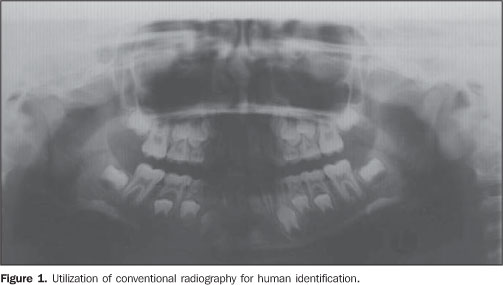
CONVENTIONAL RADIOGRAPHY The identification technique utilizing conventional radiography is based on the comparison between ante-mortem images recorded in dental offices and centers with post-mortem radiographic images. Much information can be obtained from these images. This method allows the observation of anatomical characteristics such as coronal shape and size, pulp anatomy, positioning and shape of the alveolar bone crest, besides unique and individual characteristics resulting from dental treatments(10).
DIGITAL RADIOGRAPHY Until recently, the greatest part of dental restoration materials was metal and therefore radiopaque. Peculiar characteristics of every restoration could be easily observed on conventional radiography. However, the process of identification based on conventional radiography became more difficult because of the dissemination of prophylactic dental treatments and the consequential, significant reduction in the incidence of cavities, particularly in developed countries(10). At the same time, the spectacular development of microelectronics and information technology in association with the decrease in costs of computational equipment has allowed the development of more powerful and reliable techniques for comparison of radiological images with application in forensic dentistry(10). Innumerable variations of digital radiology techniques can be found in the literature, but, essentially, the method comprises the following steps: 1) radiographic images digitization with the aid of a scanner(26), video camera(27) or, yet with images acquisition directly from a x-ray system coupled with a computer with monitor, printer and CD-ROM recorder; 2) images processing through an appropriate software, allowing comparisons based either on images superimposition(27), interposition(26) or subtraction(27).
These modern techniques allow an accurate analysis of the spatial relations of teeth roots and supporting structures on ante- and post-mortem images(29). There are softwares with resources for images rotation, translation and scaling, facilitating the correct alignment between ante- and post-mortem radiographs without the necessity of new exposures(28). It is important to observe that differences in the geometry between radiographs represent the main factor of error in this type o technique, and the above mentioned correction is essential to reduce the noise resulting from the process of image subtraction(27).
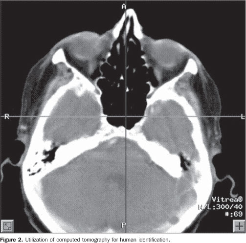
COMPUTED TOMOGRAPHY Conventional, two-dimensional or three-dimensional computed tomography (CT) is a useful imaging method in the process of human identification, and presents innumerable advantages in this field as compared with the traditional radiographic projection. Firstly, because this method is free from the problem of structures superimposition beyond the plane of interest, and also for allowing the visualization of small differences of density(30). Additionally, CT presents other advantages, such as images segmentation - an important resource in cases where internal points must be evaluated -, easy images manipulation, imaging quality with excellent color scale and transparency, obtention of volume, area and both angular and linear measurements(31). An ante-mortem CT image provides information which can be utilized in the construction of a post-mortem facsimile image, considering that craniometric points can be precisely located and measurements can be accurately performed(31). Besides, the film includes a complete description of the radiological protocol, with the positioning of the patient, angulation, slice thickness, kV, exposure time, size of the visual field, etc. Name, age and sex of the patient, as well as name of the assisting physician, name of the hospital, type of scanner utilized and other relevant information are also included. Individually, the films indicate the imaging plane and slice thickness. Currently, slice thicknesses as low as 1.0 mm can be obtained(32). Anthropologically, CT has been utilized in the study of skulls(32,33) and also, in the forensic context, as an additional resource in processes of identification(34). Additionally, studies have demonstrated the applicability of facial reconstruction by means of 3D CT for the purposes of individual identification(31).
IDENTIFICATION BY FRONTAL SINUSES PATTERN The observation of the frontal sinuses pattern is already a well-established technique of individual identification in forensic anthropology. Variations in size, shape, symmetry, border outline, and number and presence of septa and cells are compared on ante-mortem and post-mortem radiographic and tomographic images.
The frontal sinuses are not present at birth(35) and start developing between the second and third years at a rudimentary level(23), but, according to Bensimon & Eloit(36), these structures cannot be radiologically detect before the age of six years. The development of the frontal sinuses is faster in the puberty, and is complete around the 20 years of age when the growth stops according to a consensus among all the authors studies. Studies report that, statistically, the frontal sinuses are larger in men than in women, and in women the upper borders of the frontal sinuses are deeper(13,37-39). Also, other factors may affect the regular anatomy of the frontal sinuses in adult individuals, as follows: fractures, traumas, surgeries, diseases, mucoceles and some enlargement in elder individuals, all of them of rare incidence(30). The frontal sinus configuration is peculiar to each individual, as reported by the majority of investigators in this field. Schüller(37) has reported the frontal sinuses uniqueness, a characteristic that has also been testified by other authors(13,23,39,40).
GENERAL IDENTIFICATION THROUGH RADIOLOGY In cases where previous records are not available for comparison, an alternative strategy is the obtention of the largest amount of information about the deceased in order to construct a profile as an aid to personal identification(19). Identification of gender based on the dental anatomy and cephalometric radiography, as well as the determination of ethnic groups are described by Sassouni(24). Although radiology plays an invaluable role in the differentiation between human and animal bones through the analysis of the bone tissue density(1), its contribution to the determination of age can be much more significant. During life, the bone tissue develops from ossification centers and mature to a complete development. This process is continuous and is completely finished with the epiphyseal fusion. This development is followed-up and studied by radiological methods in order to establish a chronology, allowing the age estimation. The relationship between bone and chronological ages depends on variables related with the individual organism and with the environment; this explains the higher or lower error rate, depending on the method utilized(1). Sassouni(24) has reported the wide diversity of methods for age estimation based on the chronology of the third molar eruption, dentition development and sutures. Also, other parameters which contribute to the age estimation through the teeth evaluation can be mentioned, as follows: deciduous eruption, crown and root mineralization, tooth area/pulp chamber area ratio, dental erosion. These measurements of changes related to the age of dental tissues present very good results in general identification, considering that teeth are less susceptible to nutritional, hormonal and pathological changes, particularly in children. So, age estimation in children can be based on the analysis of the developmental stages of the dental elements in the permanent dentition observed on panoramic radiographic images and classified according to the table of dental mineralization chronology(10). On the other hand, age estimation in adult individuals can be achieved by radiological determination of the reduction in size of the pulp cavity resulting from a secondary dentin deposition, which is proportional to the age of the individual(10). The physico-chemical analysis of the bone demonstrates an increased carbonate deposition with aging. Also, an increased decalcification is observed with the consequential decrease in bone density. There are morphological characters on some bones that should be analyzed separately. So, an atrophic mandible related to dental loss corresponds to a characteristic suggestive of an aged individual(1). With aging, cranial sutures ossify (synostosis) and disappear, therefore this is a parameter to be taken into consideration in age estimations. So, radiology plays an extremely significant role in age estimations focusing on epiphyseal ossification centers whose investigation results in higher reliability(1).
CONCLUSIONS Innumerable radiological techniques can be utilized as an aid in human identification, including the determination of sex, ethnic group and mainly age. However, the application of any of the mentioned techniques depends on the availability of a previous image record for comparison. So, it is very important that records of radiological images acquired during treatments are kept by health care professionals. The analysis of ante-mortem and post-mortem radiographic and tomographic images has become an essential tool in processes of human identification in the field of forensic dentistry, especially with the refinement of techniques and adoption of new technologies. With the availability of a variety of radiological methods, the forensic dentistry practitioner can opt for the method that best meet the requirements for a successful identification, focusing on a careful application of the technique and an accurate interpretation of collected data.
REFERENCES 1. Vanrell JP. Odontologia legal & antropologia forense. 1ª ed. Rio de Janeiro: Guanabara Koogan; 2002. [ ] 2. Oliveira RN, Daruge E, Galvão LCC, et al. Contribuição da odontologia legal para a identificação "post-mortem". Rev Bras Odontol. 1998;55: 117-22. [ ] 3. Brasil. Lei nº 5.081, de 24 de agosto de 1966. Regulamenta o exercício da odontologia no Brasil. Brasília: Diário Oficial da União; 1966. [ ] 4. Brasil. Conselho Federal de Odontologia. Resolução nº 63, de 30 de junho de 2005. Consolidação das normas para procedimentos nos conselhos de odontologia. [acessado em 20 de março de 2007]. Disponível em: http://www.cfo.org.br [ ] 5. Amoedo O. Study of the teeth after death from a medicolegal standpoint. Dental Digest. 1903;9: 604-8. [ ] 6. Andersen L, Wenzel A. Individual identification by means of conventional bitewing film and subtraction radiography. Forensic Sci Int. 1995;72: 55-64. [ ] 7. Arbenz GO. Identidade e identificação - conceitos gerais. In: Arbenz GO. Medicina legal e antropologia forense. Rio de Janeiro: Atheneu; 1988. p.105-27. [ ] 8. Austin-Smith D, Maples WR. The reliability of skull/photograph superimposition in individual identification. J Forensic Sci. 1994;39:446-55. [ ] 9. Bernstein ML. The application of photography in forensic dentistry. Dent Clin North Am. 1983;27: 151-70. [ ] 10. Gruber J, Kameyama MM. O papel da radiologia em odontologia legal. Pesqui Odontol Bras. 2001; 15:263-8. [ ] 11. Eckert WG, Garland N. The history of the forensic applications in radiology. Am J Forensic Med Pathol. 1984;5:53-6. [ ] 12. Schüller A. Das Röntgenogram der Stirnhöle: ein Hilfsmittel für die Identitatsbestimmung von Schadeln. Monatschrift Ohrenheilkunde. 1921; 55:1617-20. [ ] 13. Culbert WL, Law FM. Identification by comparison of roentgenograms of nasal accessory sinuses and mastoid processes. JAMA. 1927;88:1634-6. [ ] 14. Singleton AC. The roentgenological identification of victims of the "Noronic" disaster. Am J Roent-genol Radium Ther. 1951;66:375-84. [ ] 15. Petersen KB. A hotel fire. Int Dent J. 1975;25: 172-8. [ ] 16. Kessler HP, Pemble CW 3rd. Forensic dental identification of casualties during Operation Desert Storm. Mil Med. 1993;158:359-62. [ ] 17. Hazebroucq V, Bonnin A, Kannapell F, et al. Apports de la radiologie pour l'identification médico-légale des corps: une technique nouvelle de radiographie des maxillaires. J Radiol. 1993; 74:671-4. [ ] 18. Oliveira SF, Gomes GMM, Cardoso LR, et al. Alterações decorrentes do envelhecimento podem impedir a identificação de indivíduos submetidos a radiografias da coluna lombar? Potencial contribuição da avaliação radiológica para a atividade forense. Radiol Bras. 2007;40:327-30. [ ] 19. Raitz R, Fenyo-Pereira M, Hayashi AS, et al. Dento-maxillo-facial radiology as an aid to human identification. J Forensic Odontostomatol. 2005;23:55-9. [ ] 20. Eastman JR, Raibley S, Schwartz L. Trabecular bone patterns in dental radiographs: a further aid to dentists involved in forensic dentistry. Ill Dent J. 1982;51:161-3. [ ] 21. Harris AM, Wood RE, Nortjé CJ, et al. The frontal sinus: forensic fingerprint? A pilot study. J Forensic Odontostomatol. 1987;5:9-15. [ ] 22. Murphy WA, Spruill FG, Gantner GE. Radiologic identification of unknown human remains. J Forensic Sci. 1980;25:727-35. [ ] 23. Yoshino M, Miyasaka S, Sato H, et al. Classification system of frontal sinus patterns by radiography. Its application to identification of unknown skeletal remains. Forensic Sci Int. 1987;34:289-99. [ ] 24. Sassouni V. A proposed method of identification of war-dead by means of roentgenographic cephalometry [thesis dissertation]. Philadelphia: University of Pennsylvania; 1958. [ ] 25. Sholl SA, Moody GH. Evaluation of dental radiographic identification: an experimental study. Forensic Sci Int. 2001;115:165-9. [ ] 26. Wood RE, Tai CC, Blenkinsop B, et al. Digitized slice interposition in forensic dental radiographic identification. An in vitro study. Am J Forensic Med Pathol. 1994;15:70-8. [ ] 27. Wenzel A, Sewerin I. Sources of noise in digital subtraction radiography. Oral Surg Oral Med Oral Pathol. 1991;71:503-8. [ ] 28. Hubar JS, Carr RF. Computed dental radiography used to reproduce antemortem film position. J Forensic Sci. 1999;44:401-4. [ ] 29. Wood RE, Kirk NJ, Sweet DJ. Digital dental radiographic identification in the pediatric, mixed and permanent dentitions. J Forensic Sci. 1999; 44:910-6. [ ] 30. Reichs KJ. Quantified comparison of frontal sinus patterns by means of computed tomography. Forensic Sci Int. 1993;61:141-68. [ ] 31. Rocha SS, Ramos DLP, Cavalcanti MGP. Applicability of 3D-CT facial reconstruction for forensic individual identification. Pesqui Odontol Bras. 2003;17:24-8. [ ] 32. Wind J, Zonneveld FW. Computed tomography of an Australopithecus skull (Mrs Ples): a new technique. Naturwissenschaften. 1989;76:325-7. [ ] 33. Conroy GC, Vannier MW. Dental development of the Taung skull from computerized tomography. Nature. 1987;329:625-7. [ ] 34. Farrell WL, Rawson RD, Steffens RS, et al. Computerized axial tomography as an aid in bite mark analysis: a case report. J Forensic Sci. 1987;32: 266-72. [ ] 35. Kullman L, Eklund B, Grundin R. The value of the frontal sinus in identification of unknown persons. J Forensic Odontostomatol. 1990;8:3-10. [ ] 36. Bensimon JL, Eloit C. Exploration radiologique du massif facial normal. In: Encyclopédie médico--chirurgicale. 30-830-A-10. Paris: Elsevier; 1992. [ ] 37. Schüller A. A note on the identification of skulls by x-ray pictures of the frontal sinuses. Med J Australia. 1943;1:554-6. [ ] 38. Buckland-Wright JC. A radiographic examination of frontal sinuses in early British populations. Man. 1970;5:512-7. [ ] 39. Marek Z, KuÑmiderski J, Lisowski Z. Radio-gramme der Stirnhöhlen als Grundlage für die Identifizierung von Katastrophenopfern und von unbekannten Skeletten. Arch Kriminol. 1983; 172:1-6. [ ] 40. Guthier D, Scott CE. Anatomy: external nose, the nasal cavity, the nasopharynx and the paranasal sinuses. In: Hajek M, editor. Pathology and treatment of the inflammatory diseases of the nasal accessory sinuses. 5th ed. St. Louis: CV Mosby; 1926. p. 35-43. [ ] Received September 19, 2008. * Study developed at Faculdade de Odontologia de Bauru da Universidade de São Paulo (FOB-USP), Bauru, SP, Brazil. |
|

Av. Paulista, 37 - 7° andar - Conj. 71 - CEP 01311-902 - São Paulo - SP - Brazil - Phone: (11) 3372-4544 - Fax: (11) 3372-4554