Radiologia Brasileira - Publicação Científica Oficial do Colégio Brasileiro de Radiologia
AMB - Associação Médica Brasileira CNA - Comissão Nacional de Acreditação
 Vol. 45 nº 3 - May / June of 2012
|
CASE REPORT
|
|
Poland's syndrome: radiologic findings |
|
|
Autho(rs): João Lourenço Bazzi Junior1; Eduardo Simões da Matta2; Luciano De Bortoli3; Felipe Raasch De Bortoli4 |
|
|
Keywords: Poland's syndrome; Chest asymmetry; Congenital malformation. |
|
|
Abstract: INTRODUCTION
Poland's syndrome is characterized by pectoralis muscle agenesis associated with ipsilateral brachydactyly(1-4). Although more rarely, other alterations may be associated with the syndrome whose early diagnosis may contribute for the therapeutic management of the condition. CASE REPORT A male, four-year-old patient with a history of no particular finding at prenatal follow-up and term birth with Apgar 9/10 was referred for study with chest computed tomography for deformity of the left anterior chest wall associated with asymmetry between his hands (smaller at left) detected at puericulture consultation. The patient did not present any motor, sensorial or neurological deficits. Clinical examination detected axillary folds asymmetry, left nipple depression, volumetric reduction of the pectoral region and of the left hand as compared with the right side (Figure 1). 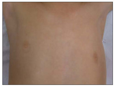 Figure 1. Asymmetry of the axillary folds and left nipple depression. Non-contrast-enhanced helical computed tomography was performed with the objective of evaluating the soft tissues and bone structures, demonstrating pectoralis major and minor muscles at the left (Figure 2). Vertebral bodies, costal arches, mediastinum and pulmonary parenchyma were found within normality parameters. A supplementary ultrasonography study (Figure 3) was performed, demonstrating the same findings. Doppler ultrasonography of the subclavian and axillary vessels demonstrated symmetrical vascular structures, with normal caliper as well as normal arterial and venous blood flow. Radiography of the hands for comparative analysis confirmed subtle hypoplasia of the left hand bones, with no sign of syndactyly or other local skeletal deformity (Figure 4). 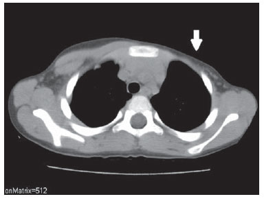 Figure 2. Computed tomography. Axial section and soft-tissue window showing agenesis of the left pectoral muscle (arrow). Compare with the right chest wall, where the major and minor pectoral muscles present a normal tomographic appearance. 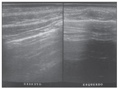 Figure 3. Ultrasonography showing agenesis of the left pectoral muscle as compared with the normal right side.  Figure 4. Comparative anteroposterior radiography of wrists demonstrating subtle hypoplasia at left. The findings are better observed on the capitate and hamate bones, as well as on the fourth and fifth metacarpal bones. The radial epiphyses are symmetrical and both ossification centers of the ulnar epiphysis are not yet present. DISCUSSION Poland's syndrome is a rare congenital disorder fundamentally characterized by unilateral agenesis of the pectoral muscles associated with ipsilateral brachydactyly(1- 4). Individuals affected by this syndrome may also present several other alterations, such as hypoplasia or agenesis of the nipple and breast parenchyma, hypoplasia or aplasia of the costal arches, shortening of upper limb due to hypoplasia of the radius and ulna, syndactyly, among other less frequently described abnormalities, such as dextrocardia, single transverse palmar crease, scapular anomalies, vertebral anomalies, and anomalies of the urinary and digestive systems(3,5-9). With a still unknown etiology, Poland's syndrome affects a higher percentage of boys than girls, with the right side of the body being most frequently affected. In spite of the eminently clinical diagnosis, the radiological evaluation is justifiable for staging of the alterations found in addition to the classical findings, thus directing the treatment. The early diagnosis may contribute to pediatric, orthopedic, psychomotor and aesthetic management (the latter, with greater impact on women with amastia), as well as to the awareness of parents and family members, and to the social and school life of the child(1,2,4,6). REFERENCES 1. Araújo ALN, França JCQ, Vieira SC, et al. Síndrome de Poland. Brasília Med. 2009;46:3958. 2. Caldas FAA, Isa HLVR, Trippia AC, et al. Síndrome de Poland: relato de caso e revisão da literatura. Radiol Bras. 2004;37:3818. 3. Dilmann JR, Sanchez R, Ladino-Torres MF, et al. Expanding upon the unilateral hyperlucent hemithorax in children. Radiographics. 2011;31:72341. 4. Pearl M, Chow TF, Friedman E. Poland's syndrome. Radiology. 1971;101:61923. 5. Chung EM, Cube R, Hall GJ, et al. From the archives of the AFIP: breast masses in children and adolescents: radiologic-pathologic correlation. Radiographics. 2009;29:90731. 6. Freitas RS, Tollazi ARD, Martins VDM, et al. Poland's syndrome: different clinical presentations and surgical reconstructions in 18 cases. Aesthetic Plast Surg. 2007;31:1406. 7. Friedman T, Reed M, Elliott AM. The carpal bones in Poland syndrome. Skeletal Radiol. 2009;38:58591. 8. Jeung MY, Gangi A, Gasser B, et al. Imaging of chest wall disorders. Radiographics. 1999;19:61737. 9. Mutlu H, Sildiroglu O, Basekim CC, et al. A variant of Poland syndrome associated with dextroposition. J Thorac Imaging. 2007;22:3412. 1. MD, Radiologist at Clínica Via Imagem, Xanxerê, SC, Brazil. 2. MD, Vascular Surgeon and Vascular Echographist at Pró Circulação - Clínica de Angiologia, Cirurgia Vascular e Ecografia Vascular, Xanxerê, SC, Brazil. 3. MD, Pediatrician at Materclínica Materno Infantil, Xanxerê, SC, Brazil. 4. Graduate Student of Medicine, Universidade Católica de Pelotas (UCPel), Pelotas, RS, Brazil. Mailing Address: Dr. João Lourenço Bazzi Jr Rua Celestino do Nascimento, 573 Xanxerê, SC, Brazil, 89820-000 E-mail: joaobazzijr@gmail.com Received October 1st, 2011. Accepted after revision February 9, 2012. * Study developed at Clínica Via Imagem, Xanxerê, SC, Brazil. |
|
Av. Paulista, 37 - 7° andar - Conj. 71 - CEP 01311-902 - São Paulo - SP - Brazil - Phone: (11) 3372-4544 - Fax: (11) 3372-4554