Radiologia Brasileira - Publicação Científica Oficial do Colégio Brasileiro de Radiologia
AMB - Associação Médica Brasileira CNA - Comissão Nacional de Acreditação
 Vol. 45 nº 2 - Mar. / Apr. of 2012
|
CASE REPORT
|
|
Intrathoracic rib: a case report |
|
|
Autho(rs): Fernando Henrique Guimarães de Carvalho1; Gesner Pereira Lopes2 |
|
|
Keywords: Intrathoracic rib; Costal anomaly; Computed tomography. |
|
|
Abstract: INTRODUCTION
Anatomic rib variants are found in 2% of the population(1) and include bone dysplasias, focal abnormalities, cervical rib, intercostal synostosis, bifid anterior extremity, either in association or not with thoracic vertebral malformations. Intrathoracic rib was first described in 1947 by Lutz, and since then it is presented as a rare, generally symptomatic congenital abnormality, most of times diagnosed as an additional finding at chest radiography(24). However, in cases where the abnormality arises from a vertebral body, the diagnosis by conventional radiography becomes more difficult and in such cases computed tomography (CT) is required, principally for diagnostic differentiation from hilar or mediastinal masses(57). CASE REPORT Female, 51-year-old patient with leftsided sharp chest pain for approximately two years with progressive development to cervicobrachialgia associated with a subtle thoracic scoliosis. After consultations with an orthopedist and recurrence of the painful condition, radiography of the chest and cervical spine was requested to elucidate the diagnosis. Chest radiography demonstrated bone density image, with bone cortex and medulla, lateral convexity, progressive reduction of craniocaudal dimensions, located in the upper and middle thirds of the left hemithorax, associated with thoracic scoliosis (Figure 1). Once the presence of intrathoracic rib was observed, CT was suggested for a better evaluation. With the aid of 3D reconstruction, it was possible to thoroughly visualize a vertical bone image joining the fourth rib posteriorly at left (Figure 2), better identified on oblique view (Figure 3). Coronal reconstruction of the thoracic spine allowed characterization of dextroconcave scoliosis in association with structural deformity at T4-T5 level (Figure 4). 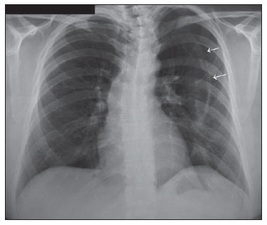 Figure 1. Chest radiography. Posteroanterior view demonstrating bone density image with bone cortex and medulla, lateral convexity, progressive reduction of craniocaudal dimensions, located in the upper and middle thirds of the left hemithorax, associated with thoracic scoliosis. 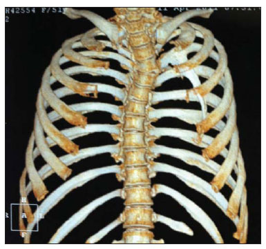 Figure 2. Computed tomography 3D reconstruction demonstrating vertical bone image joining the fourth rib posteriorly at left. 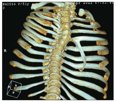 Figure 3. Computed tomography 3D reconstruction. Oblique view providing a better visualization of the bone image insertion into the anteroinferior aspect of the fourth costal arch posteriorly. 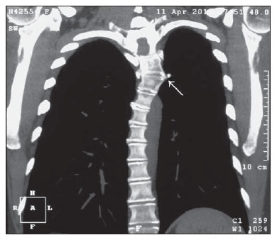 Figure 4. Computed tomography coronal reconstruction of thoracic spine showing dextroconcave scoliosis associated with structural deformity at T4-T5 level. DISCUSSION Despite the 45 cases reported in the English literature(4), the mechanism of development of such anomaly is still to be understood. The most accepted hypothesis would be that of a failure in the fusion of cephalic and caudal processes of sclerotomes between the fourth and sixth embryogenesis weeks(1,4). With no sex prevalence, most frequently at right, and with morphology similar to a normal rib extending laterally downwards in the extrapleural space, intrathoracic rib may reach the diaphragm. The range of differential diagnosis includes: exostosis, bone tumors, chest drain and calcified pleural plaques. The condition is generally asymptomatic, but chest pain and dyspnea have already been reported( 3). It is strongly suggested that the risk for pulmonary injury is enhanced if these patients are affected by chest trauma. The early diagnosis based on either an additional finding or any sign/symptom related to the malformation is extremely relevant, principally because it rules out differentially diagnosed conditions which would require follow-up and a possible definitive treatment. It can be affirmed that, in spite of being a rare malformation, intrathoracic rib is relatively easy to diagnose by means of conventional radiography in cases where it arises from an otherwise normal rib. However, in cases where the rib arises from a vertebral body, CT becomes essential for a correct diagnosis, avoiding not only unnecessary interventions, but also avoiding that differentially diagnosed conditions go untreated. REFERENCES 1. Kelleher J, O'Connell DJ, MacMahon H. Intrathoracic rib: radiographic features of two cases. Br J Radiol. 1979;52:1813. 2. Lutz,P. Über eine ungewöhnliche Rippenanomalie zugleich ein Beitrag zur distalen Rippengabelung. Wien Klin Wochenschr. 1947;59:8469. 3. Laufer L, Schulman H, Hertzanu Y. Intrathoracic rib demonstrated by helical CT with three-dimensional reconstruction. Eur Radiol. 1999;9:601. 4. Hasan I, Mahajan P, Ahamad N, et al. A rare case of left bifid intrathoracic rib associated with supernumerary rib and block vertebrae. Kuwait Radiol J. 2011;2:217. 5. Trigaux JP, Sibille Y, Van Beers B. Intrathoracic rib: CT features. J Comput Assist Tomogr. 1990;14:1335. 6. Freed C. Intrathoracic rib: a case report. S Afr Med J. 1972;46:11657. 7. Weinstein AS, Mueller CF. Intrathoracic rib. Am J Roentgenol Radium Ther Nucl Med. 1965;94:58790. 1. MD, Resident of Radiology and Imaging Diagnosis, Universidade Federal do Triângulo Mineiro (UFTM), Uberaba, MG, Brazil. 2. Master, Titular Member of Colégio Brasileiro de Radiologia e Diagnóstico por Imagem (CBR), Head of Division of Radiology and Imaging Diagnosis, Universidade Federal do Triângulo Mineiro (UFTM), Uberaba, MG, Brazil. Mailing Address: Dr. Fernando Henrique Guimarães de Carvalho Praça Frei Eugênio, 444, ap. 702, São Benedito Uberaba, MG, Brazil, 38010-280 E-mail: fhgc3@hotmail.com Received August 16, 2011. Accepted after revision October 7, 2011. Study developed at Universidade Federal do Triângulo Mineiro (UFTM), Uberaba, MG, Brazil. |
|
Av. Paulista, 37 - 7° andar - Conj. 71 - CEP 01311-902 - São Paulo - SP - Brazil - Phone: (11) 3372-4544 - Fax: (11) 3372-4554