Radiologia Brasileira - Publicação Científica Oficial do Colégio Brasileiro de Radiologia
AMB - Associação Médica Brasileira CNA - Comissão Nacional de Acreditação
 Vol. 45 nº 2 - Mar. / Apr. of 2012
|
ORIGINAL ARTICLE
|
|
Enhancement of radiological protection through an internal quality assessment cycle |
|
|
Autho(rs): Filipe Morais de Figueiredo1; Zenewton André da Silva Gama2 |
|
|
Keywords: Radiological protection; Quality enhancement; Institutional evaluation; Health care quality assurance. |
|
|
Abstract: INTRODUCTION
A quality management program can be defined as a "set of structural elements and activities with the specific purpose of promoting continuous improvement of quality"( 1,2). In what regards the three starting points of the program, three areas of different activities may be identified in the management program, namely, improvement cycles, monitoring and quality planning(1,2). The improvement cycles, corresponding to one of the activities by which the implementation of a quality management program can be started, resort to detected quality problems to analyze and develop solutions for such problems(1,2). A problem intrinsically associated with radiology services is related to the fact that the population is increasingly exposed to a greater amount of ionizing radiation originated from medical diagnosis apparatuses( 3). According to the 2006 report of the National Council on Radiation Protection and Measurements (NCRP), in 2006, the North American population was exposed to seven times more ionizing radiation originating from medical procedures than in 1980(3). According to the report of the United Nations Scientific Committee on the Effects of Atomic Radiation, in 2000, the patients were exposed to approximately 200 times more ionizing radiation than health workers, and in some countries such value may be almost 500 times higher. According to the World Health Organization, at least 3,000 patients were affected by incidents involving ionizing radiation during medical procedures over the previous 30-year period(4). Among all the diagnostic imaging techniques, conventional radiology exposes the patient to lower radiation doses for a shorter period of time, in comparison with techniques such as interventional radiology and computed tomography(5), but one should not neglect any procedure which may minimize the ionizing radiation dose to which the patient is exposed(6), as during the performance of such exams, the operator must always follow the ALARA (As Low As Reasonably Achievable) principle, i.e., utilize the lowest possible radiation dose to achieve the best diagnostic result(7). In radiology services, there are basic principles of radiological protection intended to minimize the ionizing radiation dose to which patients are exposed(8), but such principles are not always followed by the involved professionals. Thus, it is important to optimize the work procedures, since they directly affect the quality and safety in patients care(9). Some of the main radiological protection measures which can/must be adopted in the pursuit of minimizing undesirable effects of ionizing radiation, and which are many times forgotten on account of various factors, are the following: a) always utilize gonad shields and lead skirt aprons on patients, except in cases where such shielding exclude or degrade important diagnostic information(10); b) always make the best efforts to minimize the repetition of radiographic studies(4); c) utilize appropriate collimation for the area of interest in the study(9); d) optimize technical factors (acquisition time, mA and kV) to reduce the radiation dose while maintaining radiographic quality(3); e) avoid studies during pregnancy(11). Based on the above considerations, the authors have implemented a cycle of improvement of radiological protection for patients during examinations in a radiology service. The general objective of the present study was the maximization of the radiological protection of patients while reminding radiologists of the importance of radiological protection. More specifically, the objectives were the following: to evaluate the quality of radiological protection with basis on appropriate criteria; to identify the most representative problems in order to guide their solution; and assess the effectiveness of an intervention aimed at improving baseline quality. MATERIALS AND METHODS Design and scope of the study The present study approaches an internal improvement cycle characterized by the identification and prioritization of a problem of quality (radiological protection of patients during the performance of radiological examinations), its analysis, assessment of quality based on criteria, intervention to promote improvement and quality reevaluation to verify the effectiveness of the applied intervention. Such cycle of improvement was implemented during the year of 2010 in the Service of Radiology at Unidade Funcional de Olhão, Unidade de Recursos Assistenciais Partilhados do Agrupamento de Centros de Saúde (ACES) Central, which together with the ACES do Barlavento and ACES do Sotavento comprises the Administração Regional de Saúde do Algarve (Algarve Regional Health Administration), located in the Algarve region, in Portugal. In this radiology service, where only conventional radiology studies are performed, there are three radiologists, although occasionally radiologists from other ACES Central services provide their assistance in the operation of the service. Development of the quality criteria After a qualitative analysis on the causes of the inappropriate radiological protection by means of a cause-effect diagram( 12), a task group comprising three radiologists developed requirements or quality criteria related to radiological protection of patients. Definitions, exceptions, and clarification of each one of those criteria are presented on Table 1. 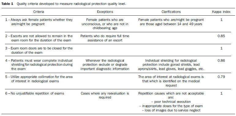 All of the defined criteria are related to the assistance process, as they comprise the activities or procedures undertaken by the health professionals to transform resources into results. Additionally, the authors have taken the precaution of analyzing the validity and reliability of the criteria. Purpose, contents and foundations for each one of the criteria were considered appropriate and, in a pilot study by means of a test-retest design (n = 30), a satisfactory reliability (kappa index) was demonstrated. Population and sample The target population of the criteria comprised all patients seeking the service for their exams, except in the case of criterion 1, which applied only to female patients. The temporal parameters for case extraction varied according to the criteria over one week (criteria 2, 3, 4 and 5), one month (criterion 1) and a quadrimester (criterion 6). The sampling of all criteria was systematic and random, and the sample included 60 cases for each criteria. Data collection Several information sources were utilized in the collection of data related to compliance with the criteria, namely, review of the clinical process (criterion 1), patient questionnaire (criterion 2), procedural compliance (criteria 3 and 4) and analysis of the images on the image treatment console (criteria 5 and 6). As regards timing, the evaluation was concurrent for criteria 3 and 4, and retrospective for criteria 1, 2, 5 and 6. The initiative of undertaking such evaluation came from the professionals themselves, that is, it was an internal process, with the professionals from the service being responsible for collecting the data and performing a cross-analysis where each professional evaluates the actions of another peer. The time elapsed between the two evaluations corresponded to approximately eight months, the period over which the improvement intervention was carried out. Improvement intervention For the development of an improvement intervention plan, the authors have resorted to a participative planning method which included and comprised the radiologists related to the processes which are object of improvements. The set of interventions that originated from the generation of ideas within the group was distributed over an affinity diagram into three groups of actions to be implemented, as follows: 1. Education of the radiologists on movement and transfer of patients, children immobilization, radiological protection and interaction with patients. 2. Changes in registration forms, adding YES and NO filling spaces on the requests for female patients asking whether they are/might be pregnant. 3. Disclosure of results: by means of a storyboard recording the progress of the activities located in a place where all professionals could see it, and awareness development actions for the follow-up of the study results. Once the improvement action was defined, the authors decided to utilize two instruments to assure and supervise the implementation of the action plan: the storyboard, utilized to record the progress of the activities at the sight of all involved professionals; and a Gantt chart, which is a graphic representing the scheduled time required for the execution of the actions, as well as the names of the responsible agents for each one of them. Data analysis Both in initial evaluations and in the reevaluation, calculations of point and interval estimates (95% confidence level) were performed on the compliance with criteria in random selected samples. In order to estimate the improvement observed between the reevaluation and the initial evaluation, absolute and relative improvements were calculated for each one of the criteria. In order to prove the statistical significance of the detected improvement, a unilateral hypothesis test was performed by means of the calculation of the Z value, considering as the null hypothesis the absence of improvement, which was rejected whenever the p-value was lower than 0.05. Additionally, the main quality defects identified at both evaluations were graphically represented. For such a purpose a before-and-after Pareto chart was utilized, for being a complete and informative representation( 2) which makes the prioritization of intervention strategies easier. In the early stage of the chart construction, a table of absolute and relative non-compliance frequencies was developed. Subsequently, the before-and-after Pareto chart was built on a three-axis Cartesian plane, where the central axis represents the absolute frequencies demonstrating the results from the two evaluations, and the left and right axes represent, respectively, the relative percentage of non-conformities in the first evaluation and in the reevaluation. The lines drawn on the chart represent the accumulated frequency of quality defects observed in each evaluation. RESULTS Basic level of quality in radiological protection Table 2 demonstrates that all six criteria presented a high rate of compliance already in the first evaluation (minimum = 85%; maximum = 93%). The highest compliance level was observed for the criterion "Escorts are not allowed to stay in the room during the performance of the exam" (criterion 2), with a compliance rate of 93% (CI 95%: 8799), followed by the criteria "Always ask female patients whether they are/might be pregnant" (criterion 1), with a compliance rate of 92% (CI 95%: 8797).  Analysis of identified quality defects and intervention priorities On the before-and-after Pareto chart, one can observe and compare the values of the corresponding non-compliances for each one of the six criteria at the two evaluations. It is also possible to identify the most problematic criteria denominated as "vital few" according to the "Pareto's principle"(3). In the first evaluation, a pair of criteria (criteria 6 and 4) stood above the others for representing 44.7% of all defects identified amongst the 100% representing all defects under the six criteria included in the present study, which allows those two criteria to be considered as the "vital few" criteria which were prioritized in the effort to obtain improvements (as previously indicated under the topic "Improvement Intervention"). In the second evaluation, criteria 3 and 6 represented approximately 62% of the noncompliance cases, i.e., being considered as "vital few" criteria. Thus the planning for a new intervention should prioritize those two criteria, not neglecting the others, as in the second evaluation all criteria presented non-conformities. Reevaluation of the level of quality and effectiveness of the improvement intervention All of the six criteria presented a high compliance rate in the reevaluation (Table 2). Additionally, in absolute terms, improvements were observed for every criteria, except for the criterion "examination room doors must be closed throughout the performance of the exam" (criterion 3), which presented a higher number of nonconformities in the reevaluation than in the first evaluation. All of the criteria which demonstrated improvements (criteria 1, 2, 4, 5, and 6) presented relative improvement rates above 30% between the two evaluations. As regards statistical significance of such improvement, the criteria "The patients must wear complete individual shielding for radiological protection during the exam" and "Utilize collimation appropriate for the area of interest in radiological examinations" (criteria 4 and 5 respectively) presented a p-value lower than 0.05, characterizing a statistically significant improvement in the level of quality regarding those criteria. Statistically significant improvements were not achieved in the remaining criteria. (Table 2). According to Table 2 and Figure 1, one can observe that the set of the six criteria totals 21 non-conformities in the second evaluation, 17 less than in the first evaluation, corresponding to an absolute improvement of approximately 45% between the evaluations (corresponding to the area highlight on the chart of the second evaluation). However, in a negative result, criterion 3 ("exam room doors must be closed throughout the performance of the exam") presented a higher rate of non-compliances in the reevaluation than in the first evaluation (absolute improvement = 2%). The statistical significance of the worsening was not calculated, as the sensitivity of the test for the improvement hypothesis was prioritized with the method of unilateral analysis described in the methodology. 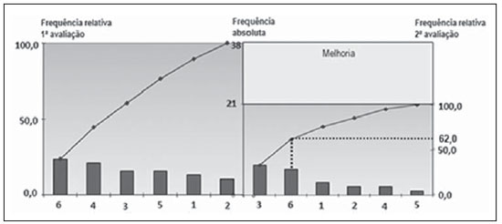 Figure 1. Pareto's chart showing the absolute, relative and accumulated noncompliance frequencies before and after the intervention. 1. Always ask female patients whether they are/might be pregnant; 2. Escorts are not allowed to remain in the exam room for the duration of the exam; 3. Exam room doors are to be closed for the duration of the exam; 4. Patients must wear complete individual shielding for radiological protection during the exam; 5. Utilize appropriate collimation for the area of interest at radiological exams; 6. No unjustifiable repetition of exams. The two criteria considered as "vital few" in the first evaluation criteria 6 and 4, on which more improvement activities were developed in the intervention presented a significant decrease in the number of non-conformities in the reevaluation, with a relative improvement of 77% for criterion 4, while criterion 6 presented a relative improvement of 33%. However, criterion 6 ("No repetition of exams"), although presenting a decrease in the number of non-conformities between evaluations, remained as the second criteria with more non-conformities (second only to criterion 3). DISCUSSION The results obtained in this improvement cycle contribute to the understanding of the effectiveness of the cycles of institutional quality assessments in radiology services. In general, the method based on the internal quality management scope was effective in changing the professionals' attitude and in improving the level of good practices in radiological protection. Although the improvement intervention planned and implemented by the professionals in the center were not completely effective in solving all of the evaluated radiological protection deficiencies, the consolidation of the philosophy and structure of quality management in this institution paved the way to the continuity of the same evaluation cycle and for the evaluation and improvement of other priority problems in the pursuit of excellence. During the improvement cycle in the present study, actions which were within the capabilities of the professionals were utilized as quality criteria, aiming at the optimization of the radiological protection of the patients and which must always be adopted during the performance of a radiological exam and therefore, ideally, nonconformities should not exist with respect to the criteria utilized for the improvement cycle in the present study(10,13,14). However, the results of the present study demonstrate that non-conformities occurred in every criterion, even in the reevaluation after the implementation of some improvement strategies. The fact that the reevaluation revealed a criterion which presented a higher number of non-conformities than in the first evaluation, and also other criteria which did not present statistically significant improvements (criteria 1, 2, and 6), demonstrates that the improvement activities implemented during the intervention did not produce optimum effects for such criteria. The cause for such facts may reside in mistakes that may have been made in analyzing the causes of the problems or in the planning and/or implementation of the intervention. Thus, the continuity of the assessment cycle is important, improving the analysis of causes and planning of the intervention, which are the key steps to achieve improvements. The continuity of the improvement cycle also allows the consolidation of the processes, methods and tools utilized in this type of activity on a theme with accumulated experience, as well as it strengthens the improvements achieved in the first cycle, helping such improvements to become sustainable(2,15). As regards the difficulties experienced during the improvement cycle, which can be similar to those in other institutions undertaking similar projects, the task group reported some difficulties in the implementation and in the form of utilization of tools and methods applied in the activities, causing some delays in the improvement cycle. Possibly, this occurred because of the fact that this was the first time in which most of the involved radiologists had a contact with systematic quality improvement activities. One of the phases where more difficulty was encountered was the implementation of improvement strategies adopted after the first evaluation, where delays occurred in relation to the timeline initially established on the Gantt chart. A positive note, considering that the improvement cycle was the first contact of all intervenients with quality improvement activities, refers to the fact that the analyzed problems were internally prioritized by the professionals themselves. This allowed them to work on a known field, where they found themselves directly involved, recognizing its relevance, thus facilitating the commitment with the quality improvement actions. CONCLUSIONS The results of the present study demonstrated that the radiological protection quality level in the evaluated service, although reasonably high, presented a margin for improvement, particularly in the criteria concerning the non-repetition of exams and the utilization of appropriate individual shielding for radiological protection. The identification of such problems has motivated and guided an intervention based on the participative principle, allowing a significant improvement in two of the aspects with greater impact on quality, demonstrating the effectiveness of the evaluation cycle in this context. The fact that an optimum quality level was not reached (absence of non-conformities with the criteria) only highlights the relevance of the continuity of the evaluation cycle with the purpose of further improving the processes and the currently prioritized criteria. Finally, it is possible to say that undertaking such an improvement cycle has been beneficial for the patients, as the optimization of their radiological protection means that they will be exposed to lower radiation doses, a benefit of utmost importance, even not being directly perceived by them. REFERENCES 1. Saturno PJ. Gestión de la calidad. Concepto y componentes de un programa de gestión de la calidad. Manual del máster en gestión de la calidad en los servicios de salud. Módulo 1: Conceptos básicos. Unidad temática 2. 2ª ed. Murcia: Universidad de Murcia; 2008. 2. Juran JM, Gryna FM, Binghan RS. Manual de control de la calidad. 2ª ed. Barcelona: Reverté; 1990. 3. National Council on Radiation Protection and Measurements. Medical radiation exposure of the U.S. population greatly increased since the early 1980s. 2009. [cited 2011 Feb 23]. Available from: http://www.ncrponline.org/Press_Rel/Rept_160_Press_Release.pdf 4. Henriques S. IAEA culture shift needed to achieve patient radiation safety. 2011. [cited 2011 May 17]. Available from: http://www.iaea.org/newscenter/news/2010/cultureshift.html 5. Wall BF, Hart P. Revised radiation doses for typical X-ray examinations. Report on a recent review of doses to patients from mrdical X-ray examinations in the UK by NRPB. Br J Radiol. 1977;70:4379. 6. Sacchetti D. IAEA offers guidance on radiation protection for patients, travelers. 2011. [cited 2011 May 23]. Available from: http://www.iaea.org/newscenter/news/2010/guidanceprotect.html 7. International Atomic Energy Agency. Heavy component replacement in nuclear power plants: experience and guidelines. IAEA Nuclear Energy Series No. NP-T-3.2. Vienna: IAEA; 2008. 8. International Atomic Energy Agency. International action plan for the radiological protection of patients. 46th IAEA General Conference (2002) Documents; GOV/2002/36-GC(46)/12; IAEA. 2002. [cited 2011 May 27]. Available from: http://www.iaea.org/About/Policy/GC/GC46Documents/English/gc46-12_en.pdf. 9. Pisco JM. Imagiologia básica texto e atlas. 2ª ed. Lisboa: Lidel Edições Técnicas; 2009. 10. Soares FAP, Pereira AG, Flôr RC. Utilização de vestimentas de proteção radiológica para redução de dose absorvida: uma revisão integrativa da literatura. Radiol Bras. 2011;44:97-103. 11. D'Ippolito G, Medeiros RB. Exames radiológicos na gestação. Radiol Bras. 2005;38:447-50. 12. Saturno PJ, Gascon JJ. Métodos de análisis de los problemas de calidad. Manual del máster en gestión de la calidad en los servicios de salud. Módulo 3: Actividades básicas para la mejora continua: métodos y herramientas para la realización de ciclos de mejora. Unidad temática 11. 1ª ed. Murcia: Universidad de Murcia; 2008. 13. Ministério da Saúde. Portugal. Despacho nº258/ 2003 (2ª série). Diário da República nº 6, II Série, de 8 de janeiro de 2003. 14. Ministério da Saúde. Portugal. Decreto-Lei nº180/ 2002. Diário da República nº 182, Série I-A, de 8 de agosto de 2002. 15. Demig WE. Calidad, productividad y competitividad. La salida de la crisis. Madrid: Ediciones Días de Santos; 1989. 1. Master of Quality Management, Universidad de Murcia, Spain, Radiology Technician at the Unit of Radiology, Centro de Saúde de Olhão, Algarve, Portugal. 2. PhD, Universidad de Murcia, Spain, Professor, Department of Collective Health, Universidade Federal do Rio Grande do Norte (UFRN), Natal, RN, Brazil, Tutor, Program of Mastership of Quality Management in Health Services, Universidad de Murcia, Spain. Mailing Address: Dr. Zenewton André da Silva Gama Departamento de Saúde Coletiva, Universidade Federal do Rio Grande do Norte Rua General Gustavo Cordeiro de Farias, s/nº, Petrópolis Natal, RN, Brazil, 59010-180 E-mail: zgama@ufrnet.br Received October 3, 2011. Accepted after revision March 1st 2012. Study developed at Universidad de Murcia, Spain. |
|
Av. Paulista, 37 - 7° andar - Conj. 71 - CEP 01311-902 - São Paulo - SP - Brazil - Phone: (11) 3372-4544 - Fax: (11) 3372-4554