Radiologia Brasileira - Publicação Científica Oficial do Colégio Brasileiro de Radiologia
AMB - Associação Médica Brasileira CNA - Comissão Nacional de Acreditação
 Vol. 44 nº 6 - Nov. / Dec. of 2011
|
ORIGINAL ARTICLE
|
|
Sonographic evaluation of esophageal transit time: influence of gender and body mass index |
|
|
Autho(rs): Makoto Sakate1; Seizo Yamashita2; Caroline Guedes Correia Toledo3; Patricia Sanches3; Carlos Roberto Padovani4; Maria Aparecida Coelho de Arruda Henry5 |
|
|
Keywords: Ultrasonography; Intra-abdominal esophagus; Body mass index. |
|
|
Abstract: INTRODUCTION
Esophageal complaints are commonly found in both men and women of every age group, reflecting characteristics or consequences of different diseases. Thus, it is important that a simple, rapid and noninvasive evaluation tool is available to suggest and indicate more complex studies. Currently, there are several methods allowing the study of esophageal disorders, such as contrast-enhanced radiography with fluoroscopy, scintigraphy, manometry, upper endoscopy and prolonged esophageal pH-metry (24 hours). Notwithstanding, simple, rapid, low-cost and noninvasive methods which do not use ionizing radiation are necessary(17). In this context, transabdominal ultrasonography presents itself as a good imaging method to evaluate the intra-abdominal esophagus with the following advantages: it is characterized as an initial study, optimizing the indication for complementary studies, and providing better quality of life for the patient since this method is more innocuous, besides allowing the evaluation of treatment outcomes in patients with dysphagia(6). Objective The present study was aimed at utilizing intra-abdominal esophagus ultrasonography during swallowing of non-solid foods (water and yogurt), with the patients in orthostatic position, as a reference parameter in esophageal motor disorders, and evaluating the influence of gender and body mass index (BMI). MATERIALS AND METHODS The present study included 89 young adult individuals aged between 17 and 27 years, 40 of them women (mean age 20.13 ± 1.62 years) and 49 men (mean 20.43 ± 2.17 years), who did not show upper gastrointestinal disorders. The body mass index (BMI) was calculated for both the female and male individuals. The esophageal transit time (ETT) was measured with the volunteer in orthostatic position swallowing water (20 ml at room temperature), with a stopwatch being started as the observer felt the thyroid cartilage elevation, during swallowing by the volunteer, and stopped at the moment the bolus passage was visualized in the intra-abdominal esophagus. After a 30-minute interval, the maneuver was repeated with yogurt (20 ml at 5°C)(Figures 1 and 2). 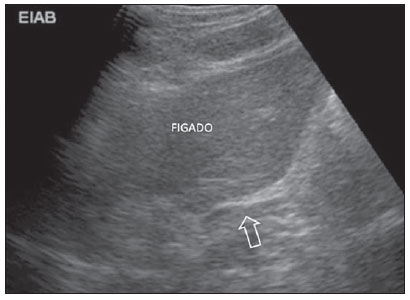 Figure 1. Sonographic image of upper abdomen demonstrating left hepatic lobe and intra-abdominal esophagus (arrow). 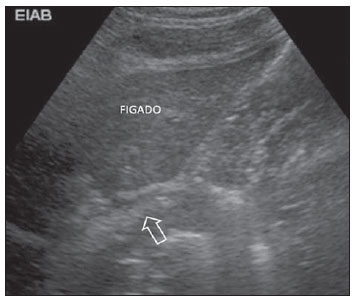 Figure 2. Sonographic image of upper abdomen demonstrating left hepatic lobe and intra-abdominal esophagus during the passage of a bolus (arrow). The ultrasonography apparatus utilized was a Toshiba Sonolayer SSH 140 A/G series (Toshiba; Tokyo, Japan) with a semi-convex 3.5 MHz transducer; and the stopwatch, a Seiko 3 BAR (Seiko; Tokyo, Japan). The experiment homogeneity as regards gender was evaluated by comparison of mean ETTs with water and yogurt. A similar statistical analysis was separately undertaken for BMI, with the two-tailed t-test, considering the impossibility of anticipating the difference significance. The comparison between ETT with water and yogurt was performed by means of the one-tailed t-test, assuming that the swallowing of less fluid substances results in longer ETT. The evaluation of associations between variables was performed with the Student's t test. Analysis of variance was utilized for a two-factor model (sex and BMI 50th percentile)(8). RESULTS The mean ETT for water was 5.84 ± 1.60 seconds in the female volunteers, and 6.66 ± 1.68 seconds in the male volunteers, with no statistically significant difference as regards gender. For yogurt, the mean ETT was 9.12 ± 1.96 seconds in the female volunteers, and 9.28 ± 1.70 seconds in the male volunteers (p > 0.05), also with no statistically significant difference as regards gender. The mean female BMI (21.53 ± 2.was significantly lower than the male BMI (23.65 ± 3.00 kg/m2)) (p < 0.001), but the association between BMI and ETT for water and yogurt was not statistically significant (p > 0,05), demonstrating that the fact of women presenting lower BMI was not a determining factor for the study of the differences (Table 1). 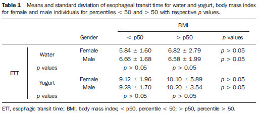 DISCUSSION In the present experiment, the volunteers, in orthostatic position, did not experience difficulty in holding the water/yogurt in their mouth, starting the swallowing only upon the observer command. All the steps of the investigation could be uneventfully performed in a simple manner, with all the volunteers comfortably positioned, in a quiet environment in the examination room, taking a short time to be completed. In the elderly with deglutition difficulties, such an examination should be avoided. In such cases, considering the higher incidence of malignant neoplasms in this age group, esophagogram, videofluoroscopy and even endoscopy would be the alternatives. The intra-abdominal esophagus could be easily reached and identified by ultrasonography, allowing the visualization of the water/yogurt passage(9,10). The ETT could be determined in both men and women, with the aid of a stopwatch started at the moment the deglutition of water or yogurt was initiated, until the substance passage was visualized through the intra-abdominal esophagus(10). The sonographic evaluation of the thoracic esophagus by external approach is impaired by the access difficulty, both through the anterior and posterior mediastina because of the presence of air in the lungs and bone tissue in the dorsal spine through which sound waves cannot appropriately propagate(11). The cervical esophagus can be sonographically evaluated, considering its lateral location in relation to the trachea, and because of its proximity to the surface and interposition of soft tissue such as the left thyroid lobe(12). In healthy individuals, the intra-abdominal segment of the esophagus is easily accessible by ultrasonography because of its posterior positioning in relation to the left hepatic lobe acting as an access window for ultrasound imaging (a relatively homogeneous organ that displaces the bowel loops with gas)(13-15). In the present study, the method demonstrated that there was no statistically significant difference between ETTs with water and yogurt and between genders, which was not observed with the individuals in dorsal decubitus(10), although a possible cause could be the individuals positioning (orthostasis). As regards the BMI, the values for the female volunteers were significantly lower than for the male volunteers, although not representing a determining factor to enhance differences in the ETTs with water and yogurt. With the advent of new technologies, new methods have been introduced allowing the evaluation of the esophagus, such as nuclear medicine, fiberoptic endoscopy, manometry, pH-metry and endoscopic ultrasonography. All of them emphasize the identification of anatomical and functional aspects of the esophagus(1618), but are invasive or utilize ionizing radiation with highly expensive equipment. The present study has allowed a less invasive, cost-effective and probably more easily available technique as compared with radiographic, manometric or scintigraphic methods, considering that this method requires only a conventional ultrasonography unit and a stopwatch to be reproduced. Other advantage of the method is the possibility of performing the examination out of the hospital environment. CONCLUSION The experimental method for evaluating the esophageal transit time with external approach by ultrasonography with the patient in orthostatic position, establishing correlation between genders and BMI is feasible, simple, non-invasive and do not require ionizing radiation. In the present study, the authors have observed that the ETT for water and yogurt has not presented statistically significant differences for both genders, even with the BMI significantly lower in women than in men. REFERENCES 1. Defagó M, Kuschnir E, Ninci CM, et al. Evaluation of methods for the study of gastroesophageal reflux. Rev Fac Cien Med Univ Nac Córdoba. 1985;43:913. 2. Penas ME. Motilidade esofagiana: ensaio iconográfico sobre cintilografia dinâmica do esôfago. Radiol Bras. 2007;40:4237. 3. Machado MM, Rosa ACF, Barros N, et al. Ultra-sonografia endoscópica (USE) do esôfago, estômago, cólons e reto. Radiol Bras. 2002;35:21923. 4. Giannini EG, Zentilin P, Dulbecco P, et al. Management strategy for patients with gastroesophageal reflux disease: a comparison between empirical treatment with esomeprazole and endoscopy-oriented treatment. Am J Gastroenterol. 2008;103:26775. 5. Henry MACA. Distúrbios motores do esôfago. In: Fraga GP, Sevá-Pereira G, Lopes LR. Atualidades em clínica cirúrgica. São Paulo, SP: Atheneu; 2011. p. 293306. 6. Henry MACA, Harbermann MC, Rocha OM. Esophageal motor disturbances in progressive systemic sclerosis. Dis Esophagus. 1999;12:513. 7. Nacif MS, Jauregui GF, Rocha VB, et al. Análise crítica da seriografia do esôfago, estômago e duodeno em um serviço de radiologia de um hospital geral. Radiol Bras. 2004;37:4259. 8. Zar JH. Biostatistical analysis. Upper Saddle River, NJ: Prentice-Hall; 1996. 9. Sakate M, Silveira GL, Muzio BP, et al. Refluxo gastroesofágico: estudo comparativo da receptividade e sensibilidade entre seriografia e ultrassonografia. Radiol Bras. 2009;42:2458. 10. Sakate M, Teixeira AS, Yamashita S, et al. Um novo método de avaliação do "tempo esofágico" com ultra-sonografia por abordagem externa. Radiol Bras. 2008;41:30912. 11. Penas ME. Motilidade esofagiana: ensaio iconográfico sobre cintilografia dinâmica do esôfago. Radiol Bras. 2007;40:4237. 12. Westra SJ, Derkx HHF, Taminiau JAJM. Symptomatic gastroesophageal reflux: diagnosis with ultrasound. J Pediatr Gastroenterol Nutr. 1994;19:5864. 13. Esposito F, Lombardi R, Grasso AC, et al. Transabdominal sonography of the normal gastroesophageal junction in children. J Clin Ultrasound. 2001;29:32631. 14. Johnson MC. The esophagus. Prim Care. 2001;28:45985. 15. Costa CD, Zomignan HP, Rocha JIP, et al. Refluxo gastroesofágico em pediatria: estudo radiológico. Pediatria (São Paulo). 1986;8:13640. 16. Henry MACA, Prado RG. Emprego da toxina botulínica no tratamento do espasmo difuso esofagiano. Relato de caso. Arq Gastroenterol. 1998;35:2747. 17. Foley LC, Slovis TL, Campbell JB, et al. Evaluation of the vomiting infant. Am J Dis Child. 1989;143:6601. 18. Cerri GG. Vísceras ocas. In: Rocha DC, Cerri GG, Prando A, et al. Ultra-sonografia abdominal. São Paulo, SP: Sarvier; 1988; p. 16986. 1. PhD, Assistant Professor at Faculdade de Medicina de Botucatu Universidade Estadual Paulista (FMB-Unesp), Botucatu, SP, Brazil. 2. Assistant Professor at Faculdade de Medicina de Botucatu Universidade Estadual Paulista (FMB-Unesp), Botucatu, SP, Brazil. 3. Biomedical Scientists, Students in the Course of Improvement in Photobiology and Radiobiology, Department of Dermatology and Radiotherapy, Faculdade de Medicina de Botucatu Universidade Estadual Paulista (FMB-Unesp), Botucatu, SP, Brazil. 4. Full Professor, Department of Biostatistics, Instituto de Biociências de Botucatu Universidade Estadual Paulista (IBB-Unesp), Botucatu, SP, Brazil. 5. Full Professor, Department of Surgery and Orthopedics, Faculdade de Medicina de Botucatu Universidade Estadual Paulista (FMB-Unesp), Botucatu, SP, Brazil. Mailing Address: Dr. Makoto Sakate Rua Aleixo Varoli, 651, Jardim Paraíso Botucatu, SP, Brazil, 18610-295 E-mail: msakate@fmb.unesp.br Received July 20, 2011. Accepted after revision October 28, 2011. * Study developed at Faculdade de Medicina de Botucatu Universidade Estadual Paulista (FMB-Unesp), Botucatu, SP, Brazil. |
|
Av. Paulista, 37 - 7° andar - Conj. 71 - CEP 01311-902 - São Paulo - SP - Brazil - Phone: (11) 3372-4544 - Fax: (11) 3372-4554