Radiologia Brasileira - Publicação Científica Oficial do Colégio Brasileiro de Radiologia
AMB - Associação Médica Brasileira CNA - Comissão Nacional de Acreditação
 Vol. 40 nº 5 - Sep. / Oct. of 2007
|
ORIGINAL ARTICLE
|
|
Agreement between magnetic resonance imaging and ultrasonography in the classification of schistosomal periportal fibrosis, according to Niamey's criteria |
|
|
Autho(rs): Eduardo Scortegagna Junior, Alberto Ribeiro de Souza Leão, José Eduardo Mourão Santos, Danilo Moulin Sales, David Carlos Shigueoka, Luciane Aparecida Kopke de Aguiar, Paulo Eugênio Brant, Ramiro Colleoni Neto, Durval Rosa Borges, Giuseppe D'Ippolito |
|
|
Keywords: Schistosomiasis, Periportal fibrosis, Ultrasound, Magnetic resonance imaging |
|
|
Abstract:
INTRODUCTION Schistosomiasis is a millenarian disease affecting more than 200 million people in about 76 countries in Africa, Asia and Americas, and represents a significant problem of public health in Brazil(1,2). Periportal fibrosis is the main cause of complications resulting from schistosomiasis and occurs in 4% to 8% of the patients who develop chronic infection. Hepatomegaly reflects the presence of granulomatous inflammation and occurs early in the disease progression. Periportal collagen deposition leads to a progressive portal vein occlusion, portal hypertension, and a possible progression to varices, high digestive hemorrhage, splenomegaly and hypersplenism. Periportal fibrosis is characteristic of the disease and may be identified by means of several diagnostic methods(2). Ultrasonography (US) has been the method most frequently utilized in this group of patients, demonstrating a typical pattern of abnormalities(3), especially including thickening along the walls of the portal vein and its branches characterized by periportal hyperechogenic bands(4). Aiming at establishing standardized protocol for diagnosing and quantifying periportal fibrosis in schistosomal patients, ultrasonographic criteria were initially defined in a World Health Organization consensus meeting held in Cairo in 1991(5), and later reviewed in the meeting on "Ultrasound Schistosomiasis" held in 1996 in Niamey, resulting in the evolution of the standard ultrasound scoring protocol to include qualitative criteria, considering the hepatic texture as a whole, besides the previous quantitative criteria utilizing the objective measurement of the portal branch wall thickening(4). US, although widely adopted in the assessment of patients with schistosomiasis, has demonstrated a moderate reproducibility in the classification of portal fibrosis(6). On the other hand, some studies have demonstrated the usefulness of magnetic resonance imaging (MRI) in the evaluation of hepatosplenic and vascular alterations in schistosomal patients, with high reproducibility(7). Additionally, the MRI capacity in the diagnosis of hepatic fibrosis has been well demonstrated(7). However, the role of MRI in the characterization and quantification of periportal fibrosis in schistosomal patients is still to be established. Considering its high reproducibility, anatomical detailing and high spatial resolution, some authors believe that MRI may be a more sensitive method than US for indicating the disease progression, grading and therapeutic response, as a result of a more comprehensive evaluation of the abdominal cavity(8). Therefore, it is important to establish the MRI value in the evaluation of periportal fibrosis. The present study was aimed at evaluating the MRI reproducibility and the agreement between US and MRI in the grading of periportal fibrosis in schistosomal patients, according to qualitative criteria defined in the ultrasound consensus meeting of Niamey for assessing periportal fibrosis.
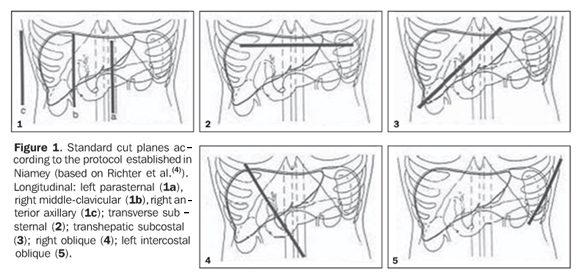
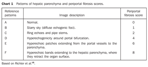
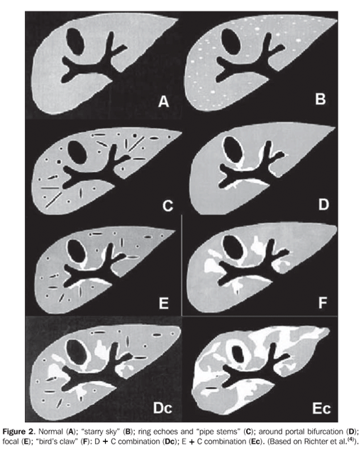
MATERIALS AND METHODS In the period between February/2005 and June/2006, a prospective, transversal, observational, double-blinded study was developed with 20 patients (10 men, and 10 women in the age range between 24 and 60 years, mean 42.75 years) referred by the clinic of schistosomiasis, Discipline of Clinical Gastroenterology, Universidade Federal de São Paulo/Escola Paulista de Medicina (Unifesp/EPM), with diagnosis of schistosomiasis mansoni. Such diagnosis had been obtained either by means of rectal biopsy or consistent clinical laboratory (signs of portal hypertension and/or positive fecal parasitologic examination) or epidemiological evidence (contact with contamined water of lakes, ponds, rivers in endemic areas), and subclassified into the hepatosplenic form of the disease because of the finding of portal hypertension and splenomegaly. Exclusion criteria were: patients with contraindication for MRI (cardiac pacemaker, cochlear implants, claustrophobia, cerebral aneurysm clips, allergy to paramagnetic contrast agents), patients with a previous history of alcoholism (ingestion of more than 160 g of ethanol/week), positive serology for hepatitis B or C viruses, with a previous history of proven autoimmune disease, and use of hepatotoxic drugs. The patients underwent US and MRI examinations with a minimum seven-day interval between studies. The results interpretation was performed by two independent observers, both residents in imaging diagnosis (A.R.S.L. and D.M.S.) and with at least two-year experience in the diagnostic methods utilized, with training in ultrasonography specifically for assessment of schistosomal patients. In a later moment, the observers met to reach a consensus as regards the sonographic classification of periportal fibrosis which later on this study was utilized as a gold standard in the comparison between readings and MRI studies. Ultrasound scan US scans were performed with a Philips EnVisor® model with a convex, multi-frequency, 2.5-4.5 MHz transducers, in patients after eight-hour fasting, according to the Niamey standard protocol(4) , with longitudinal (left paraesternal, right hemiclavicular, right anterior axillary), substernal, subcostal, Intercostal left and right oblique scans (Figure 1). The patterns observed were classified according the specific protocol (Chart 1 and Figure 2).
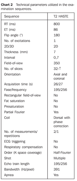
Magnetic resonance imaging MRI examinations were performed in a Siemens Sonata model system operating in a high field (1.5 T), with synergy coil and MRI sequences configured for periportal fibrosis assessment(9) (Chart 2).
The periportal fibrosis classification was performed similarly to the one performed by US, according to the previously mentioned criteria of the Niamey protocol adapted for MRI. The observers had no previous information on the sonographic classification. The statistical analysis was based on the calculation of the interobserver agreement between MRI and the consensual gold standard obtained by sonographic evaluation.
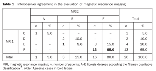
RESULTS The analysis of the MRI results by observers 1 and 2 demonstrated a global interobserver agreement of 70% (14/20 patients) (Table 1).
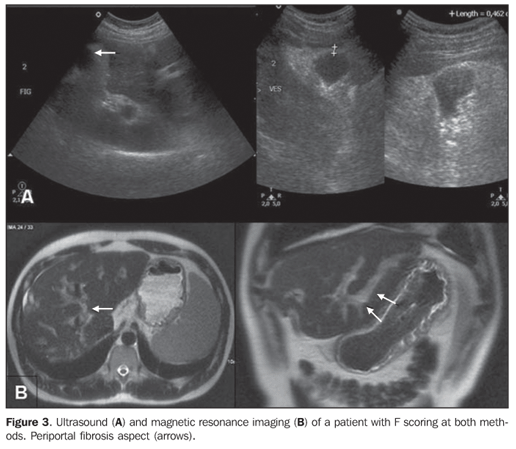
A poor agreement was found in a comparison between MRI results analyzed by the observer 1 and US results considered as gold standard. Agreement was found in only six cases (30%) (Figure 3), with discrepancies in the classification of the 14 remaining patients (70%) (Table 2).
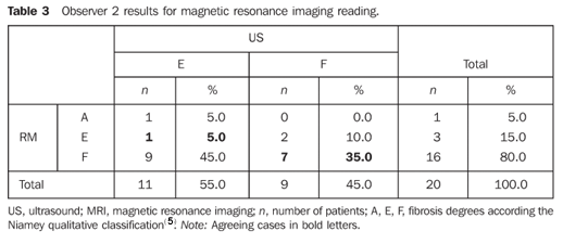
The results of the observer 2 were not very different. Agreement was found only in eight patients (40%) (Table 3).
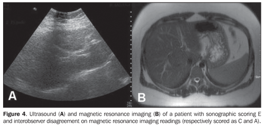
The highest variation in the classification level occurred with a patient classified as level E at US and Levels A and C at MRI (Figure 4).
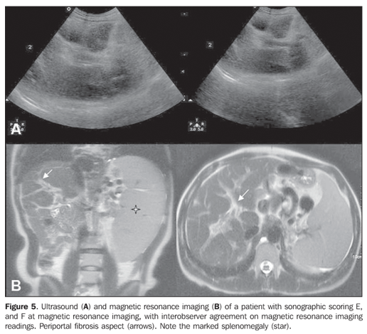
The other differences occurred from a classification level to its subsequent level, i.e., D to E in two cases, and from E to F in three cases (Figure 5).
DISCUSSION Schistosomiasis is extremely important both at national and international levels, because of its high prevalence and morbidity in patients at more advanced stages of disease(2,10). Generally, its treatment leads to a decrease in the levels of infection and improvement in some clinical findings such as hepatosplenomegaly and periportal fibrosis. The reduction of periportal fibrosis possibly prevents the emergence of portal hypertension and may result event in decrease of the portal pressure(11), a fact that highlights the significance of methods with high reliability and reproducibility for the disease follow-up. Ultrasonography has been widely utilized for evaluation of this disease, considering its non-invasiveness, low cost and wide availability, constituting the ideal method for assessment of the disease in large population groups. This method can be utilized for detecting portal fibrosis and hypertension (dilatation of portal and splenic veins and portosystemic collaterals). Evidently, US is more reliable than clinical methods for the diagnosis of hepatosplenic diseases. Periportal fibrosis, the essential lesion observed in schistosomiasis, is generally identified years after the infection onset, but it has already been report at earlier stages of infestation(4). On the other hand, ultrasonography has demonstrated just a moderate reproducibility in the assessment of schistosomal patients(6). MRI, on its turn, has progressively emerged as an extremely useful method in the evaluation of focal or diffuse hepatopathies(12), and with the high reproducibility(13) necessary for consistent results, stimulating the method adoption. Similarly, MRI has demonstrated to be capable of accurately identifying hepatic fibrosis(7,12). Notwithstanding, until the present moment there is no study in the literature comparing the performances of MRI and US in the evaluation of periportal fibrosis, so, as far as we are concerned, the present study is the first one to do it. The portal fibrosis aspects have already been described. On T1-weighted sequences, fibrosis presents as a hypointense band in relation to the liver, while on T2-weighted sequences it presents a high signal(11), the T2-weighted images being the most comprehensively utilized with this purpose according to the literature(8,9,13,14), a fact observed in the present study, although with a poor agreement with sonographic findings. The present study presents some limitations that must be pointed out. All of the patients receive Niamey sonographic reference patterns E or F, characterizing an advanced stage of periportal fibrosis and limiting the evaluation of the method for this group of patients. The method behavior in the assessment of patients with milder degrees of periportal fibrosis is still to be known. This also has prevented the kappa calculation of interobserver agreement for MRI results because there was no patient with all the levels of fibrosis according to the Niamey criteria. On the other hand, the patients who most benefited from a comprehensive assessment like that provided by MRI, would be those with more advanced stages of the disease with higher risks for developing complications. This was precisely the group included in the present study. The adaptation of the Niamey ultrasonographic criteria to MRI demonstrated a good reproducibility of the method (70%), however, a satisfactory relation between US and MRI classifications. Agreement between both methods was found in less than 50% of studied cases. A possible explanation for this results may be found in the subtle morphological variation between E and F reference patterns, whose the only difference is the parenchymal retraction in F, a finding that, like the others, presents a certain subjectivity. It is important to note that, even utilizing ultrasonography, the interobserver variability is considerable, which has been demonstrated in another study developed by the authors(6). Another factor to be considered is that the present study is based on the assumptions that US is the gold standard in the evaluation of periportal fibrosis, despite the absence of a consensus on this matter. Correlation between US, MRI and anatomopathological analysis of periportal fibrosis is still to be established to define which method presents the higher efficacy. Additional studies will be necessary to establish a proposal of a simplified classification model for periportal fibrosis to meet the needs of the professionals involved in the follow-up of this group of patients. Also studies involving a higher number of patients in early stages of the disease will be necessary to validate an eventual MRI usefulness. Finally, in the evaluation of periportal fibrosis according to the criteria defined in Niamey and adapted for MRI, the method has presented a good reproducibility, but low rate of agreement with US results, so its applicability is still controversial, particularly in schistosomal patients.
REFERENCES 1.Alves Jr A, Fontes DA, Melo VA, Machado MCC, Cruz JF, Santos EAS. Hipertensão portal esquistossomótica: influência do fluxo sangüíneo portal nos níveis séricos das enzimas hepáticas. Arq Gastroenterol 2003;40:203–208. [ ] 2.Ross AG, Bartley PB, Sleigh AC, et al. Schistosomiasis. N Engl J Med 2002;346:1212–1220. [ ] 3.Homeida M, Ahmed S, Dafalla A, et al. Morbidity associated with Schistosoma mansoni infection as determined by ultrasound: a study in Gezira, Sudan. Am J Trop Med Hyg 1988;39: 196–201. [ ] 4.Richter J, Hatz C, Campagne G, Berquist NR, Jenkins JM. Ultrasound in schistosomiasis. A practical guide to the standardized use of ultrasonography for the assessment of schistosomiasis-related morbidity. Second International Workshop, Niamey, Niger, October 22–26, 1996. [ ] 5.International conference on schistosomiasis. Cairo, Egito, 1995. [ ] 6.Santos GT, Sales DM, Shigueoka DC, et al. Reprodutibilidade da classificação ultra-sonográfica de Niamey na avaliação da fibrose periportal na esquistossomose mansônica. Radiol Bras 2006;39 (Supl 2):57. [ ] 7.Bezerra ASA, D'Ippolito G, Caldana RP, Cecin AO, Szejnfeld J. Avaliação hepática e esplênica por ressonância magnética em pacientes portadores de esquistossomose mansônica crônica. Radiol Bras 2004;37:313–321. [ ] 8.Patel SA, Castillo DF, Hibbeln JF, Watkins JL. Magnetic resonance imaging appearance of hepatic schistosomiasis, with ultrasound and computed tomography correlation. Am J Gastroenterol 1993;88:113–116. [ ] 9.Kashitani N, Kimoto S, Tsunoda M, et al. Portal blood flow in the presence or absence of diffuse liver disease: measurement by phase contrast MR imaging. Abdom Imaging 1995;20:197–200. [ ] 10.Hatz CF. The use of ultrasound in schistosomiasis. Adv Parasitol 2001;48:225–284. [ ] 11.Homeida MMA, Eltoum IA, Ali MM, et al. The effectiveness of annual versus biennial mass chemotherapy in reducing morbidity due to schistosomiasis: a prospective study in Gezira-Managil, Sudan. Am J Trop Med Hyg 1996;54:140–145. [ ] 12.Vitellas KM, Tzalonikou MT, Bennett WF, Vaswani KK, Bova JG. Cirrhosis: spectrum of findings on unenhanced and dynamic gadolinium-enhanced MR imaging. Abdom Imaging 2001;26: 601–615. [ ] 13.Lambertucci JR, Silva LC, de Queiroz LC, Pinto-Silva RA. Magnetic resonance imaging and ultrasound in hepatosplenic schistosomiasis mansoni. Rev Soc Bras Med Trop 2004;37:333–337. [ ] 14.Herborn CU, Akkoyunlu B, Ruehm G. Schistosoma mansoni-Befall der Leber – Darstellung mittels Magnetresonanztomographie (MRT). Rofo 2002;174:490–494. [ ]
Received December 15, 2006. Accepted after revision February 8, 2007.
* Study developed in the Department of Imaging Diagnosis and Disciplines of Clinical and Surgical Gastroenterology at Universidade Federal de São Paulo/Escola Paulista de Medicina (Unifesp/EPM), São Paulo, SP, Brazil. |
|


Av. Paulista, 37 - 7° andar - Conj. 71 - CEP 01311-902 - São Paulo - SP - Brazil - Phone: (11) 3372-4544 - Fax: (11) 3372-4554