Radiologia Brasileira - Publicação Científica Oficial do Colégio Brasileiro de Radiologia
AMB - Associação Médica Brasileira CNA - Comissão Nacional de Acreditação
 Vol. 40 nº 5 - Sep. / Oct. of 2007
|
ORIGINAL ARTICLE
|
|
Uterine embolization for management of symptomatic fibroids: quality-of-life impact |
|
|
Autho(rs): Nestor Kisilevzky |
|
|
Keywords: Therapeutic embolization, Uterus, Leiomyoma, Quality of life, Interventional radiology |
|
|
Abstract:
Nestor Kisilevzky Master in Surgery - Universidade Estadual de Campinas (Unicamp), Campinas, SP, Interventional Radiologist at Endovascular Clínica Médica Ltda., São Paulo, SP, Brazil
INTRODUCTION Uterine myomas, also called leiomyomas or fibroids, are the most common benign gynecologic tumors, and may be present in up to 40% of women in child-bearing age(1). Most frequently, myomatosis affects nulliparous, obese, black women, or those with family history of myomatosis or hyperestrogenic syndrome(2). Despite the absolutely benign nature of myomas, complaints of uncomfortable symptoms such as menorrhagia (abnormal menstrual bleeding), dysmenorrhea (painful menstruation), a felling of pressure in the pelvis, increase in the urinary frequency, pain, infertility or increase in abdominal volume and palpable pelvic mass are very frequent(3). Clinical presentation is variable and is particularly related to the size, location and number of myomas, but certainly myomatosis symptoms, when present, result in a remarkable impairment of the patients' quality of life. The most frequent symptom is an abnormal uterine bleeding (menorrhagia) that usually is characterized by increased menstrual duration and blood flow volume, sometimes leading to anemia(4). Generally, women complain of a progression in the intensity, that is to say, a gradual increase in the menstrual blood flow obliging them to more frequent tampon or pad changes, or even to the use of diapers during the period. It is in these circumstances where women report that they avoid leaving home, scheduling professional or social activities because of the discomfort caused by the menstruation, or in order to avoid embarrassing or upsetting situations. Until recently, only two therapeutic modalities were available for treating symptomatic myomatosis: the surgical approach or hormone therapy. Definitely, hysterectomy is the most commonly performed procedure for the management of myomatosis. It is estimated that myomatosis accounts for one third of almost 400,000 hysterectomies performed in the United States of America(5). Despite the advantage of being definitely curative, hysterectomy is a formal surgical procedure that requires hospital stay for some days and a variable period of postoperative recovery and convalescence. Also, hysterectomy may be associated to a considerable blood loss, ureter injury, prolapse and other complications(6). Besides, it eliminates any possibility of fertility, which represents a deep loss for nulliparous women. Starting in 1991, a team of French doctors started the clinical utilization of uterine embolization as an alternative for treating uterine fibroids. The early results of these experiments were published in 1995 by the prestigious journal The Lancet, em 1995, suggesting that this was a highly efficient method for the management of uterine myomatosis symptoms(7). Since that time, numerous clinical experiments have been reported worldwide, demonstrating the validity of this promising percutaneous procedure(8-15). Uterine embolization is a minimally invasive modality of interventional radiology that consists in intentionally blocking the arteries that supply blood to the fibroids, causing their ischemia and shrinkage, resulting in the resolution of their symptoms. For this purpose, a thin catheter is inserted under local anesthesia, by means of puncture of the femoral artery in the inguinal region, and, under fluoroscopic vision generated by a digital angiography apparatus, the catheter is inserted and advanced to the uterine arteries. Gelatin microspheres measuring about 500-µ is then injected through the angiographic catheter until stasis of the branches that supply blood to the fibroids(16). Since we started applying this technique in Brazil, in 1999, we have already treated 450 patients. Technical and clinical outcomes from this initial experiment have been previously published(17). The present study reports outcomes from a group of patients submitted to uterine embolization, utilizing a system for evaluation and comparison of their quality of life before and after the treatment.
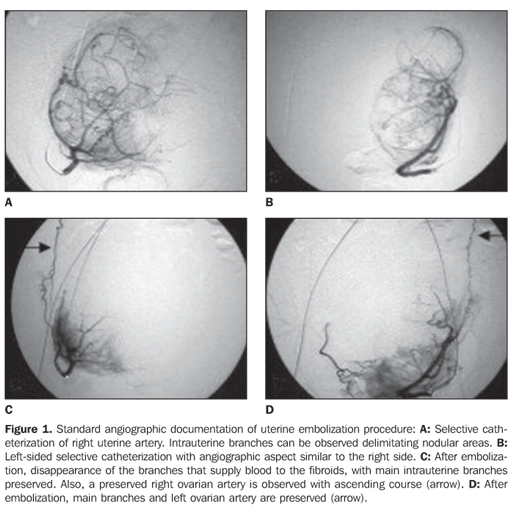
MATERIALS AND METHODS The present study evaluated data obtained from images and questionnaires answered by 40 women with symptomatic uterine myomatosis who were submitted to uterine embolization in the period between July/2004 and June/2006, in the Division of Interventional Radiology at Hospital Santa Catarina in São Paulo, SP, Brazil. The analysis included data from women with complaints resulting from the presence of uterine fibroids who, during the preoperative clinical evaluation and the period of postoperative clinical follow-up had answered a questionnaire about quality of life related to the disease treated. The mean age of patients was 38 years (age range = 22 - 46 years). As regards the patients' race, 31 women were Caucasian, five were black, and four Asian. The patients' gestational history showed that 28 of them were multiparous, and 12 were nulliparous. Amongst the patients included in the present study, 28 reported a job or professional activity, and 12 defined their activity as "housewives". The main complaint leading to the treatment was increased menstrual bleeding, with or without anemia in 29 patients, and pain or compressive symptoms secondary to uterine increase in 11. The diagnosis of uterine myomatosis was based on data collected from the clinical history, clinical examination and pelvic magnetic resonance imaging or ultrasound. All of the pre- and postoperative image studies were performed in an ambulatorial setting in different divisions of the hospital where the patients were submitted to the treatment. All the patients presented with increased uterine volume (mean = 666 cm3, ranging between 245 cm3 and 1.930 cm3). The method included puncture of the right femoral artery, angiographic study of uterine arteries, and embolization with calibrated 500-700-µ spheres (Embosferas®). The procedures were documented by means of pre- and post-embolization angiographic studies (Figure 1).
All the patients remained hospitalized 24 hours for rest and observation. At the time of the preprocedural clinical evaluation, all the patients answered a questionnaire for evaluation of their quality of life related to the presence of fibroids (Chart 1). This questionnaire was specifically developed for this purpose in the Georgetown University, Washington, USA(18), and translated into Portuguese by a professional translator. It includes 37 questions, and is divided into two parts. The first one includes eight questions about the intensity or severity of symptoms reported by the patient. Each of these questions offer five options corresponding to intensity: "no", "little", "reasonable", "much", "very much", with a corresponding scoring from 1 to 5. The final scoring is converted into a corrected score by means of a mathematical formula. The second part of the questionnaire includes 29 questions about the frequency in which the fibroids symptoms affect aspects of the patients' daily lives. These questions are divided into groups corresponding to six aspects: preoccupation, activity, mood/energy, self-control, embarrassment, and sexuality. Each of these questions offers five answering options to measure frequency: "at no time", "few times", "sometimes", "most of time" and "all the time", with a corresponding scoring from 1 to 5. The final score is converted into a corrected score by means of a mathematical formula. In the first part, the questionnaire presents the evaluation of symptoms intensity stratified from 0 to 100, meaning that 100 corresponds to the highest intensity or severity of the patients complaint. The second part evaluates the quality-of-life itself, i.e. the health condition expressed by a stratified scoring from 0 to 100, where 100 corresponds to the best level of quality-of-life in general, and for each of the specific aspects investigated by the questionnaire. During the 12-week follow-up period following the procedure, the patients were submitted to a pelvic magnetic resonance imaging for evaluating the uterine size as compared to the similar study previously performed. Also, they were asked to, again, answer the quality-of-life questionnaire. Data obtained from questionnaires answers as well as those concerning the uterine volumes evaluated by magnetic resonance imaging were transcribed into a Microsoft Excel worksheet for statistical analysis. Initially, all of the variables were descriptively analyzed. As for quantitative variables, this analysis was performed through observation of minimum and maximum values, as well as calculation of median and standard deviation. As for qualitative variables, absolute and relative frequencies were calculated. The paired Student's t test was utilized for analysis of premoment x postmoment equality hypothesis; when the data normality assumption was rejected, the non-parametric Wilcoxon test was utilized. The significance level utilized for these tests was 5%. The present study was submitted to the Committee for Ethics in Research of do Hospital Santa Catarina, whose approval (Process CEP019/06) has established that the study was conducted in compliance with the Resolution 196/96 of Conselho Nacional de Saúde (National Council of Health).
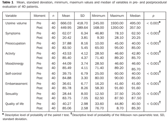
RESULTS All of the statistical variables evaluated before and after the procedure are shown on Table 1.
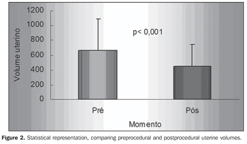
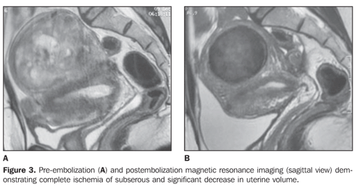
Mean uterine volume measured by post-embolization magnetic resonance imaging was 450 cm3, corresponding to a statistically significant decrease in volume of 32.5% (Figures 2 and 3).
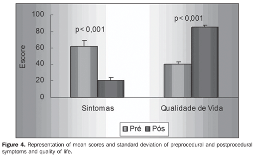
The mean score related to symptoms intensity reported in the preprocedural quality-of-life questionnaires was 62.07. The postprocedural quality-of-life questionnaires demonstrated a statistically significant decrease in the mean score to 20.42, representing a 67.1% improvement in symptoms. The analysis of total quality of life (health condition) related to the patients' myomatosis showed postprocedural score of 40.26, that changed substantially after the treatment, achieving 85.06. This was statistically significant, representing an improvement of 52.6% in the patients' quality of life (Figure 4).
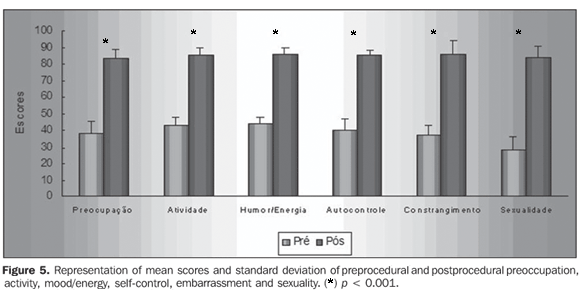
All the items evaluated by the questionnaire demonstrated changes after the treatment (Figure 5).
Mean score related to "preoccupation" in the preprocedural questionnaires was 37.87. In the post procedural questionnaires this score changed to 83.5, showing a statistically significant difference, and corresponding to an improvement of 54.6% in this item. Mean score related to "activity" in the preprocedural questionnaires was 43.53. In the postprocedural questionnaires, this score changed to 85.49, showing a statistically significant difference, and corresponding to an improvement of 49% in this item. Mean score related to "mood/energy" in the preprocedural questionnaires was 44.01. In the postprocedural questionnaires, this score changed to 85.39, showing a statistically significant difference, and corresponding to an improvement of 48.52% in this aspect. Mean score related to "self-control" in the preprocedural questionnaires was 39.75. In the postprocedural questionnaires, this score changed to 84.87, showing a statistically significant difference, and corresponding to an improvement of 53.31% in this item. Mean score related to "embarrassment" in the preprocedural questionnaires was 36.60. In the postprocedural questionnaires, this score changed to 85.78, showing a statistically significant difference, and corresponding to an improvement of 57.33% in this item. Mean score related to "sexuality" in the preprocedural questionnaires was 28.43. In the postprocedural questionnaires, this score changed to 83.55, showing a statistically significant difference, and corresponding to an improvement of 65.97%.
DISCUSSION Since the publication of the first scientific study on uterine embolization in 1995, much has been learned about this theme. The huge amount of papers published and studies presented in international congresses in the last ten years constitute unequivocal scientific evidence that uterine embolization is an effective and safe method for treating symptomatic fibroids, representing a dominant therapy for symptomatic myomatosis. Up to the present moment, it is estimated that more than 200,000 patients have already been treated worldwide by means of uterine embolization. Besides being safe and effective in the management of myomatosis symptoms, the method has already proved to present some additional advantages. Considering that this is a minimally invasive, percutaneous method performed under local anesthesia, it allows a rapid clinical recuperation and quick recovery of normal daily activities of the patients. A study developed in Canada and published in 2003, including more than 550 women, demonstrated that 82% of patients submitted to uterine embolization have a single-day hospital stay(19). Another study developed in the United States of America and published in 2004 reported that 94% of patients submitted to uterine embolization lost less than ten working days, and that about 90% of women fully recovered their activities within two - three weeks following the procedure(20). Uterine embolization advantages become more evident in a comparison between results from hysterectomy and uterine embolization. A randomized study developed in Spain, comparing results from hysterectomy and uterine embolization, has evidenced that embolization results in shorter hospital stay, quicker clinical recovery, and lower incidence of complications(21). Besides the mentioned advantages, the embolization impact on the patients' quality of life must be taken into consideration. The questionnaire utilized in the present study was based on a survey involving both healthy women and other affected by symptomatic myomatosis. So, the idea was to create a simple tool for evaluating the impairment to the quality of life from the point-of-view of the patients. This study is the first to report the application of this type of questionnaire to Brazilian women. Besides being evident per se, the results are very similar to those presented by international studies. A North-American study involving has shown a 35% improvement in symptoms and quality of life in 64 patients(22). The largest multicentric study ever developed in the world including more than 2 thousand patients submitted to uterine embolization has demonstrated that the symptoms intensity score evaluated by the quality-of-life changed from 59 before the procedure to 20 after the treatment. The same study has shown that the patients' quality of life improved from 47 to 87 points(23). Uterine embolization benefits and advantages are translated into a very high rate of satisfaction reported by patients submitted to this treatment. A recently published Dutch study has shown that, 36% of 158 women submitted to embolization declared to be "satisfied", and 57% "very satisfied" with this modality of treatment(24).
CONCLUSION Uterine embolization is a minimally invasive and effective method for alleviating the uncomfortable symptoms caused by fibroids. The utilization of a quality-of-life questionnaire has resulted in a simple and effective tool for demonstrating the improvement in symptoms and in quality of life as a whole. Uterine embolization becomes a highly significant method for treating women who wish to preserve their uteri, or those who need to quickly recover their normal activities after the treatment.
REFERENCES 1. Cramer SF, Patel A. The frequency of uterine leiomyomas. Am J Clin Pathol 1990;94:435–438. [ ] 2. Brosens IA, Lunenfeld B, Donnez J. Pathogenesis and medical management of uterine fibroids. London, UK: Parthenon Publishing Group, 1999. [ ] 3. Buttram VC Jr, Reiter RC. Uterine leiomyomata: etiology, symptomatology, and management. Fertil Steril 1981;36:433–445. [ ] 4. American College of Obstetricians and Gynecologists. An educational aid to obstetrician-gynecologist: uterine leiomyomata. ACOG Technical Bulletin 1994;192:863–870. [ ] 5. Lepine LA, Hillis SD, Marchbanks PA, et al. Hysterectomy surveillance – United States, 1980–1993. MMWR CDC Surveill Summ 1997;46:1–15. [ ] 6. Harris WJ. Complications of hysterectomy. Clin Obstet Gynecol 1997;40:928–938. [ ] 7. Ravina JH, Herbreteau D, Ciraru-Vigneron N, et al. Arterial embolisation to treat uterine myomata. Lancet 1995;346:671–672. [ ] 8. Ravina JH, Merland JJ, Herbreteau D, Houdart E, Bouret JM, Madelenat P. Preoperative embolization of uterine fibroma. Preliminary results (10 cases). Presse Med 1994;23:1540. [ ] 9. Goodwin SC, McLucas B, Lee M, et al. Uterine artery embolization for the treatment of uterine leiomyomata: midterm results. J Vasc Interv:Radiol 1999;10:1159–1165. [ ] 10. Spies JB, Scialli AR, Jha RC, et al. Initial results from uterine fibroid embolization for symptomatic leiomyomata. J Vasc Interv Radiol 1999;10: 1149–1157. [ ] 11. Walker W, Green A, Sutton C. Bilateral uterine artery embolisation for myomata: results, complications and failures. Min Invas Ther Allied Technol 1999;8:449–454. [ ] 12. Worthington-Kirsch RL, Popky GL, Hutchins FL Jr. Uterine arterial embolization for the management of leiomyomas: quality-of-life assessment and clinical response. Radiology 1998;208:625–629. [ ] 13. Hutchins FL, Worthington-Kirsch RL, Berkowitz RP. Selective uterine artery embolization as primary treatment for symptomatic leiomyomata uteri. J Am Assoc Gynecol Laparosc 1999;6:279–284. [ ] 14. Spies JB, Ascher SA, Roth AR, Kim J, Levy EB, Gomez-Jorge J. Uterine artery embolization for leiomyomata. Obstet Gynecol 2001;98:29–34. [ ] 15. Pelage JP, LeDref O, Soyer P, et al. Fibroid-related menorrhagia: treatment with superselective embolization of the uterine arteries and midterm follow-up. Radiology 2000;215:428–431. [ ] 16. Spies JB, Benenati JF, Worthington-Kirsch RL. Initial experience with use of tris-acryl gelatin microspheres for uterine artery embolization for leiomyomata. J Vasc Interv Radiol 2001;12:1059–1063. [ ] 17. Kisilevzky N. Embolização uterina para tratamento de mioma sintomático: experiência inicial e revisão da literatura. Radiol Bras 2003;36:129–140. [ ] 18. Spies JB, Coyne K, Guaou-Guaou N, Boyle D, Skyrnaz-Murphy K, Gonzalves SM. The UFS-QOL, a new disease-specific symptom and health-related quality of life questionnaire for leiomyomata. Obstet Gynecol 2002;99:290–300. [ ] 19. Pron G, Mocarski E, Bennett J, et al. Tolerance, hospital stay, and recovery after uterine artery embolization for fibroids: the Ontario Uterine Fibroid Embolization Trial. J Vasc Interv Radiol 2003;14:1219–1222. [ ] 20. Bruno J, Sterbis K, Flick P, et al. Recovery after uterine artery embolization for leiomyomas: a detailed analysis of its duration and severity. J Vasc Interv Radiol 2004;15:801–807. [ ] 21. Pinto I, Chimeno P, Romo A, et al. Uterine fibroids: uterine artery embolization versus abdominal hysterectomy for treatment – a prospective, randomized, and controlled clinical trial. Radiology 2003;226:425–431. [ ] 22. Smith WJ, Upton E, Shuster EJ, Klein AJ, Schwartz ML. Patient satisfaction and disease specific quality of life after uterine artery embolization. Am J Obstet Gynecol 2004;190:1697–1703; discussion 1703–1706. [ ] 23. Spies JB, Myers ER, Worthington-Kirsch R, Mulgund J, Goodwin S, Mauro M; FIBROID Registry Investigators. The FIBROID Registry: symptom and quality-of-life status 1 year after therapy. Obstet Gynecol 2005;106:1309–1318. [ ] 24. Lohle PNM, Boekkooi FP, Smeets AJ, et al. Limited uterine artery embolization for leiomyomas with tris-acryl gelatin microspheres: 1-year follow-up. J Vasc Interv Radiol 2006;17(2 Pt 1): 283–287. [ ]
Received November 13, 2006. Accepted after revision April 17, 2007.
* Study developed at Unidade de Radiologia Intervencionista do Hospital Santa Catarina, São Paulo, SP, Brazil. |
|
Av. Paulista, 37 - 7° andar - Conj. 71 - CEP 01311-902 - São Paulo - SP - Brazil - Phone: (11) 3372-4544 - Fax: (11) 3372-4554
