Radiologia Brasileira - Publicação Científica Oficial do Colégio Brasileiro de Radiologia
AMB - Associação Médica Brasileira CNA - Comissão Nacional de Acreditação
 Vol. 40 nº 4 - July / Aug. of 2007
|
ORIGINAL ARTICLE
|
|
Radiographic image of fecal loading in the cecum as a diagnostic sign of acute appendicitis |
|
|
Autho(rs): Andy Petroianu |
|
|
Keywords: Appendicitis, Radiography, Cecum, Fecal loading, Diagnosis |
|
|
Abstract:
Titular Professor, Department of Surgery at Faculty of Medicine – Universidade Federal de Minas Gerais (UFMG), Belo Horizonte, MG, Associate Professor in Surgical Technique and Experimental Surgery, Universidade Federal de São Paulo/Escola Paulista de Medicina (Unifesp/EPM), São Paulo, SP, Associate Professor in Surgical Gastroenterology, Faculdade de Medicina de Ribeirão Preto – Universidade de São Paulo (FMRP-USP), Ribeirão Preto, SP, PhD in Physiology and Pharmacology, Instituto de Ciências Biológicas – Universidade Federal de Minas Gerais (UFMG), Belo Horizonte, MG, Brazil, Researcher IA for CNPq
INTRODUCTION Abdominal pain in the right lower quadrant is probably one ofthe most challenging problems in medicine(1).In the majority of patients, acute appendicitis is diagnosed onthe basis of clinical examination, leukocyte count, abdominalradiographic studies, and abdominalultrasound(1,2). However, possible inaccuracyof these studies and tests may lead to a high rate ofmisdiagnosis which, according to the literature, results in up to15% of unnecessary appendicectomies(1,3,4). Inorder to avoid misdiagnosis, more expensive and sophisticateddiagnostic methods such as computed tomography and scintigraphyhave been proposed although not presenting any diagnosticadvantage in cases of the so called right-sidedpain(2). In the presence of acute abdomen, the plain abdominalradiograph on anteroposterior view, remains considered asrelevant and extremely useful, but little significance has beenattributed to this method as a complementary study for thediagnosis of acute appendicitis. Main findings of this diagnosticmethod are: adynamic ileus (51–81% of cases), increase insoft-tissue density in the right-lower quadrant (12–33%),appendicoliths (7–14%), and deformity of the cecum(4–5%)(1–3,5–9). The objective of the present study was to evaluate a newradiographic sign observed in patients with acute appendicitis:the image of fecal loading in the cecum.
MATERIALS AND METHODS The present study was approved by the Committee of Ethics ofFaculty of Medicine Surgery Department, and by the Committee ofEthics in Human Research of Universidade Federal de Minas Gerais(UFMG). The sample of the present study included 100 consecutivepatients (62 men and 38 women) with ages ranging between 10 and73 years, submitted to surgery for acute appendicitis confirmedby transoperatory study and anatomopathological evaluation. Thesepatients were attended at the Emergency Sector of UFMG ClinicsHospital, Instituto Alfa de Gastroenterologia of UFMG ClinicsHospital, and Hospital Júlia Kubitschek in BeloHorizonte. The research consisted in analyzing plain abdominalradiographies (anteroposterior view), obtained as a routine inthose institutions for investigating patients with acute abdomen.All of these studies were performed in a less-than24-hour periodbefore the surgical procedure, and with no previous intestinalpreparation, to investigate the presence of hypotransparentimages of intracecal mass intermixed with hypertransparentbubbles characteristic of fecal loading. This sign was consideredas positive when the hypotransparent image extended throughoutthe cecum and, sometimes, also the ascending colon. The size ofthis image was not taken into consideration since in some casesthe cecum presented with normal dimensions, while in othersituations it was found dilated due to the fecal loading.
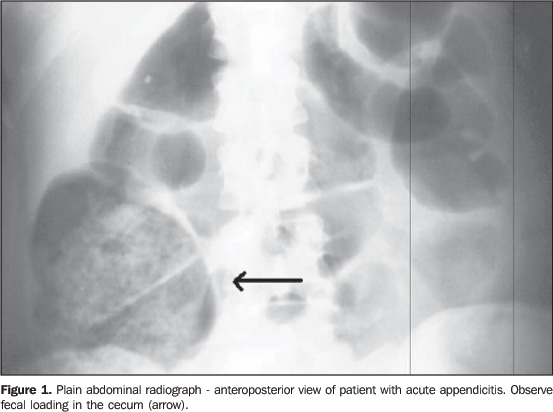
RESULTS There was no difficulty in the development of the presentstudy, since radiographies were part of the routine diagnosticworkup of patients with acute abdomen. The stage of appendicularinvolvement ranged from an initial edematous appendicitis toadvanced appendicitis, with necrosis, perforation andperitonitis. The presence of radiographic image of fecal loading in the cecum was observed in 97% of the cases, independently of the disease stage. In some cases, dilatation of the cecum was found as a result of the higher amount of feces in the cecum (Figure 1). In some cases, the image of the fecal loading extended up to the hepatic angle of the colon.
DISCUSSION Although a few studies suggest that plain abdominalradiography is not helpful in the diagnosis of acute abdomen,this diagnostic method remains as part of the routine practice inthe majority of medical emergencyservices(1–3,5–9). However, radiographic signsdescribed in the literature are not steadily found orcharacteristic of appendicitis. This lack of specificity raisesdoubts about the inclusion of the abdominal radiography in theroutine diagnostic workup in the suspect of acuteappendicitis(1,2). Also, it must be noted thatultrasound and computed tomography started being considered asmore sensitive methods for diagnosing acuteappendicitis(10,11). In the present prospective study with consecutive patients,the image of fecal loading in the cecum presented a sensitivityof 97%. The frequency of this sign is higher than the frequencyof other signs included as part of the clinical, laboratory andeven imaging workups for patients with acute appendicitis. The results of the present study seem to demonstrate that thefinding of fecal loading in the cecum on plain abdominalradiographs may be a sign associated with acute appendicitis.Further research is necessary to confirm the sensitivity of thissign, and to determine its specificity for acute appendicitis inrelation to other inflammatory, right-sided abdominalconditions. Acknowledgment The author would like to thank Doctor Luiz Ronaldo Alberti –Hospital Júlia Kubitschek for his help in the datacollection for the present study.
REFERENCES 1. Boleslawski E, Panis Y, Benoist S, Denet C, Mariani P, Valleur P. Plain abdominal radiography as a routine procedure for acute abdominal pain of the right lower quadrant: prospective evaluation. World J Surg 1999;23:262–264. [ ] 2. Rao PM, Rhea JT, Rao JA, Conn AKT. Plain abdominal radiography in clinically suspected appendicitis: diagnostic yield, resource use, and comparison with CT. Am J Emerg Med 1999;17: 325–328. [ ] 3. Thorpe JAC. The plain abdominal radiograph in acute appendicitis. Ann R Coll Surg Engl 1979; 61:45–47. [ ] 4. Petroianu A, Oliveira Neto JE. Prevalence of acute appendicitis in a mixed population. Dig Surg 1997;14:195–197. [ ] 5. Shimkin PM. Radiology of acute appendicitis. AJR Am J Roentgenol 1978;130:1001–1004. [ ] 6. Marincek B. Nontraumatic abdominal emergencies: acute abdominal pain: diagnostic stategies. Eur Radiol 2002;12:2136–2150. [ ] 7. Bondrager KL. Abdome e procedimentos contrastados comuns. In: Bondrager KL, editor. Técnica radiológica e bases anatômicas. 3ª ed. Rio de Janeiro, RJ: Guanabara Koogan, 2003;211–250. [ ] 8. Sivit CJ. Imaging the child with right lower quadrant pain and suspected appendicitis: current concepts. Pediatr Radiol 2004;34:447–453. [ ] 9. Oncel M, Degirmenci B, Demirhan N, Hakyemez B, Altuntas YE, Aydinli M. Is the use of plain abdominal radiographs (PAR) a necessity for all patients with suspected acute appendicitis in emergency services? Curr Surg 2003;60:296–300. [ ] 10. Pinto Leite N, Pereira JM, Cunha R, Pinto P, Sirlin C. CT evaluation of appendicitis and its complications: imaging techniques and key diagnostic findings. AJR Am J Roentgenol 2005;185:406–417. [ ] 11. Mun S, Ernst RD, Chen K, Oto A, Shah S, Mileski WJ. Rapid CT diagnosis of acute appendicitis with IV contrast material. Emerg Radiol 2006;12:99–102. [ ]
Received August 11, 2006. Accepted after revision October 11, 2006.
* Study developed at Instituto Alfa de Gastroenterologia – Hospital das Clínicas da Universidade Federal de Minas Gerais (UFMG), Belo Horizonte, MG, Brazil. |
|

Av. Paulista, 37 - 7° andar - Conj. 71 - CEP 01311-902 - São Paulo - SP - Brazil - Phone: (11) 3372-4544 - Fax: (11) 3372-4554