Radiologia Brasileira - Publicação Científica Oficial do Colégio Brasileiro de Radiologia
AMB - Associação Médica Brasileira CNA - Comissão Nacional de Acreditação
 Vol. 40 nº 2 - Mar. / Apr. of 2007
|
ORIGINAL ARTICLE
|
|
Patients exposure and imaging quality in chest radiographs: a critical evaluation |
|
|
Autho(rs): Adelaja Otolorin Osibote, Ana Cecília Pedrosa de Azevedo, Antonio Carlos Pires Carvalho, Helen Jamil Khoury, Sergio Ricardo de Oliveira, Marcos Otaviano da Silva, Carla Marchon |
|
|
Keywords: Chest x-ray, Quality control, Dosimetry |
|
|
Abstract:
IPhysicist, Doctorship Student, Fundação Oswaldo Cruz (Fiocruz), Escola Nacional de Saúde Pública Sergio Arouca-CESTEH, Rio de Janeiro, RJ, Brazil
INTRODUCTION Medical clinics and hospitals which utilize ionizing radiationhave been searching to be in compliance with the radiologicalprotection and quality control requirements of the Order(Portaria) No. 453/98 "Diretrizes de proteçãoradiológica em radiodiagnóstico médico eodontológico" ("Radiological protection guidelines inmedical and odontological radiodiagnosis"), published in 1998 bythe Ministry of the Health – National Sanitary VigilanceAgency(1). In the states of Rio de Janeiro and Pernambuco, severalhospitals and clinics have been the target of academic researchesin the field of radiological protection and quality control indiagnostic radiology. These studies are coordinated by the Groupof Radiological Protection and Quality Control ofFundação Oswaldo Cruz – Escola Nacional deSaúde Pública Sergio Arouca – CESTEH, and Group ofDosimetry and Instrumentation of Universidade Federal dePernambuco Department of Nuclear Energy. This study presents the partial results of the interactionbetween these two institutions, with a evaluation of doses andimages quality in chest x-rays performed in eight hospitals inseven cities in the states of Rio de Janeiro and Pernambuco.
MATERIAL AND METHODS This study was developed in eight hospitals in the followingcities: Angra dos Reis, Cabo Frio, Campos dos Goytacazes,Itaperuna, Niterói, Recife and Rio de Janeiro. Thesehospitals are randomly denominated A, B, C, D, E, F, G e H. Entrance skin dose (ESD) and effective dose (ED) wereevaluated in 520 posteroanterior/anteroposterior (PA/AP) and 215lateral chest x-rays of adult patients. A parallel criticalanalysis of images quality was made in 44 x-ray films, adoptingthe European guidelines on quality criteria for diagnosticradiographic images of the Commission of EuropeanCommunities(2). 1. Doses calculation Aiming at speeding up the process of patient dose measurementa software called DoseCal, running on Windows platform wasemployed(3). The DoseCalsoftware(4) calculates the ESD, the body organdose (BOD), and the ED, based on values of the radiographictechnique employed, the x-ray tube output, and the patients'anthropometric data. The DoseCal software has been developed inthe Radiological Protection Center at Hospital Saint Georges(London) and plays an essential role in the evaluation ofradiation doses for a great number of patients. This software hasbeen kindly provided for the present project in Brazil. For a correct operation of the software, it is necessary to enter the x-ray tube output in mGy/mAs; this data may be easily obtained with a calibrated ionization chamber. In the present study, we have utilized a Nero 8000-Inovision and a Radcheck Plus 06-526. Once output values, current, kilovoltage, exposure time and focus-skin distance (FSD) are known, the following equation (1) will demonstrate the ESD.
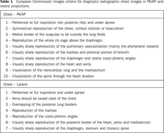
where: Output is the x-ray tube performance expressed inmGy/mAs, at 80 kV, and at a distance of 1 m normalized for 10mAs; kV is the potential applied to the tubr (in kilovolts); mAsis the product from current × exposure time; FSD isexpressed in centimeters (cm); and BSF is the backscatter factor.The DoseCal software utilizes the conversion factors included intables NRPB-SR262(5) applied for ESD, BOD andED calculations. 2. Images criteria Based on the premise that "the best image will provide abetter diagnosis" the European Union has formed a commission todevelop quality criteria for diagnostic radiographic images.Other criteria such as general principles associated with goodimaging performance and guidelines on radiation dose to thepatient have been included. The most recent version of thisdocument(2) was published in 1996 (EUR 16260EN-European Guidelines on Quality Criteria for DiagnosticRadiographic Images), including criteria for imaging the chest,skull, lumbar spine, pelvis, urinary tract, and breast. Thesecriteria were basically defined considering or not the presenceof anatomical structures of the focused region, as well as theirvisualization degree. The criteria are the following:visualization — anatomical characteristics are detected butare not totally reproduced; reproduction — anatomicaldetials are identified, but are not clearly defined; visuallysharp reproduction — anatomical details are clearlydefined. The images criteria for chest in PA/AP and lateral projections, according to the European Communities, are shown on Table 1.
RESULTS The Table 2 includes the statistics (mean, first and second quartiles) of ESD (in mGy) in PA/AP and lateral chest x-rays. It may be oserved that, on PA/AP projections, mean values range between 0.07 mGy (hospitals E and H) and 0.64 mGy (hospital C) (mean value, 0.24 mGy). Values on lateral projection ranged between 0.14 mGy and 1.02 mGy (mean value, 0.47 mGy). As regards ED, values ranged between 0.01 mSv (hospitals B, D, E and H) and 0.06 mSv (hospital C) (mean value, 0.03 mSv) for PA/AP projections, and between 0.01 mSv and 0.7 mSv (mean value, 0.2 mSv) on lateral projections.
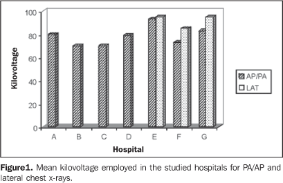
Table 3 and Figure 1 show values of radiographic techniques employed and patients' anthropometric data. Mean kilovoltage values ranged between 70 kV (hospitals B and C) and 93 kV (hospital E) (mean value, 78 kV) on PA/AP projections. On lateral projections, values ranged between 85 kV and 95 kV (mean value, 90 kV). As regards milliamperage, values ranged between 3 mAs (hospital E) and 36 mAs (hospital C) (mean value, 12 mAs) on PA/AP projections. On lateral projections, the values ranged between 7 mAs and 24 mAs (mean value, 15 mAs). The FSD ranged between 120 cm (hospital C) and 162 cm (hospital G) (mean value, 139 cm) on PA/AP projections. On the lateral projections, values ranged between 109 cm and 157 cm. Patients' mean age was 48 years for PA/AP projections, and 49 years for lateral projections. Mean weight was 67 kf for PA/AP and lateral projections.
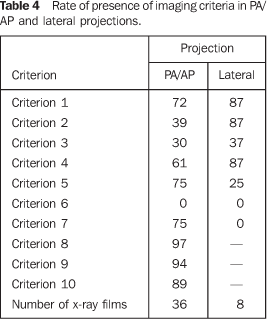
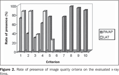
As regards image criteria, the results are shown on the Table 4 and Figura 2. The criteria with highest compliance rates were criterion 8 present in 97% of x-ray studies, and criterion 9, in 94%, both for PA/AP projections. On the other hand, criterion 6 (for both projections), and criterion 7 (for lateral projection) were absent in all the images.
DISCUSSION ESD values demonstrate high variation among the hospitalsevaluation. A difference of more than nive times was found in ESDvalues, and more than six times in ED values. These differencesreflect the disparity of radiographic techniques employed in eachinstitution. It is possible to observe that hospital C, withhighest ESD value (0.64 mGy), was the one utilizing the lowestmean kilovoltage (70 kV) and highest milliamperage (36 mAs).Additionally, hospital C employs an extremely low focus-filmdistance (120 cm). These factors contribute for an increase inthe radiation dose to patient. Several factors also contribute for the variation of doses;the most significant are: technicians training, system ofradiographic films processing, the luminance of the negatoscopeutilized for images evaluation and x-ray beam filtration. As regards images quality criteria, the criterion 6 in PA/APprojections ("Visually sharp reproduction of trachea and proximalportion of bronchi") was absent in all of the images, indicatingthe impossibility of a visally sharp reproduction of this regionin these projections. Also, criterion 6 in lateral projection("Visually sharp reproduction of the llposteiror border of theheart, aorta and the mediastinum") could not be detected in anyof the images. The criterion 7 ("Visually sharp reproduction ofthe diaphragm, sternum and thoracic spine"), for lateralprojection, also was not present in any of the images analyzed.The mean rate ofpresence of criteria was 55% (63% for PA/APprojections, and 46% for lateral projection). Also, based on data, it could be observed that on PA/AP chestx-rays, the criteria 2 and 3 presented the lowest rate ofpresence. These criteria refer to the symmetrical reproduction ofthe chest and medial border of the scapulae to be outside of thelung fields, and therefore, to the patient positioning. Thisreuslt demonstrate the relevance of the necessity of techniciansqualifying.
CONCLUSION The doses standardization/reduction may be achieved by meansof easy-to-implement (almost always very simple) measures. Thesemeasures should be included in a program of quality control andassurance to be implemente in every service of diagnosticradiology. The appropriate training of technicians, the x-rayequipment and automatic films processor performance, as well asthe employment of high kilovoltage techniques may be extremelyuseful for reduction of radiation dose to patients and obtentionof high quality images. As regards the quality criteria established by the EuropeanCommunities, certainly a x-ray in compliance with all thecriteria will result in a better diagnosis. Basically, a goodx-ray film depends on an adequate training of the technician,who, in the absence of a radiologist, must be able to decidewhether the image is adequate or not. This will be easier if theimage criteria are known. The mean rate of presence of theEuropean criteria on the images in the present study (55%)demonstrates that maybe such images have not the quality requiredfor a more reliable and adequate diagnosis. Acknowledgements We would like to thank Centro de VigilânciaSanitária-SES/RJ, Fundação Oswaldo Cruz(Fiocruz), Universidade Federal de Pernambuco, Third WorldOrganization for Women in Science, and International Centre forTheoretical Physics-ICTP de Trieste, Italy.
REFERENCES 1. Brasil, Ministério da Saúde, Secretaria de Vigilância Sanitária. Diretrizes de proteção radiológica em radiodiagnóstico médico e odontológico. Portaria 453/98, de 1/6/1998. Diário Oficial da União:Brasília 103, 2/6/1998. [ ] 2. Commission of European Communities. European guidelines on quality criteria for diagnostic radiographic images. Report EUR 16260EN. Bruxelas: European Communities/Union, 1996. [ ] 3. Kyriou JC, Newey V, Fitzgerald MC. Patient doses in diagnostic radiology at the touch of a button. London, UK: The Radiological Protection Center, St. George's Hospital, 2000. [ ] 4. Azevedo ACP, Mohamadain KEM, Osibote AO, Cunha ALL, Pires Filho A. Estudo comparativo das técnicas radiográficas e doses entre o Brasil e a Austrália. Radiol Bras 2005;38:343–346. [ ] 5. Hart D, Jones DG, Wall BF. Normalized organ doses for medical x-ray examinations calculated using Monte Carlo techniques. NRPB-SR262. Chilton: NRPB, 1994. [ ]
Received April 18, 2006.
* Study developed at Fundação Oswaldo Cruz (Fiocruz), Rio de Janeiro, RJ, Brazil. |
|
Av. Paulista, 37 - 7° andar - Conj. 71 - CEP 01311-902 - São Paulo - SP - Brazil - Phone: (11) 3372-4544 - Fax: (11) 3372-4554


