Radiologia Brasileira - Publicação Científica Oficial do Colégio Brasileiro de Radiologia
AMB - Associação Médica Brasileira CNA - Comissão Nacional de Acreditação
 Vol. 41 nº 5 - Sep. / Oct. of 2008
|
ORIGINAL ARTICLE
|
|
A new method for evaluating the esophageal transit time with external approach by ultrasonography |
|
|
Autho(rs): Makoto Sakate, Altamir Santos Teixeira, Seizo Yamashita, Thais Ricardo Medeiros, Pedro Gabriel da Silva, Maria Aparecida Coelho de Arruda Henry |
|
|
Keywords: Ultrasonography, Esophageal transit time, Abdominal esophagus |
|
|
Abstract:
IPhD, Assistant Professor, Division of Radiodiagnosis - Department of Tropical Diseases and Imaging Diagnosis at Universidade Estadual Paulista Júlio de Mesquita Filho (Unesp), Botucatu, SP, Brazil
INTRODUCTION Over the last decades, huge development in medicine have led to the introduction of increasingly complex imaging methods which, however, are hardly accessible by the general population because of the high costs involved in the utilization of these techniques. Imaging methods such as chest radiography have been routinely performed in hospitals and, considering the high incidence of normal results (70%(1) to 77%(2)), it is suggested that a more careful selection of cases is made in order to avoid unnecessary procedures. A study has been developed with other complementary diagnostic methods such as the technique of gastric emptying by means of ultrasonography(3), to avoid that patients are excessively exposed to ionizing radiation. In the case of esophageal complaints, contrast-enhanced esophageal radiography, esophageal manometry, scintigraphy, intraesophageal ultrasonography are routinely performed, all of them with precise indications(4-8). Radioscopy and scintigraphy are the methods indicated for evaluating the esophageal transit(9-11). However, these methods involve either a higher or lower degree of radiation exposure to the patients, despite routine implementation of quality control and guarantee systems(12). Manometry, endoscopy and intraesophageal ultrasonography have been utilized as a means for reducing ionizing radiation exposure. These techniques, although invasive, allow the investigation of peristalsis, mucosal lesions and diseases affecting the thickness of esophageal wall and adjacent structures. There is a concern in finding other faster methods for preliminary evaluation in cases of mild esophageal complaints in order to optimize the indication for more complex complementary studies. "Esophageal transit time" corresponds to the time interval required for the swallowed content to pass through the esophagus into the stomach. Esophageal transit time, in the present study was measured by means of ultrasonography, from the moment of the glottis movement (entry of the bolus into the esophagus) up to the visualization of the swallowed bolus passing through the intra-abdominal esophagus. No similar study involving the utilization of ultrasonography for this purpose has been found in the literature. The first objective of the present experimental study was utilizing ultrasonography as a method for evaluating the esophageal transit time, and the second, estimating the esophagus capability of differentiating between liquid (water) and pasty (yoghurt) substances ingested.
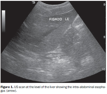
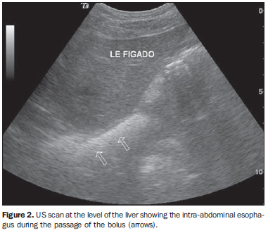
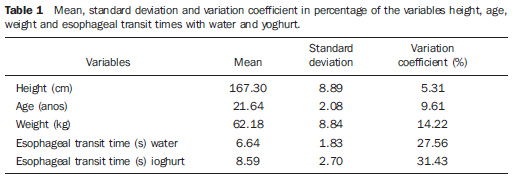
MATERIALS AND METHODS The sample of the present study included 22 young, healthy volunteers of both sexes, with ages ranging between 19 and 26 years (mean 21.64 ± 2.08 years), heights ranging between 155 and 184 cm (mean 167 ± 8.89 cm) and weighting between 48 and 82 kg (mean 62.18 ± 8.84 kg). Exclusion criteria were the following: symptoms associated with the high digestive tract and diseases that could interfere with the data collection. A Toshiba Sonolyer series SSH 140 A/G (Toshiba; Tokio, Japan) US unit with a semi-convex 3.5 MHz transducer, maintained at a controlled temperature of 22ºC was utilized in the present study. Also a Seiko 3 BAR stopwatch (Seiko; Tokio, Japan) with a resolution of up to 1/100 seconds was utilized. Mineral water maintained at room temperature (approximately 20 ml/volunteer) and yoghurt (approximately 20 ml or 20 g/volunteer ) maintained in a refrigerator at 5º, were administered during the examinations. After fasting for three hours, every volunteer, received water in the oral cavity, and was positioned in dorsal decubitus on the examination table, with the epigastric region exposed. Subsequently, the investigator, holding the stopwatch with the left hand, positioned the fifth finger of this hand on the epiglottis of the patient and the right hand holding the US probe positioned on the left lateral region of the xiphoid appendix. The ultrasound beam was cranially directed until the intra-abdominal esophagus was visualized. At this moment, the volunteer was asked to swallow the water, so the investigator could feel the glottis movement and immediately activate the stopwatch that was deactivated upon visualization, by means of US, of the passage of the liquid bolus through the intra-abdominal esophagus. The esophageal transit time for water was recorded and, after a 30-minute interval, the same volunteer was given yoghurt and, again, positioned in dorsal decubitus; and the procedure was repeated (Figures 1 and 2).
The 44 values for esophageal transit time were obtained, constituting paired treatments, with every of the 22 volunteers forming a pair of values for esophageal transit time after receiving water and yoghurt. The present study was approved by the Committee for Ethics in Research of the Institution where the study was developed. All of the volunteers were given an explanation about the methodology utilized and signed a term of free and informed consent. The experiment homogeneity was evaluated by means of a comparison between mean esophageal transit times according to sex, separately for water and yoghurt, utilizing the two-tailed t-test, considering the impossibility of anticipating the significance of the difference. The comparison between esophageal transit time with water and yoghurt was made through the one-tailed t-test(13), considering the hypothesis that the swallowing of less fluid substances results in a longer esophageal transit time. For the purposes of statistical analysis, the significance level was fixed in 5%, and the confidence interval in 95% for mean esophageal transit time with both water and yoghurt.
RESULTS The lowest value obtained for esophageal transit time with water was 3.74 seconds, and with yoghurt, 4.71 seconds; and the highest values were, respectively 9.85 seconds and 16.6 seconds. In four cases, the esophageal transit time was longer with water ingestion. No significant difference was observed as regards esophageal transit time in male and female volunteers (twater = 0.8576, gl = 20, p = 0.4012; tyoghurt = 0.6031, gl = 20, p = 0.5532). The mean esophageal transit time for water was 6.64 ± 1.83 seconds, and for yoghurt, 8.59 ± 2.70 seconds (Table 1).
The analysis of the difference (yoghurt-water) between esophageal transit times has indicated that the mean esophageal transit time with water ingestion was statistically lower as compared with yoghurt ingestion (t = 2.9905, gl = 21, p = 0.0039).
DISCUSSION In the present experiment, the volunteers had no difficulty keeping water or yoghurt in the oral cavity. All the steps of the investigation could be accomplished in a calm environment for all the individuals inside the examination room. The intra-abdominal esophagus could be easily accessed and identified at US, allowing the visualization of the progression of the water/yoghurt bolus through this region. The esophageal transit time could be determined with the aid of the stopwatch in all the volunteers, by measuring the time interval between the moment where the device was activated once the deglutition of water or yoghurt was initiated, up to the visualization of the bolus transit through the intra-abdominal esophagus. All the examinations could be easily and simply performed, with the patients comfortably positioned and taking a short time to be completed. The method could differentiate the esophageal transit time for water that was statistically shorter than for yoghurt (p < 0.0039), demonstrating that both substances present a defined time for passing through the esophagus, and that both can be utilized in an initial screening for conditions affecting the peristalsis of this organ and, consequently, the delay in the bolus transit through the esophagus. The evaluation of the thoracic esophagus with external approach by ultrasonography is impaired by the access difficulty, both through the anterior and the posterior mediastinum because of the presence of air in the lungs and bone tissue in the dorsal spine through which sound waves cannot be transmitted, with the exception of the segment posterior to the aorta and the heart(14). The cervical esophagus can be evaluated by ultrasonography, considering its lateral location in relation to the trachea, proximity with the surface and interposition of soft tissue such as the left thyroid lobe(15). Given its posterior positioning in relation to the left hepatic lobe, the intra-abdominal segment of the esophagus is easily accessible by ultrasonography, acting as an access window for ultrasound imaging (a relatively homogeneous organ that displaces the bowel loops with gas)(16-19). With the advent of new technologies, new method have been introduced for evaluating the esophagus such as nuclear medicine, optic fiber endoscopy, manometry with pH-metry, and endoscopic ultrasonography. All these methods demonstrate the location and anatomical and functional aspects of the esophagus(20-22), but are invasive or utilize ionizing radiation with highly expensive equipment. Therefore, the method evaluated in the present study has allowed a less invasive, a cost-effective and probably more easily available technique for the patients as compared with esophageal radiography or scintigraphy, considering that this method requires only a conventional ultrasonography unit and a stopwatch to be reproduced.
CONCLUSION The new experimental method for evaluating the esophageal transit time with external approach by ultrasonography is a simple, non-invasive technique that provides significant information without requiring ionizing radiation. This method gives us information on the differences between the esophageal transit time for liquid and pasty food. It is widely available and can be simply performed, with no contraindication and playing a significant role in the initial evaluation of conditions that may affect the esophagus. It is a safe, reliable and discriminative method including bedside capability (suitable for utilization in residences, hospitals and infirmaries), allowing the obtention of fast results with a good cost-benefit ratio.
REFERENCES 1. Ramos JH, Santos MB, Tavares-Neto J. Estudos dos critérios clínicos para requisição de radiografias de tórax em um hospital universitário (Salvador, Bahia). Radiol Bras. 1999;32:243-6. [ ] 2. Song KS, Song HH, Park SH, et al. Impact of clinical history on film interpretation. Yonsei Med J. 1992;33:168-72. [ ] 3. Valadares CP, Silva RAP, Tavares Jr WC, et al. Apresentação da técnica de estudo do tempo de esvaziamento gástrico por meio da ultra-sonografia. Radiol Bras. 2006;39:15-8. [ ] 4. Meschan I. Oropharynx, laryngopharynx, and esophagus. In: Meschan I, editor. Roentgen signs in diagnostic imaging. Volume 1 - abdomen. Philadelphia: WB Saunders; 1984. p. 487-560. [ ] 5. Sakate M et al. Parkman HP, Maurer AH, Caroline DF, et al. Optimal evaluation of patients with nonobstructive esophageal dysphagia. Manometry, scintigraphy, or videoesophagography? Dig Dis Sci. 1996;41:1355-68. [ ] 6. Henry MA, Harbermann MC, Rocha OM. Esophageal motor disturbances in progressive systemic sclerosis. Dis Esophagus. 1999;12:51-3. [ ] 7. Machado MM, Rosa ACF, Barros N, et al. Ultra-sonografia endoscópica (USE) do esôfago, estômago, cólons e reto. Radiol Bras. 2002;35:219-23. [ ] 8. Ling TC, Johnston BT. Esophageal investigations in connective tissue disease: which test are most appropriate? J Clin Gastroenterol. 2001;32:33-6. [ ] 9. Nakajima K, Kawano M, Kinuya K, et al. The diagnostic value of oesophageal transit scintigraphy for evaluating the severity of oesophageal complications in systemic sclerosis. Nucl Med Commun. 2004;25:375-81. [ ] 10. Nassif MS, Jauregui GF, Rocha VB, et al. Análise crítica da seriografia do esôfago, estômago e duodeno em um serviço de radiologia de um hospital geral. Radiol Bras. 2004;37:425-9. [ ] 11. Penas ME. Motilidade esofagiana: ensaio iconográfico sobre cintilografia dinâmica do esôfago. Radiol Bras. 2007;40:423-7. [ ] 12. Costa MMB, Canevaro LV, Azevedo ACP, et al. Valores típicos do "produto dose-área" obtidos durante o estudo videofluoroscópico da deglutição. Radiol Bras. 2003;36:17-20. [ ] 13. Zar JH. Bioestatistical analysis. New Jersey: Prentice-Hall, 1996. [ ] 14. Westra SJ, Derkx HHF, Taminiau JAJM. Symptomatic gastroesophageal reflux: diagnosis with ultrasound. J Pediatr Gastroenterol Nutr. 1994;19:58-64. [ ] 15. Esposito F, Lombardi R, Grasso AC, et al. Trans-abdominal sonography of the normal gastroesophageal junction in children. J Clin Ultrasound. 2001;29:326-31. [ ] 16. Johnson MC. The esophagus. Gastroenterology. 2001;28:459-85. [ ] 17. Costa CD, Zomignan HP, Rocha JIP, et al. Refluxo gastroesofágico em pediatria: estudo radiológico. Pediatria (S. Paulo). 1986;8:136-40. [ ] 18. Defagó M, Kuschnir E, Ninci CM, et al. Evaluation of methods for the study of gastroesophageal reflux. Rev Fac Cienc Med Univ Nac Córdoba. 1985;43:9-13. [ ] 19. Foley LC, Slovis TL, Campbell JB, et al. Evaluation of the vomiting infant. Am J Dis Child. 1989;143:660-1. [ ] 20. Cerri GG. Vísceras ocas. In: Rocha DC, Cerri GG, Prando A, et al. Ultra-sonografia abdominal. São Paulo: Sarvier; 1988; p. 169-86. [ ] 21. Naik DR, Bolia A, Moore DJ. Comparison of barium swallow and ultrasound in diagnosis of gastro-oesophageal reflux in children. Br Med J (Clin Res Ed). 1985;290:1943-5. [ ] 22. Tovar JA, Eizaguirre I. Advances in the diagnosis of gastroesophageal reflux. An Esp Pediatr. 1992;36 Suppl 48:288-91. [ ] Received November 17, 2007. Accepted after revision May 6, 2008. * Study developed at Faculdade de Medicina de Botucatu, Universidade Estadual Paulista Júlio de Mesquita Filho (Unesp), Botucatu, SP, Brazil. |
|

Av. Paulista, 37 - 7° andar - Conj. 71 - CEP 01311-902 - São Paulo - SP - Brazil - Phone: (11) 3372-4544 - Fax: (11) 3372-4554