Radiologia Brasileira - Publicação Científica Oficial do Colégio Brasileiro de Radiologia
AMB - Associação Médica Brasileira CNA - Comissão Nacional de Acreditação
 Vol. 42 nº 6 - Nov. / Dec. of 2009
|
ORIGINAL ARTICLE
|
|
Do children need fasting before abdominal ultrasonography? |
|
|
Autho(rs): Luiza Alina Almeida Araújo Rabelo, Ilka Rocha Florêncio, Iggor Medeiros Pirauá, Silvio Cavalcanti de Albuquerque, João Vicente Ribeiro Neto, Eduardo Just da Costa e Silva |
|
|
Keywords: Ultrasonography, Children, Abdominal, Fasting |
|
|
Abstract:
IMDs, Radiologists at Instituto de Medicina Integral Professor Fernando Figueira, Recife, PE, Brazil
INTRODUCTION Ultrasonography is among the most frequently utilized imaging methods for evaluating abdominal diseases in children, playing a relevant role in the detection and characterization of a wide range of conditions(1-4). Many times, it is demonstrated that ultrasonography is the only imaging method that may be required for such evaluation(1,5), with advantages including portability, low cost, accuracy and absence of ionizing radiation(6). However, many factors may affect the quality of sonographic images. Presence of intestinal gas is considered as a limiting factor in the evaluation of the pancreas, gall bladder and gastrointestinal tract(7). An appropriate distension of the gallbladder and reduced presence of gas in the digestive tract are considered as optimum conditions for the acquisition of appropriate images(8). Many authors recommend that the examination is preceded by fasting, in the expectation that the gastrointestinal contents are reduced and an appropriate distension of the gallbladder is achieved(7,9-12). Four-to six-hour fasting is routinely recommended for children(9,13). However, this practice may be very discomfortable and poorly tolerated by many patients. Children with hunger may become quite irritated, affecting the quality of the study and causing anxiety for parents. Additionally, small children may even develop hypoglycemia episodes after short fasting periods. The benefits of fasting for the sonographic images quality has been discussed by some authors(7,8). According to the authors, there is no study approaching this topic specifically in relation to children. The objective of the present study is to compare the quality of images acquired during abdominal ultrasonography in children previously submitted to fasting with those from children fed immediately before the examination.
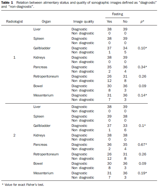
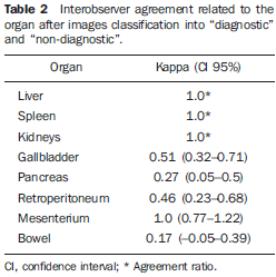
MATERIALS AND METHODS The present study was previously approved by the Committee for Ethics in Research of the Institution, and a term of free and informed consent was signed by all the parents and guardians after an explanation about the nature and objective of the research. This is a prospective study including children up to 12 years with clinical complaints not related to the abdomen. Convenience was a key factor in the sampling for allowing the inclusion of patients present in the hospital at the moment where the two radiologists participating in the study were available in the department, allowing comparison between images acquired in the same conditions. Because of the present study nature, including children with no abdominal complaint, the selected samples did not present any particular characteristic that might have contributed for influencing such selection. All the individuals included in the present study were inpatients so their meals timetable could be managed accordingly. A list including all the inpatients either with no abdominal complaint or previous history of abdominal surgery was obtained. The patients were divided into two groups as follows: group 1, including fasting children, and group 2, including children evaluated 60 minutes after their usual meal. The studies were sequentially performed by two medical sonographers involved in a daily practice of pediatric pediatric ultrasonography, one of them with six-year- and the other with three-year-experience. The standard technique was utilized, with the patients in dorsal decubitus, with all the examinations being performed in a Sonosite Titan unit (Sonosite Inc.; Bothell, USA), with linear (L25, 5-10 MHz) and convex (C11, 5-8 MHz) transducers. The sonographers did not know the patients and were not authorized to ask questions to their parents or guardians. Images of the liver, gallbladder, spleen, kidneys, pancreas, retroperitoneum, mesenterium (vessels, fat and lymph nodes) and bowels were obtained and classified according to a method described in a previous study(8), as follows: score 1 (non-visualized or partially visualized, inappropriate for diagnosis); score 2 (sufficient for diagnosis); score 3 (excellent, appropriate for classes of sonographic anatomy). Statistical analysis The statistical significance of the differences between the groups was evaluated by means of the Mann-Whitney test. Subsequently, images classified as 3 or 2 were grouped and named as "diagnostic", the other images, classified as 1, being named "non-diagnostic". The statistical significance of the differences observed with this new scoring system was evaluated by the chi-squared method. The interobserver agreement was evaluated by the kappa coefficient of agreement(10), with the sole objective of having a gross evaluation of the method applied in the calcification of the images quality, with no direct relation with the objective of the present study. The significance level adopted was 95%.
RESULTS The sample of the present study included 77 patients - 47 (61%) boys and 30 (39%) girls - with ages ranging from one week to 12 years (mean age, 2.7 years). Fasting has shown to be advantageous only in the evaluation of the gallbladder by the sonographer 2 (p = 0.032), this advantage being not observed by the sonographer 1. No statistically significant difference was observed between the groups of "diagnostic" and "non-diagnostic" images (Table 1). Ages did not demonstrate any significant correlation with the scores observed in fasting and nonfasting patients.
The interobserver agreement in relation to the classification of images into "diagnostic" and "non-diagnostic" is shown on Table 2.
DISCUSSION The prescription of any type of previous preparation for an imaging study is aimed at making the procedure safer (like in the case where an antiallergenic preparation is prescribed before imaging studies requiring iodinated contrast injection), or improving the images quality, providing higher safety and diagnostic efficacy(14). However, any prescription of either a drug or simply fasting must be based on studies that demonstrate an actual benefit from its practice. The prescription of fasting is usual in many hospital procedures, including ultrasonography studies. The moment to start the fasting period is always based on the expected time of the imaging study, although this timetable is not always can be accomplished. In large hospitals, imaging studies are performed in a high number of patients, and hence the occurrence of crowded waiting rooms and long waiting periods for examinations. Usually, the presence of an emergency department leads to an increase in the number of request for imaging studies, particularly ultrasonography in cases of abdominal emergency(15,16). Such phenomenon contributes for an extension of the waiting time for the previously scheduled inpatients, considering that, usually, patients coming from the emergency department are examined at the intervals between previously scheduled studies. The results of the present study demonstrate that previous fasting in children to be submitted to abdominal ultrasonography does not contribute to the acquisition of higher quality images. A higher score was observed only on sonographic images of the gallbladder, but solely by one of the sonographers. Nevertheless, no difference was observed as the images were classified into "diagnostic" or "non-diagnostic". Studies on this topic approaching specifically children are not available in the literature, but the results of the present study are similar to those observed with adults. Windler et al. have observed that the weight/height ratio was the most relevant determining factor in the evaluation of abdominal sonographic images quality in their study(7). Also, they have observed that fasting contributed for higher quality images only in the evaluation of the biliary tract. Additionally, images of the right kidney achieved higher scores in nonfasting patients, a finding that has not been observed in the present study. The inclusion of adult individuals in the mentioned study may explain this small discrepancy between the results. Abdominal sonographic images of children are usually better because of the small dimensions of the pediatric abdomen. Sinan et al. have not found any difference in scores among adult individuals submitted to previous fasting and those previously fed(8). The number of "diagnostic" images in such study, however, was lower than in the present one, a finding that also may be attributed to the inclusion of adult individuals. Fasting may be problematic in some cases. This practice should not be adopted if good quality images can be obtained in nonfasting individuals. At the unit of ultrasonography of large hospitals, high number of patients coming from the emergency department and inpatients with complications requiring other further imaging studies replace previously scheduled patients, extending considerably the waiting time, much more than expected since the number of unexpected examinations may be high. Hunger may be extremely distressing for children, and may lead to hypoglycemia and dehydration. Weep and irritability may even impair the study performance. Many patients may even refuse to wait, giving up submitting to the examination, which may lead to delayed diagnosis. The present study had several limitations. The evaluation of sonographic images through scores does not reflect the actual accuracy of the method. Ideally, it would be necessary to evaluate the performance of the method in the detection of specific diseases such as retroperitoneal lymphadenomegaly, cholelithiasis and thickening of intestinal loops. Also, clinically relevant situations, such as detection and staging of neoplasms and evaluation of inflammatory bowel diseases should be included. A situation where fasting could be useful is the evaluation of neonatal cholestatic icterus, since the evaluation of the gallbladder is required in this case. A previous meal may impair the study of this organ because of the vesical contractility(12). Another limitation is the absence of data regarding body mass index or body weight that presumably could influence the results. Additionally, the adopted scoring system is subjective and may affect the study reproducibility. Although the present study has not been designed to evaluate the quality of sonographic scoring systems, the interobserver agreement obtained indicates the necessity of developing other systems for evaluating the quality of sonographic images. However, the system adopted has been utilized in several studies with similar objectives(7,8,17,18). Studies approaching interobserver agreement on any scoring system are not available in the literature. Such studies are required in addition to the development of better-defined systems for evaluating the quality of sonographic images.
CONCLUSION The authors conclude that the practice of fasting was not essential for the acquisition of quality abdominal sonographic images in the evaluated children. Further studies evaluating other variables such as age and body mass index, besides usual clinical situations are required. Also, it is important to note the need for developing better methods for evaluating the quality of abdominal sonographic images.
REFERENCES 1. Strouse PJ. Sonographic evaluation of the child with lower abdominal or pelvic pain. Radiol Clin North Am. 2006;44:911-23. [ ] 2. Levy JA, Noble VE. Bedside ultrasound in pediatric emergency medicine. Pediatrics. 2008; 121:e1404-12. [ ] 3. Haber HP. Cystic fibrosis in children and young adults: findings on routine abdominal sonography. AJR Am J Roentgenol. 2007;189:89-99. [ ] 4. Rocha SMS, Ferrer APS, Oliveira IRS, et al. Determinação do tamanho do fígado de crianças normais, entre 0 e 7 anos, por ultrassonografia. Radiol Bras. 2009;42:7-13. [ ] 5. Eppich WJ, Zonfrillo MR. Emergency department evaluation and management of blunt abdominal trauma in children. Curr Opin Pediatr. 2007;19:265-9. [ ] 6. Costa JD, Leão ARS, Santos JEM, et al. Quantificação do fluxo portal em indivíduos sadios: comparação entre ressonância magnética e ultrasom Doppler. Radiol Bras. 2008;41:219-24. [ ] 7. Windler EE, Lempp FL. US of the upper abdomen: factors influencing image quality. Radiology. 1985;157:513-5. [ ] 8. Sinan T, Leven H, Sheikh M. Is fasting a necessary preparation for abdominal ultrasound? BMC Med Imaging. 2003;3:1. [ ] 9. Devos AS, Meradji M, Blickman JG. The small bowel. In: Devos AS, Blickman JG, editors. Radiological imaging of the digestive tract in infants and children. Berlin: Springer; 2008. p. 167-91. [ ] 10. Sommer G, Filly RA, Laing FC. Use of simethicone as a patient preparation for abdominal sonography. Radiology. 1977;125:219-21. [ ] 11. Scortegagna Junior E, Leão ARS, Santos JEM, et al. Avaliação da concordância entre ressonância magnética e ultra-sonografia na classificação de fibrose periportal em esquistossomóticos, segundo a classificação de Niamey. Radiol Bras. 2007;40:303-8. [ ] 12. Teixeira MS, Coelho CAR, Teixeira AS. Avaliação da contratilidade da vesícula biliar com leite materno e leite de vaca em lactentes. Radiol Bras. 2004;37:163-6. [ ] 13. Siegel MJ. Fígado. In: Siegel MJ, editor. Ultrasonografia pediátrica. 3ª ed. Rio de Janeiro: Guanabara Koogan; 2003. p. 189-244. [ ] 14. Trindade R, Sumi DV, Kravetz WL, et al. Avaliação do conhecimento de médicos não-radiologistas sobre reações adversas aos contrastes iodados. Radiol Bras. 2007;40:321-8. [ ] 15. Cavalcanti AF, Menezes MR. Radiologia de emergência: perspectivas. Radiol Bras. 2001;34:v-vi. [ ] 16. Vabo KA, Torres Neto G, Santos AASMD, et al. Achados ultra-sonográficos abdominais em pacientes com dengue. Radiol Bras. 2004;37:159-62. [ ] 17. Elam EA, Hunter TB, Hunt KR, et al. The lack of sonographic image degradation after barium upper gastrointestinal examination. AJR Am J Roentgenol. 1989;153:993-4. [ ] 18. Friedman DL, Hunter TB, Elam EA, et al. Sonographic image degradation after barium enema. Invest Radiol. 1993;28:295-6. [ ] Received June 21, 2009. * Study developed at Instituto de Medicina Integral Professor Fernando Figueira, Recife, PE, Brazil. |
|

Av. Paulista, 37 - 7° andar - Conj. 71 - CEP 01311-902 - São Paulo - SP - Brazil - Phone: (11) 3372-4544 - Fax: (11) 3372-4554