ABSTRACT
Glenohumeral cartilage lesions represent a significant challenge in both diagnosis and management due to the complex anatomy of the joint and the variability in lesion presentation. In this review, we elucidate the anatomical considerations of the glenohumeral joint, discuss multimodal diagnostic imaging strategies, and highlight imaging features critical for the effective management of focal and diffuse cartilage lesions in the glenohumeral joint, especially in the preoperative context. We also present the role of advanced magnetic resonance imaging techniques and the crucial parameters that radiologists must report in preoperative planning, in order to optimize outcomes in clinical practice.
Keywords:
Shoulder joint; Osteoarthritis; Cartilage, articular; Diagnostic imaging; Preoperative care.
RESUMO
Lesões da cartilagem glenoumeral representam um desafio tanto para o diagnóstico quanto para o seu apropriado manejo, devido à complexidade anatômica articular e à variabilidade de apresentação dessas lesões. Nesta revisão, abordamos a avaliação por imagem dessas lesões, destacando aspectos imaginológicos importantes para o manejo de lesões condrais glenoumerais focais e difusas, especialmente no contexto pré-operatório. Também trazemos informações sobre técnicas de imagem avançadas e parâmetros importantes de serem relatados no laudo dos exames para a obtenção de melhores resultados clínicos.
Palavras-chave:
Articulação do ombro; Osteoartrite; Cartilagem articular; Diagnóstico por imagem; Cuidados pré-operatórios.
INTRODUCTION
The glenohumeral joint is formed by the articulation between the shallow glenoid cavity of the scapula and the hemispherical head of the humerus in a configuration that allows great range of movement – as it is the most mobile joint in the human body. Its inherent instability derives primarily from the discrepancy between the articular surfaces of these joint components, because the humeral head is much larger than the glenoid cavity(1,2).
Variations in glenoid morphology, such as hypoplasia and retroversion, may predispose the joint to instability(3,4). Soft tissue stabilizers, both static and dynamic, compensate for that mismatch. When those structures fail, internal imbalances may lead to cartilage damage(1,5).
Multiple other factors such as exercise and occupation, as well as aging, inflammation, obesity and genetics, accelerate cartilage degeneration(6–8). Glenohumeral cartilage lesions lead to progressive joint degeneration (osteoarthritis), which has substantial impact on affected individuals; in severe cases, osteoarthritis requires surgery for joint replacement(7–9).
This review discusses the intricate anatomy of the glenohumeral joint and the spectrum of chondral lesions. We also highlight the latest diagnostic imaging strategies that can contribute to patient management and improve the quality of radiologic reports, especially in the preoperative setting.
GLENOHUMERAL JOINT STABILIZERS
Glenohumeral stability is maintained by static and dynamic stabilizers, which work synergistically to preserve joint congruency, as well as to prevent excessive translation and rotation(1,2).
Static stabilizers include the glenoid labrum, the joint capsule and the glenohumeral ligaments. The joint capsule is thick and strong, attaching to the humeral neck and scapula just beyond the glenoid labrum. However, it is lax inferiorly, forming a larger axillary recess (1,2). It is lined internally by the synovial membrane and displays focal thickened bands anteriorly—properly referred to as the superior, middle, and inferior glenohumeral ligaments—which provide additional resistance against anterior and inferior translation(1,10). The coracohumeral ligament, extending from the coracoid process to the humeral tuberosities, provides additional support superiorly, limiting joint flexion and extension. The joint capsule itself is also a significant static stabilizer, particularly in terms of limiting the extremes of motion(1,2,5).
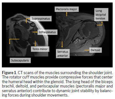
Dynamic stabilization of the glenohumeral joint is primarily attributed to the rotator cuff muscles (supraspinatus, infraspinatus, teres minor, and subscapularis), which provide compressive forces that center the humeral head within the glenoid (Figure 1). The long head of the biceps brachii, deltoid, and other periscapular muscles also contribute to dynamic joint stability by balancing forces during shoulder movements. Effective coordination among these muscles is essential for maintaining shoulder stability and preventing pathological lesions(1,11,12).
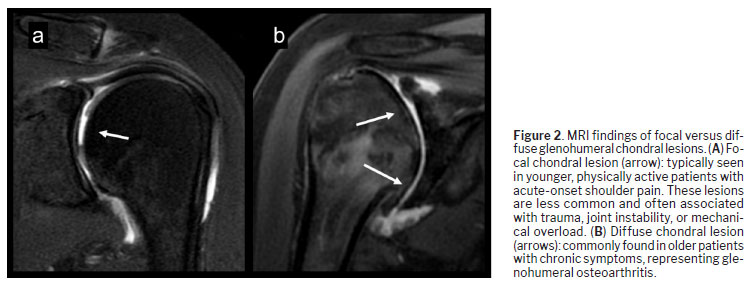
Glenohumeral chondral lesions are categorized as focal or diffuse, each with distinct etiologies, clinical implications, and management strategies. Focal lesions are more commonly associated with acute trauma or repetitive microtrauma, as illustrated in Figure 2A, whereas diffuse lesions often result from degenerative processes such as osteoarthritis
(6,13-16) , as depicted in Figure 2B).
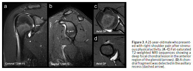
Focal lesionsFocal chondral lesions are localized cartilage defects that can appear as fissures, flaps, or full-thickness loss (Figure 3). The reported incidence of symptomatic glenohumeral chondral defects ranges from 5% to 17%
(15). Their etiology comprises previous surgical and traumatic conditions, including iatrogenic damage during arthroscopy, repetitive microtrauma and overuse. Younger individuals and athletes are the most commonly affected
(6,13,15-17).
Clinical manifestations of focal chondral lesions are nonspecific, including pain, clicking and locking, and may overlap with other shoulder pathologies. Therefore, arthroscopy remains the gold standard for their diagnosis
(13,15,17). Because either or both articular surfaces may be involved, specificities regarding their isolated development are provided further below.
Glenoid chondral defects often coexistent with labral tears—the so-called “chondrolabral injuries”—related to mechanisms such as glenohumeral impaction, subluxation and dislocation, contributing to or resulting from joint instability. The spectrum of chondrolabral lesions encompasses glenoid labral articular disruption, glenoid articular rim divot, glenoid labral articular flap, and glenoid labral articular teardrop, among other variants, all of them involving the anteroinferior segment of the glenoid labrum and the adjacent glenoid cartilage lining
(18).
In contrast, isolated nontraumatic humeral chondral lesions are less common. Factors such as humeral osteonecrosis, subchondral fractures and unstable osteochondral fragments may be implicated in the genesis of humeral cartilage detachments
(19). Humeral cartilage lesions may also occur in overhead-throwing athletes, and their location in the posterosuperior humeral head aspect is frequently related to posterosuperior internal impingement; defects in superior humeral head segment may also be related to subacromial impingement
(20,21).
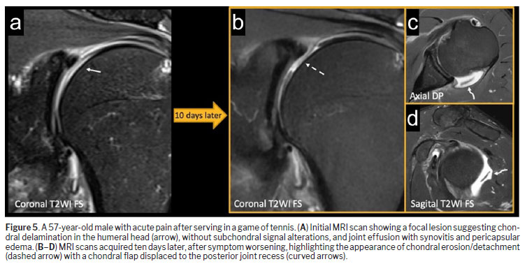
Imaging diagnosis of focal chondral defectsMagnetic resonance imaging (MRI) and magnetic resonance arthrography (MRA) are crucial imaging modalities for the diagnosis of focal cartilage lesions because they allow detailed visualization of cartilage, labrum, and other soft tissue structures
(22). However, the thin glenohumeral cartilage lining and the curved articular contours make MRI less sensitive than arthroscopy for the detection of subtle chondral lesions
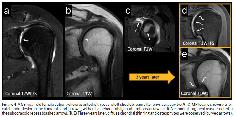
(23,24). Active searches for indirect findings such as subchondral bone marrow changes, synovitis, joint effusion, and intra-articular loose bodies may improve reading performance by radiologists
(25), as illustrated in Figures 4 and 5.
Using scanners with field intensities ≤ 1.5 T, Momenzadeh et al.
(24) reported that MRI had an overall sensitivity and specificity of 90% for detecting humeral and glenoid cartilage lesions, although both indications varied according to the chondral lesion site. Similarly, MRA showed moderate performance in detecting glenohumeral chondral defects, with a slightly higher sensitivity (approximately 77%) for detecting glenoid cartilage lesions
(26). The use of MRA also improves the detection of labroligamentous and tendinous abnormalities, making it particularly valuable in cases of glenohumeral instability and sports-related injuries
(27–29).
Technological advances in MRI, with greater commercial availability of 3.0-T scanners, have improved image resolution, therefore reducing the need for MRA in clinical practice. However, the latter is still recommended for the investigation of shoulder instability in young patients (particularly those who are overhead-throwing athletes), as well as of relapsing shoulder instability, in the assessment of postoperative labral tears, or in case of persistent clinical suspicion despite unremarkable MRI findings, given that MRA still demonstrates statistically better detection of shoulder lesions compared with conventional 3.0-T MRI, including anterior labral tears and superior labrum anterior to posterior tears
(30,31).
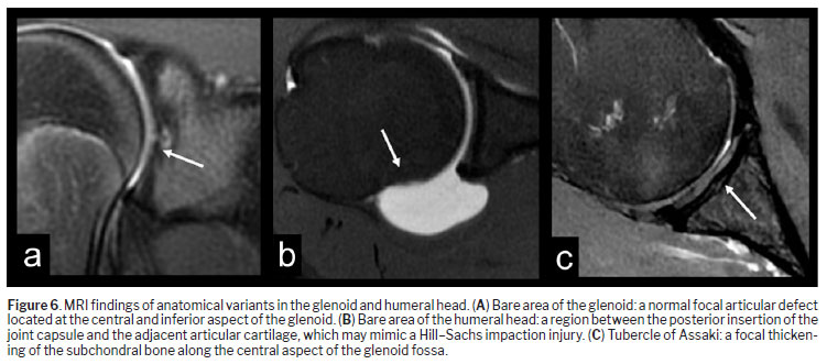
Anatomical variations can pose diagnostic challenges. For instance, the “bare areas” of the glenoid or humeral head (Figure 6), which are normal variants, may be misinterpreted as a pathological lesion if not carefully assessed
(10,32).
Advanced MRI techniques are emerging as noninvasive tools for shoulder cartilage assessment. Delayed Gadolinium-Enhanced MRI of Cartilage (dGEMRIC) assesses glycosaminoglycan depletion, an early indicator of cartilage degeneration
(33–35). This technique involves gadolinium contrast administration, allowing diffusion into cartilage over a period of time before T1 relaxation times are measured. Lower glycosaminoglycan levels correspond to higher gadolinium uptake, indicating early biochemical changes that precede structural cartilage damage
(33,35).
By focusing on structural integrity, as well as quantifying changes in cartilage water content and collagen organization, T2 mapping complements dGEMRIC. Signal changes on T2 mapping also precede gross morphological alterations visible on conventional MRI. Combining biochemical and structural assessments through dGEMRIC and T2 mapping offers a comprehensive approach to cartilage evaluation, particularly in early-stage osteoarthritis
(35,36).
Emerging modalities such as T1ρ mapping and sodium MRI show promise as alternatives for patients who cannot receive gadolinium-based agents, further enhancing diagnostic capabilities
(34).
Lastly, zero-echo time MRI of the shoulder is useful for complementary assessment of bone pathology that may be associated with glenohumeral chondral defects, avoiding the need for computed tomography (CT) and, consequently, exposure to radiation
(37,38).
ManagementThe management of focal chondral lesions varies depending on the depth, extension and location of the chondral defects, as well as of any other shoulder lesions. Other factors, such as patient age, intensity of symptoms, impairment of quality of life, and physical activity level, also need to be considered.
Nonsurgical measures, including activity modification, physical therapy, oral nonsteroidal anti-inflammatory drugs (NSAIDs), and intra-articular injections (with corticosteroids, local anesthetics or hyaluronic acid) are often the first-line treatments
(15,39).
For patients with refractory symptoms, joint-preserving procedures such as arthroscopic debridement, microfracture, or cartilage transplantation may be indicated depending on the defect size and patient profile
(15,17). These procedures are aimed at relieving pain, restoring function, and prolonging joint integrity by stabilizing cartilage surfaces and removing unstable flaps
(14). In cases of extensive chondral defects, resurfacing techniques, including nonbiological or biological interposition arthroplasty, may serve as alternatives to delay definitive arthroplasty
(39,40). Lastly, correction of coexistent lesions of other shoulder structures, such as labral tears, must be included in the surgical planning and may even hasten the need for surgical treatment.
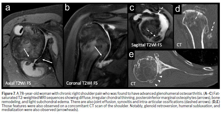
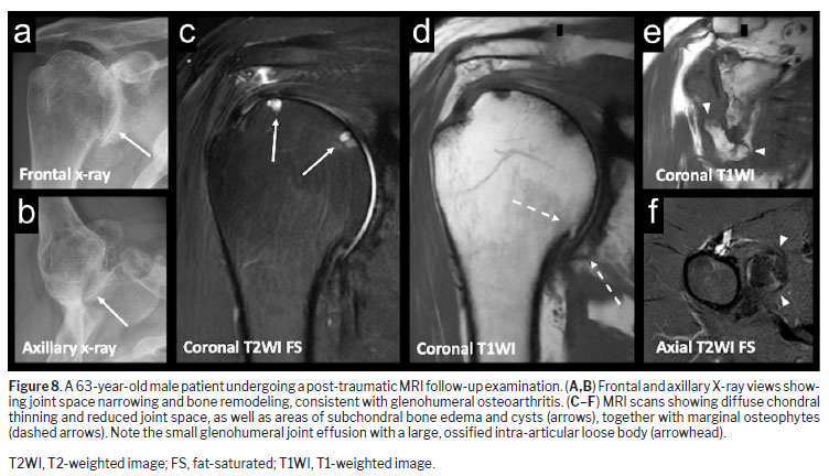
Diffuse lesionsDiffuse lesions, such as those observed in glenohumeral osteoarthritis, involve widespread degeneration of articular cartilage, typically affecting the humeral head and the glenoid, and may result in pain, loss of function, and diminished quality of life. Such lesions are characterized by cartilage thinning, subchondral bone sclerosis, and osteophyte formation. Involvement of the soft tissues (synovium, joint capsule, ligaments, and rotator cuff tendons) may play a role in the progression of cartilage degeneration, either as a causative factor for cartilage loss or as its consequence
(41), as shown in Figures 7 and 8.
Unlike focal lesions, which are typically localized and associated with specific mechanical insults, diffuse lesions are commonly present in the context of glenohumeral osteoarthritis, either primary or secondary, affecting the articular surfaces of the humeral head and of the glenoid. In cases of primary glenohumeral osteoarthritis, diffuse lesions frequently occur in older populations and are closely linked to systemic factors such as inflammation, genetic predisposition, and hormonal imbalances
(8,41).
Although glenohumeral osteoarthritis is less common than is knee or hip osteoarthritis, it has a considerable impact on affected individuals. Studies have shown that cartilage lesions are found in 5–15% of routine arthroscopy studies
(41). Population-level studies estimate the prevalence to be as high as 32.8% in individuals over 60, though asymptomatic cases likely result in underreporting
(8,41). Gender differences are also notable, with a predominance of men in younger cohorts, whereas postmenopausal women predominate in older cohorts, possibly because of hormonal changes that influence cartilage metabolism
(41).
EtiologyThe development of diffuse lesions in the glenohumeral joint is multifactorial, involving primary and secondary mechanisms. These mechanisms highlight the complexity of osteoarthritis and the variety of factors that influence its progression
(6,7,41,42).
Primary osteoarthritisPrimary osteoarthritis is mainly associated with aging and the natural degeneration of articular cartilage without preceding trauma. As individuals age, the density of chondrocytes within the hyaline cartilage decreases, as does responsiveness to anabolic growth factors, which renders the cartilage more susceptible to wear and tear. Over time, the progressive loss of cartilage and subsequent mechanical imbalance lead to joint dysfunction. The prevalence of glenohumeral osteoarthritis rises sharply with age, affecting up to 27.5% of individuals over 80 years of age
(6,42).
Genetic predisposition also plays a significant role in primary osteoarthritis. Complex polygenic inheritance patterns contribute to variations in cartilage resilience and susceptibility to degenerative processes. In addition, systemic factors such as obesity exacerbate the mechanical and biochemical stress on cartilage. Adipose tissue releases inflammatory cytokines that promote low-grade systemic inflammation, further accelerating cartilage breakdown
(6,32,42).
Secondary osteoarthritisSecondary osteoarthritis arises from identifiable causes that disrupt the biomechanics or structural integrity of the joint.
Among the most common etiologies, post-traumatic shoulder disorders due to previous fractures and dislocations can directly damage the articular cartilage or alter joint alignment and lead to accelerated degeneration. Proximal humeral fractures and glenoid fractures, for instance, can result in chronic incongruence and joint instability, significantly increasing the risk of secondary osteoarthritis
(6,7). Likewise, in the setting of chronic glenohumeral instability, recurrent shoulder dislocation or subluxation imposes abnormal stress on the glenoid and humeral cartilage, contributing to chondral damage and, eventually, osteoarthritis. Studies indicate that individuals with recurrent dislocations are at a 10- to 20-times greater risk of developing radiological signs of osteoarthritis
(5,6).
Nontraumatic etiologies also play an important role in the development of secondary shoulder osteoarthritis. Autoimmune disorders such as rheumatoid arthritis and other inflammatory arthropathies (e.g., systemic lupus erythematosus and psoriatic arthritis) induce systemic inflammatory cascades that target synovial tissue and cartilage. Rheumatoid arthritis, in particular, frequently affects the glenohumeral joint, with bilateral involvement and determining central glenoid remodeling. Pain in these cases often correlates more strongly with inflammatory synovitis than with joint destruction itself
(2,6).
Septic arthritis caused by bacterial pathogens (e.g.,
Staphylococcus aureus or
Neisseria gonorrhoeae) can rapidly destroy joint cartilage if untreated. Early diagnosis and management are critical to prevent irreversible damage
(2).
Avascular necrosis (AVN) of the humeral head results from an interruption of the blood supply, leading to localized bone collapse and secondary cartilage loss. The development of AVN can be post-traumatic or unrelated to trauma, due to prolonged corticosteroid therapy, alcohol abuse and systemic conditions such as sickle cell anemia. It is estimated that AVN accounts for 5% of all cases of secondary glenohumeral osteoarthritis
(6,41).
Extra-articular disorders can also generate joint imbalances and contribute to diffuse chondral damage. For instance, long-standing rotator cuff tears can lead to superior migration of the humeral head, causing abnormal contact with the glenoid and progressive cartilage deterioration. Secondary osteoarthritis is often accompanied by remodeling of the coracoacromial arch and erosion of the superior glenoid, characteristic of cuff-tear arthropathy
(6,7). More rarely, neurological disorders, such as cervical syringomyelia and diabetic neuropathy, can impair joint proprioception and nociception, leading to unchecked joint destruction and instability
(6).
DiagnosisThe clinical diagnosis of glenohumeral osteoarthritis is limited because of the complex anatomy of the shoulder and the clinical similarities with primary differential diagnoses such as rotator cuff syndrome, subacromial bursitis, and adhesive capsulitis
(6,11). Thorough medical history taking remains essential, with the typical chief complaints including progressive pain, crepitation, reduced range of motion, and symptoms exacerbated by activity
(2,6). Clinical features that suggest a secondary etiology for glenohumeral osteoarthritis must be actively sought: acute-onset pain along with exacerbated inflammatory changes favor the possibility of underlying inflammatory pathology, such as septic arthritis or microcrystalline disorders, whereas chronic pain accompanied by morning stiffness is a feature of rheumatoid arthritis
(2,6).
The diagnostic approach to diffuse lesions includes a combination of plain radiography, CT, and MRI.
Radiography is usually the first imaging modality required for evaluating glenohumeral lesions. Although it has limitations in detecting early cartilage damage, it remains essential for identifying structural bony changes, such as joint space narrowing, osteophyte formation, and subchondral sclerosis
(6,43,44). The radiographic shoulder series in a nontraumatic setting includes anteroposterior (AP) view in internal rotation, as well as true AP, axillary, and lateral Y views
(45,46). The most common radiographic abnormality is the presence of osteophytes, which can be an earlier marker of degeneration, making osteophyte identification clinically relevant
(44,46). The axillary view is particularly useful for visualizing joint space narrowing, which may be obscured in standard AP views, and for detecting glenohumeral subluxation and asymmetric glenoid remodeling
(44).
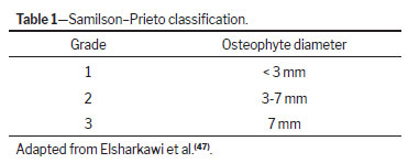
The Samilson–Prieto classification is commonly used in order to grade glenohumeral osteoarthritis based on the presence and size of osteophytes on AP views of the shoulder
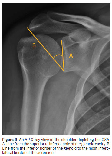
(47), as detailed in Table 1. Another important radiographic parameter, albeit not diagnostic, is the critical shoulder angle (CSA), as depicted in Figure 9, which is associated with degeneration risk
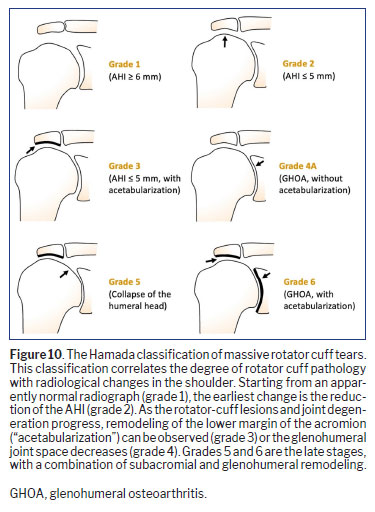
(48): an increased CSA (> 35°) correlates with rotator cuff pathology, whereas a decreased CSA (< 30°) is linked to concentric osteoarthritis. The Hamada grading system helps describe radiographic progression of glenohumeral degenerative changes related to chronic rotator cuff tears, ranging from acromiohumeral interval (AHI) narrowing to humeral head collapse and cuff-tear arthropathy
(49,50), as shown in Figure 10.
In the assessment of glenoid morphology, bone stock, and preoperative surgical planning, CT plays a crucial role. Compared with conventional radiography, CT provides superior cortical bone definition and is particularly useful for evaluating glenoid version, subluxation of the humeral head, and the extent of glenoid bone loss in detail
(43,51). Three-dimensional (3D) CT reconstructions have become fundamental for surgical planning, allowing detailed spatial analysis and improving intraoperative precision. It is especially useful for preoperative planning in shoulder arthroplasty, enabling the best quantification of glenoid bone stock
(43,44,52).Finally, MRI is useful in the context of glenohumeral osteoarthritis for assessing the soft tissues in the shoulder, providing a detailed evaluation of the articular cartilage, labrum, shoulder muscles and rotator cuff tendons
(53). Preoperative knowledge on the status of these structures is fundamental for determining the appropriate surgical technique, as explained below.
ManagementTreatment for diffuse lesions spans from nonoperative approaches to advanced surgical interventions, depending on the severity of joint degeneration and patient-related factors. Similarly to what is done in the setting of focal chondral lesions, the management of glenohumeral osteoarthritis is initially conservative, relying on physical therapy for strengthening the rotator cuff and scapular stabilizers, as well as on oral NSAIDs and intra-articular injections to provide symptomatic relief
(6,17,41). These strategies have proven to be useful for symptom control and for the restoration of function, at least in the short-term
(54,55).
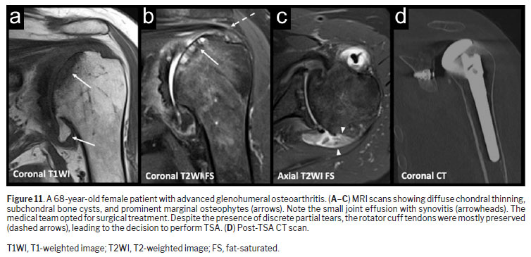
In refractory cases, surgical options for glenohumeral osteoarthritis are less extensive, because arthroscopic debridement may be effective only in early-stage lesions, making it inadequate for addressing advanced disease. Total shoulder arthroplasty (TSA) is the gold standard for end-stage glenohumeral osteoarthritis (Figure 11), benefiting from modern prosthetic designs, addressing glenoid bone loss, improving patient outcomes, and delivering better postprocedure results than does hemiarthroplasty
(9,43). Reverse shoulder arthroplasty is preferred in patients with severe rotator cuff dysfunction or significant joint deformities
(56).
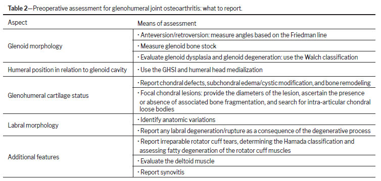
Preoperative imaging assessmentAccurate preoperative imaging assessment is vital for planning surgical interventions in glenohumeral cartilage lesions. It requires detailed analysis of glenoid morphology, bone stock, version, and soft tissue status, to optimize surgical outcomes. Must-report findings are organized in Table 2 and discussed below.
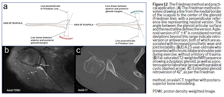
Glenoid versionDetermining the glenoid version, defined as its orientation relative to the scapular axis, is crucial for surgical planning as it impacts implant positioning and joint stability
(43). The Friedman method, widely regarded as the most reliable approach, measures the glenoid version by using a scapular reference line to determine deviations from neutral alignment
(57), as detailed in Figure 12.
The use of advanced imaging techniques, particularly 3D CT reconstruction, enhances the accuracy of the glenoid version measurement, providing clearer views of glenoid morphology and aiding in surgical planning. Variations such as significant retroversion (> 15°) or anteversion (> 5°) often necessitate specialized implants or bone grafting to restore the natural joint alignment
(43,51).
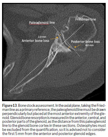
Bone stockBone stock assessment is crucial for ensuring the stability of the arthroplasty and longevity of the glenoid implant
(52). It enables qualitative and quantitative assessment of the glenoid bone resorption process in glenohumeral osteoarthritis, guiding orthopedic surgeons in determining whether there is need for glenoid augmentation in TSA planning for better postoperative results. Taking the Friedman line and the paleoglenoid line as references in the axial CT plane, bone stock should be measured in the anterior, central, and posterior portions of the glenoid cavity (Figure 13). The Pico method (“best-fit circle”) for measuring glenoid bone loss is more suitable in the setting of post-traumatic shoulder dislocations than in that of the preoperative period before elective surgery for glenohumeral osteoarthritis
(43,44,58).
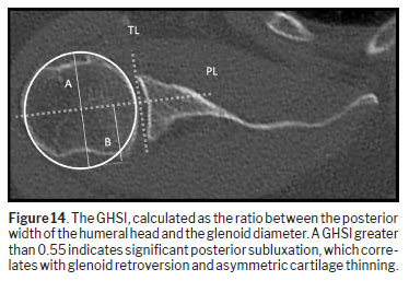
Glenohumeral subluxation assessmentThe glenohumeral subluxation index (GHSI) quantifies the posterior displacement of the humeral head relative to the glenoid, probably owing to capsuloligamentous laxity and labral lesions (Figure 14). The use of 3D imaging techniques improves the accuracy of GHSI determination by enabling precise segmentation of the humeral head and glenoid structures
(43,51,59).
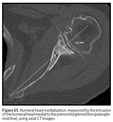
Humeral head medializationHumeral head medialization evaluates the degree of joint line displacement resulting from advanced osteoarthritis (Figure 15). Significant medialization often indicates severe cartilage and bone loss, limiting the surgical options
(43,51).
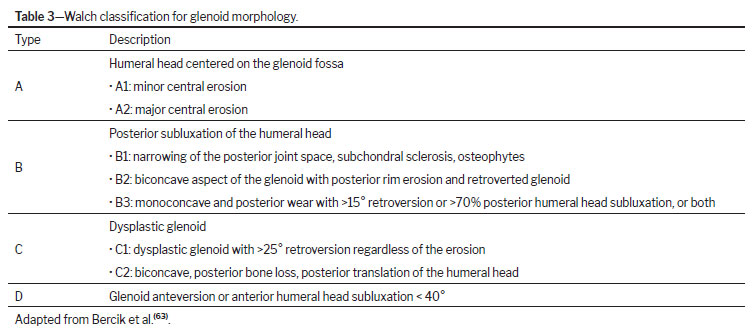
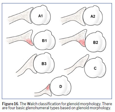
Walch classificationThe Walch classification (Table 3 and Figure 16) provides a systematic approach to categorizing glenoid morphology in osteoarthritis, with important implications for surgical planning and prognosis. Introduced in 1999, it remains the most widely used system to describe glenoid pathology in the context of shoulder replacement, helping guide decisions on prosthetic design, reaming, and bone grafting
(4,6,60–62). Walch type A represents symmetric glenoid morphology with no posterior subluxation. Due to symmetric load distribution in this glenoid morphology, there is adequate stability for joint replacement purposes
(4,6,43,60). Walch type B is common and represents asymmetric glenoid morphology, with predominant posterior glenoid wear related to progressive posterior humeral head subluxation. In this configuration, load distribution imbalances may predispose to prosthetic failure
(4,43,60). Walch type C is defined by accentuated glenoid retroversion, typically associated with dysplasia rather than erosion. This configuration is often congenital and predisposes the joint to severe dysfunction
(6,43).
Last, Walch type D represents pathologic anteversion, frequently accompanied by anterior subluxation of the humeral head. Although less common, this subtype poses unique challenges for surgical correction
(43,61).
Advanced imaging techniques, particularly 3D CT reconstructions, have significantly improved the reproducibility of the Walch classification. Studies show better interobserver and intraobserver agreement when using 3D imaging compared with traditional CT scans, emphasizing its importance in preoperative planning. In addition, the classification provides critical insights into the relationship between glenoid morphology and joint biomechanics, particularly its association with muscle fatty infiltration and outcomes in TSA
(6,64).
Muscle status assessmentThe condition of periarticular muscles, especially the rotator cuff, is critical in determining treatment outcomes for glenohumeral osteoarthritis
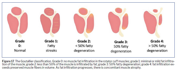
(41,43). The Goutallier grading system, as depicted in Figure 17, is particularly valuable in preoperative planning, allowing qualitative assessment of the rotator cuff muscles. It has been demonstrated that higher grades of fatty infiltration are strongly associated with poor surgical outcomes
(65). In cases of severe (grade 3 or 4) fatty degeneration, reverse shoulder arthroplasty is often required to compensate for the compromised function of the rotator cuff muscles
(36,41). Deltoid muscle trophism should also be described in the report, because its preservation is fundamental for good postoperative outcomes of reverse arthroplasty
(66).
CONCLUSIONComprehensive evaluation and management of focal glenohumeral chondral lesions and glenohumeral osteoarthritis requires a multidisciplinary approach that integrates anatomical understanding, adequate imaging techniques and clinical expertise. Advancements in imaging technologies and regenerative joint therapies hold promise for earlier detection and more conservative management of glenohumeral osteoarthritis. As these technologies continue to evolve, they offer opportunities to delay disease progression and improve long-term functional outcomes for patients.
Radiologists and orthopedic surgeons play a central role in this process by leveraging these diagnostic tools to identify key imaging parameters for optimal patient care. In addition, understanding the integration of labral degeneration, rotator cuff integrity, and secondary features like synovitis or deltoid muscle atrophy also improves patient selection for procedures such as TSA.
Continued research and collaboration across disciplines will be crucial to further enhance diagnostic accuracy and refine surgical and nonsurgical interventions, ultimately improving quality of life for patients affected by this complex condition.
REFERENCES1. Gasbarro G, Bondow B, Debski R. Clinical Anatomy and Stabilizers of the Glenohumeral Joint. Ann Joint. 2017;2(10):58.
2. Goldstein B. Shoulder Anatomy and Biomechanics. Phys Med Rehabil Clin N Am. 2004;15(2):313-49.
3. Baca M, King R, Bancroft L. Glenoid Hypoplasia. Radiol Case Rep. 2016;11(4):386-90.
4. Vo K, Hackett D, Gee A, Hsu J. Classifications in Brief: Walch Classification of Primary Glenohumeral Osteoarthritis. Clin Orthop Relat Res. 2017;475(9):2335-40.
5. Omoumi P et al. Glenohumeral Joint Instability. Magn Reson Imaging. 2010;33(1):2-16.
6. Ibounig T et al. Glenohumeral Osteoarthritis: An Overview of Etiology and Diagnostics. Scand J Surg. 2020;110(3):441-51.
7. Macías-Hernández S et al. Glenohumeral Osteoarthritis: Overview, Therapy, and Rehabilitation. Disabil Rehabil. 2016;39(16):1674-82.
8. Chillemi C, Franceschini V. Shoulder Osteoarthritis. Arthritis. 2013;2013:1-7.
9. Singh J et al. Surgery for Shoulder Osteoarthritis: A Cochrane Systematic Review. J Rheumatol. 2011;38(4):598-605.
10. Dunham K, Bencardino J, Rokito A. Anatomic Variants and Pitfalls of the Labrum, Glenoid Cartilage, and Glenohumeral Ligaments. Magn Reson Imaging Clin N Am. 2012;20(2):213-28.
11. Millett P, Gobezie R, Boykin R. Shoulder Osteoarthritis: Diagnosis and Management. Am Fam Physician. 2008;78(5):605-11.
12. Morag Yet al. MR Imaging of Rotator Cuff Injury: What the Clinician Needs to Know. Radiographics. 2006;26(4):1045-65.
13. Elser F, Dewing C, Millett P. Chondral and Osteochondral Lesions of the Humerus: Diagnosis and Management. Oper Tech Sports Med. 2008;16(4):178-86.
14. Elser F et al. Glenohumeral Joint Preservation: Current Options for Managing Articular Cartilage Lesions in Young, Active Patients. Arthroscopy. 2010;26(5):685-96.
15. DePalma A, Gruson K. Management of Cartilage Defects in the Shoulder. Curr Rev Musculoskelet Med. 2012;5(3):254-62.
16. Fiegen A et al. Articular Cartilage Defects of the Glenohumeral Joint: A Systematic Review of Treatment Options and Outcomes. Cartilage. 2019;13(1_suppl):401S-13S.
17. Gomoll A et al. Treatment of Chondral Defects in the Shoulder. Oper Tech Orthop. 2006;16(4):232-43.
18. Lederman E et al. The Glenoid Labral Articular Teardrop Lesion: A Chondrolabral Injury With Distinct Magnetic Resonance Imaging Findings. Arthroscopy. 2017;34(2):407-11.
19. Hernigou P, Hernigou J, Scarlat M. Shoulder Osteonecrosis: Pathogenesis, Causes, Clinical Evaluation, Imaging, and Classification. Orthop Surg. 2020;12(5):1340-9.
20. Carroll K, Helms C, Speer K. Focal Articular Cartilage Lesions of the Superior Humeral Head. AJR Am J Roentgenol. 2001;176(2):393-7.
21. Goes P et al. Shoulder and Elbow Injuries in Adult Overhead Throwers: Imaging Review. Radiographics. 2023;43(12):e230094.
21. Savage-Elliott I et al. Magnetic Resonance Imaging-Arthroscopy correlation for Chondral Pathology in Commonly Imaged Joints: A Narrative Review. J Cartil Jt Preserv. 2024;4(1):100154.
23. Sanders T, Morrison W, Miller M. Imaging Techniques for the Evaluation of Glenohumeral Instability. Am J Sports Med. 2000;28(3):414-34.
24. Momenzadeh O et al. Assessment of Correlation Between MRI and Arthroscopic Pathologic Findings in the Shoulder Joint. Arch Bone Jt Surg. 2015;3(4):286-90.
25. Markhardt B et al. Interpretation of Cartilage Damage at Routine Clinical MRI: How to Match Arthroscopic Findings. Radiographics. 2022;42(5):1457-73.
26. Guntern D et al. Articular Cartilage Lesions of the Glenohumeral Joint: Diagnostic Effectiveness of MR Arthrography and Prevalence in Patients with Subacromial Impingement Syndrome. Radiology. 2003;226(1):165-70.
27. Rhee R et al. MR and CT Arthrography of the Shoulder. Semin Musculoskelet Radiol. 2012;16(01):003-14.
28. Jarraya M et al. MR-Arthrography and CT-Arthrography in Sports-Related Glenolabral Injuries: A Matched Descriptive Illustration. Insights Imaging. 2016;7(2):167-77.
29. O’Brien J et al. Frequency of Glenoid Chondral Lesions on MR Arthrography in Patients with Anterior Shoulder Instability. Eur J Radiol. 2012;81(11):3461-5.
30. Guermazi A et al. MR Arthrography of the Shoulder: Optimizing Pulse Sequence Protocols for the Evaluation of Cartilage and Labrum. Eur J Radiol. 2014;83(8):1421-8.
31. Magee T. 3-T MRI of the Shoulder: Is MR Arthrography Necessary? AJR Am J Roentgenol. 2009;192(1):86-92.
32. Motamedi D et al. Pitfalls in Shoulder MRI: Part 1—Normal Anatomy and Anatomic Variants. AJR Am J Roentgenol. 2014;203(3):501-7.
33. Bittersohl B et al. T2* Mapping and Delayed Gadolinium-Enhanced Magnetic Resonance Imaging in Cartilage (dGEMRIC) of Glenohumeral Cartilage in Asymptomatic Volunteers at 3T. Eur Radiol. 2012;23(5):1367-74.
34. Rehnitz C et al. Delayed Gadolinium-Enhanced MRI of Cartilage (dGEMRIC) and T2 mapping at 3T MRI of the Wrist: Feasibility and Clinical Application. J Magn Reson Imaging. 2016;45(2):381-9.
35. Cao G, Gao S, Xiong B. Application of Quantitative T1, T2 and T2* Mapping Magnetic Resonance Imaging in Cartilage Degeneration of the Shoulder Joint. Sci Rep. 2023;13(1):4558.
36. Wuennemann F et al. 3-T T2 Mapping Magnetic Resonance Imaging for Biochemical Assessment of Normal and Damaged Glenoid Cartilage: A Prospective Arthroscopy-Controlled Study. Sci Rep. 2020;10(1):14396.
37. Breighner R et al. Technical Developments: Zero Echo Time Imaging of the Shoulder: Enhanced Osseous Detail by Using MR Imaging. Radiology. 2018;286(3):960-6.
38. Ensle F et al. Enhanced Bone Assessment of the Shoulder Using Zero-Echo Time MRI with Deep-Learning Image Reconstruction. Skeletal Radiol. 2024;53(12):2597-606.
39. Saltzman B, Leroux T, Cole B. Management and Surgical Options for Articular Defects in the Shoulder. Clin Sports Med. 2017;36(3):549-72.
40. Bhatia S et al. Surgical Treatment Options for the Young and Active Middle-Aged Patient with Glenohumeral Arthritis. Adv Orthop. 2012;2012:1-8.
41. Boselli K, Ahmad C, Levine W. Treatment of Glenohumeral Arthrosis. Am J Sports Med. 2010;38(12):2558-72.
42. Herrero-Beaumont G et al. Systemic Osteoarthritis: The Difficulty of Categorically Naming a Continuous Condition. Aging Clin Exp Res. 2024;36(1):45.
43. Sharifi A, Siebert M, Chhabra A. How to Measure Glenoid Bone Stock and Version and Why It Is Important: A Practical Guide. Radiographics. 2020;40(6):1671-83.
44. Silva FD, Ramachandran S, Chhabra A. Glenohumeral Osteoarthritis: What the Surgeon Needs from the Radiologist. Skeletal Radiol. 2022;52(11):2283-96.
45. Goud A et al. Radiographic Evaluation of the Shoulder. Eur J Radiol. 2008;68(1):2-15.
46. Kerr R et al. Osteoarthritis of the Glenohumeral Joint: A Radiologic-Pathologic Study. AJR Am J Roentgenol. 1985;144(5):967-72.
47. Elsharkawi M et al. Reliability of Radiologic Glenohumeral Osteoarthritis Classifications. J Shoulder Elbow Surg. 2013;22(8):1063-7.
48. Moor B et al. Is There an Association Between the Individual Anatomy of the Scapula and the Development of Rotator Cuff Tears or Osteoarthritis of the Glenohumeral Joint? Bone Jt J. 2013;95-B(7):935-41.
49. Hamada K et al. A Radiographic Classification of Massive Rotator Cuff Tear Arthritis. Clin Orthop Relat Res. 2011;469(9):2452-60.
50. Hasegawa A et al. Relationship Between the Hamada Grade and Underlying Pathological Conditions in the Rotator Cuff and Long Head of Biceps in Symptomatic Patients with Rotator Cuff Tears. JSES Int. 2022;6(3):488-94.
51. Yoo Y et al. Glenoid Orientation and Profile in Atraumatic or Microtraumatic Posterior Shoulder Instability: Morphological Analysis Using Computed Tomography Arthrogram. Orthop J Sports Med. 2021;9(2):2325967120982965.
52. Al-Omairi S et al. Glenoid Bone Loss in Shoulder Arthroplasty: A Narrative Review. Ann Joint. 2024;9:8.
53. de Mello R et al. Three-Dimensional Zero Echo Time Magnetic Resonance Imaging Versus 3-Dimensional Computed Tomography for Glenoid Bone Assessment. Arthroscopy. 2020;36(9):2391-400.
54. Karimi A et al. The Role of Conservative Treatment of Glenohumeral Joint Osteoarthritis: A Systematic Review. Semin Arthroplasty. 2024;34(1):34-43.
55. Yamamoto N et al. Non-Operative Management of Shoulder Osteoarthritis: Current Concepts. J ISAKOS. 2023;8(5):289-95.
56. Somerson J et al. What Factors Are Associated With Clinically Important Improvement After Shoulder Hemiarthroplasty for Cuff Tear Arthropathy? Clin Orthop Relat Res. 2016;474(12):2682-8.
57. Friedman RJ, Hawthorne KB, Genez BM. The use of computerized tomography in the measurement of glenoid version. J Bone Joint Surg. 1992;74(7):1032-1037.
58. Sheean A et al. Shoulder Instability: State of the Art. J ISAKOS. 2016;1(6):347-57.
59. Tung G, Hou D. MR Arthrography of the Posterior Labrocapsular Complex: Relationship with Glenohumeral Joint Alignment and Clinical Posterior Instability. AJR Am J Roentgenol. 2003;180(2):369-75.
60. Walch G et al. Morphologic Study of the Glenoid in Primary Glenohumeral Osteoarthritis. J Arthroplasty. 1999;14(6):756-60.
61. Neyton L et al. Mid- to Long-Term Follow-Up of Shoulder Arthroplasty for Primary Glenohumeral Osteoarthritis in Patients Aged 60 or Under. J Shoulder Elbow Surg. 2019;28(9):1666-73.
62. Jawa A, Shields M. The Evolution of the Walch Classification for Primary Glenohumeral Arthritis. J Am Acad Orthop Surg. 2021;29(13):e635-45.
63. Bercik M et al. A Modification to the Walch Classification of the Glenoid in Primary Glenohumeral Osteoarthritis Using Three-Dimensional Imaging. J Shoulder Elbow Surg. 2016;25(10):1601-6.
64. Donohue K et al. The Association Between Rotator Cuff Muscle Fatty Infiltration and Glenoid Morphology in Glenohumeral Osteoarthritis. J Bone Joint Surg. 2018;100(5):381-7.
65. Tsuchiya S et al. The Relationship Between Preoperative Goutallier Stage and Retear Rates Following Posterosuperior Rotator Cuff Repair: A Systematic Review. J Shoulder Elbow Surg. 2023;32(2):435-43.
66. de Boer F, et al. Long-Term Relation Between Deltoid Muscle Volume and Clinical Outcomes in a Reverse Shoulder Arthroplasty. Semin Arthroplasty. 2022;32(4):664-70.
1. Department of Musculoskeletal Radiology, Fleury Medicina e Saúde, São Paulo, SP, Brazil
2. Department of Radiology and Biomedical Imaging, University of California, San Francisco, CA, USA
3. Department of Orthopedic Surgery, Shoulder and Elbow Unit, Grupo Vita, São Paulo, SP, Brazil
a.
http://orcid.org/0000-0001-7474-1934b.
https://orcid.org/0009-0007-6338-1364c.
https://orcid.org/0000-0002-5075-5896d.
https://orcid.org/0000-0002-6458-3642e.
https://orcid.org/0000-0003-2815-9999.Correspondence:Dâmaris Versiani Caldeira Gonçalves
MD. Fleury Medicina e Saúde.
Rua Mato Grosso, 306, Lj 01. Higienópolis, São Paulo, SP, Brazil. 01239-040.
Email:
damaris.goncalves@grupofleury.com.brEditor in charge: Dr. Valdair Francisco Muglia
Data availability: Not applicable
Received in
January 12 2026.
Reviewed in
February 18 2026.
Accepted em
May 10 2026.
Publish in
August 28 2026.

 |
|







 PDF English
 Print
Print
 Send this article by email
 How to cite this article
 Submit a comment
 Mendeley
 Pocket
 Share on Facebook
 Share on Twitter