ABSTRACT
Neuro-Behçet’s disease (NBD) is a rare but serious manifestation of Behçet’s disease, categorized into parenchymal and non-parenchymal forms, each with distinct clinical and imaging characteristics. Parenchymal NBD primarily affects the brainstem, basal ganglia, and diencephalon. On magnetic resonance imaging (MRI), acute or subacute lesions appear hyperintense on T2-weighted or fluid-attenuated inversion recovery sequences and isointense to hypointense on T1-weighted sequences, whereas chronic lesions may present as asymmetrical atrophic changes. Non-parenchymal NBD may present as cerebral venous thrombosis, arterial involvement, and meningeal inflammation. Here, we provide a pictorial essay on MRI central nervous system studies, mostly following an international consensus classification of NBD. Although NBD is rare, recognizing its characteristic imaging features is crucial for early diagnosis and treatment, potentially improving prognosis and reducing long-term neurological complications.
Keywords:
Behçet syndrome; Magnetic resonance imaging; Vasculitis, central nervous system.
INTRODUCTION
Behçet’s disease (BD) is a variable vessel vasculitis characterized by recurrent oral and genital ulcers, often accompanied by skin and ocular manifestations. Although the etiology of BD remains unknown, studies have suggested a complex interplay between environmental and genetic factors, specifically human leukocyte antigen alleles, such as HLA-B*51. The disease can affect both sexes but tends to present more severely in men and is rare in children(1).
During the course of BD, central nervous system (CNS) involvement occurs in 9.4% of cases, leading to significant morbidity and mortality(2). Among children, the rate of CNS involvement is 3.6%(3). This neurological involvement is termed Neuro-Behçet’s disease (NBD) and is classified into two main forms(4,5): parenchymal and non-parenchymal, reportedly accounting for 75–80% and 15–20% of cases, respectively. It is most commonly observed in young adult men(2). The two subtypes exhibit distinct clinical, pathological, and prognostic features. Parenchymal NBD is thought to result from inflammation affecting small to medium-sized veins, particularly in the brainstem, basal ganglia, and diencephalon, whereas non-parenchymal NBD is primarily attributed to conditions involving larger vessels, such as cerebral venous thrombosis (CVT). Co-occurrence of parenchymal and non-parenchymal NBD forms is uncommon(2,6).
In BD, involvement of the CNS parenchyma manifests as a wide range of neurological symptoms. Although pyramidal signs, hemiparesis, headache, and dysarthria are the most common neurological features of NBD, other presentations, including dementia, psychiatric disorders, cranial nerve palsies, and cerebellar ataxia, may be observed(7). In contrast, non-parenchymal involvement typically includes benign intracranial hypertension accompanied by papilledema, meningitis, or dural sinus thrombosis(4,5). Headache is the most prevalent neurological symptom in non-parenchymal NBD, whereas pyramidal signs are more common in parenchymal disease(8). Although CVT is a common finding, aseptic meningitis is quite rare(4,6). The diagnosis of NBD is established when patients meet the 1990 International Study Group Criteria for BD (or any other accepted classification criteria), with objective neurological involvement attributed to BD. This diagnosis is supported by neuroimaging or cerebrospinal fluid analysis in the absence of a better explanation for the neurological findings, such as mimics of NBD, which include CNS infections, neoplasms, and BD treatment toxicity(4).
Magnetic resonance imaging (MRI) is the gold standard neuroimaging tool for evaluating NBD, revealing characteristic features of the disease(4). International consensus recommendations support the use of MRI, including contrast-enhanced studies and magnetic resonance venography, for the diagnosis, monitoring, and differential diagnosis of NBD(4). Computed tomography can be used as a complement to the vascular assessment, although its contribution to parenchymal evaluation is limited(6). In a multicenter retrospective study conducted between 1988 and 2008 in Japan, suspected cases of neurological BD were categorized into acute NBD (ANBD), chronic progressive NBD (CPNBD), and non-NBD conditions(9). On T2-weighted imaging (T2WI), the authors observed hyperintense lesions in approximately 60% of the patients with ANBD, in 54.2% of those with CPNBD, and in 42.4% of those with a non-NBD condition, indicating that these signal abnormalities are not specific to NBD. Those hyperintense lesions most commonly involved the pons, midbrain, and basal ganglia. Conversely, brainstem atrophy was identified in 7.5% of the patients with ANBD, in 71.4% of those with CPNBD, and in 9.0% of those with a non-NBD condition, demonstrating a strong association between brainstem atrophy and CPNBD(9). Collectively, these findings suggest that ANBD is characterized by episodic inflammatory changes within the cerebral parenchyma, meninges, or both, typically appearing as hyperintense lesions on T2WI or fluid-attenuated inversion recovery (FLAIR) sequences. Although MRI abnormalities indicative of ANBD show modest sensitivity and specificity, ANBD is generally marked by recurrent inflammatory changes in the parenchyma, meninges, or both, typically identified as hyperintense foci on T2WI or FLAIR sequences. However, brainstem atrophy is a common finding in CPNBD, and may be relevant for future diagnostic criteria. These data were integrated into the Japanese National Research Committee for Behçet’s Disease recommendations for the management of NBD(10).
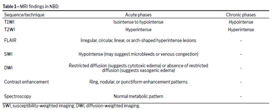
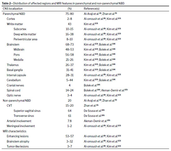
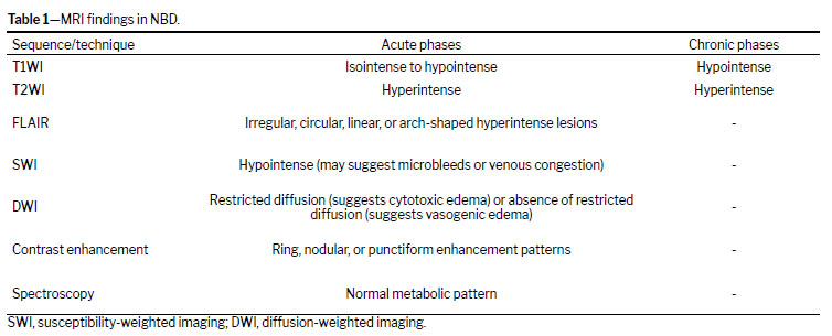
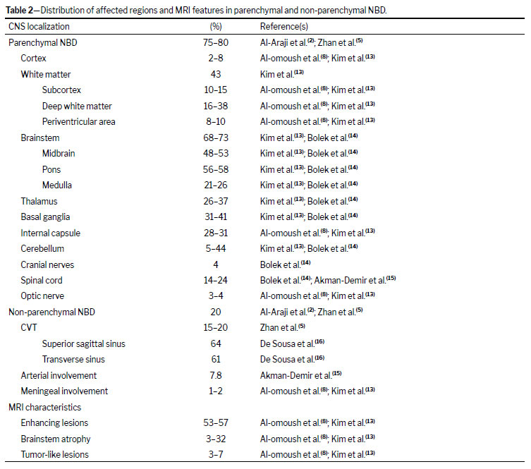
In parenchymal NBD, acute or subacute lesions typically appear as asymmetric, medium-sized (4–10 mm), multiple, hyperintense lesions on T2WI and FLAIR sequences, with corresponding isointensity or hypointensity on T1-weighted imaging (T1WI). In chronic disease, MRI may reveal small, scattered, non-enhancing lesions on T1WI, as well as slightly hyperintense lesions on T2WI, often accompanied by brainstem atrophy(4,5), as shown in Table 1. Contrast-enhancing lesions tend to predominate in acute presentations, whereas brainstem atrophy is observed more commonly in chronic disease stages(8). The brainstem is the region most commonly affected in parenchymal NBD (Table 2), and the presence of upper brainstem lesions extending unilaterally to the thalamus and basal ganglia strongly supports a diagnosis of parenchymal NBD(5). Susceptibility-weighted imaging may demonstrate hemorrhagic foci in parenchymal NBD, providing additional diagnostic support, especially in acute or subacute presentations(11). In addition, vasogenic edema (a lesion without restricted diffusion) is commonly observed in acute disease and tends to regress after corticosteroid and immunosuppressive therapy. These findings support the hypothesis that parenchymal NBD is primarily an inflammatory venous process rather than an arterial disease(5,12). However, in some cases, restricted diffusion due to cytotoxic edema may be observed. For the assessment of larger-caliber vessels, MRI venography and arteriography (or even high-resolution vessel wall imaging) are crucial for identifying CVT or arterial involvement(4,6).
The aim of this study was to present a pictorial essay of MRI findings in patients diagnosed with NBD, highlighting parenchymal and non-parenchymal involvement. While not a formal systematic review, the essay is in accordance with the international consensus classification of NBD
(4). Although NBD is a rare entity, recognition of its characteristic imaging features can assist radiologists and non-radiologists in achieving an earlier diagnosis, optimizing therapeutic strategies, and improving patient outcomes.
PARENCHYMAL NBDBrainstemThe brainstem is the region most commonly affected in parenchymal NBD. However, isolated brainstem involvement occurs in 25% of cases
(15,17). Brainstem lesions are most likely located in the midbrain and pons, with contrast-enhancing lesions being more common in acute cases
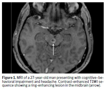
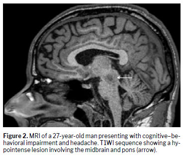
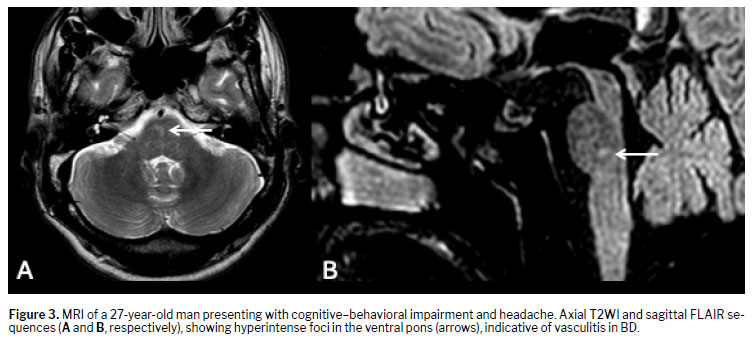
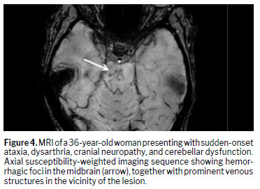
(8). In the acute phase, hypointense lesions and ring-shaped contrast enhancement can be seen on T1WI (Figures 1 and 2). On T2WI and FLAIR sequences, those lesions are hyperintense (Figure 3). Hemorrhagic lesions may also be seen, particularly on susceptibility-weighted imaging (Figure 4).
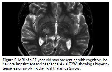
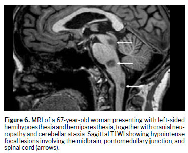
Multifocal/diffuse involvementIn multifocal or diffuse involvement, there is a concurrent involvement of various areas, including the brainstem, cerebrum, and spinal cord. The mesodiencephalic junction is described as the most frequently involved site, with lesions extending upward to the diencephalon or downward to the pontobulbar region
(6,18), as illustrated in Figures 5 and 6.
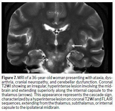
A characteristic finding in parenchymal NBD is the cascade sign, typically observed on coronal MRI during acute presentations. The cascade sign appears as a vertically oriented lesion extending from the midbrain to the thalamus
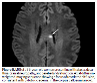
(19), as depicted in Figure 7. As illustrated in Figure 8, lesions in the periventricular, juxtacortical, and corpus callosum regions are less common
(6).
Spinal cord involvementThe spinal cord lesions seen in NBD are asymmetric, can be single or multiple, and may present as longitudinally extensive transverse myelitis affecting the posterolateral segments
(18,19). Inflammatory lesions in the cervical or thoracic spinal cord are often associated with concurrent brainstem, basal ganglia, or cerebral lesions. Isolated spinal cord involvement is rare, occurring in < 7% of cases
(15,18).
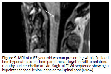
The MRI findings typically include noncontiguous multifocal lesions, which appear hypointense to isointense on T1WI (Figure 9) and hyperintense on T2WI, sometimes with contrast enhancement
(17). Two distinct MRI patterns have been described in spinal cord involvement of NBD
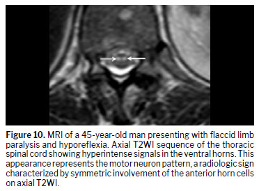
(20,21): the bagel sign, defined as a central lesion with a hypointense core and hyperintense rim on axial T2WI; and the motor neuron pattern, characterized by symmetric involvement of the anterior horn cells (Figure 10).
Cerebral involvementCerebral involvement in parenchymal NBD typically presents as multiple small white matter lesions, predominantly in subcortical regions. The basal ganglia and internal capsule are commonly affected
(22). Hemispheric white matter lesions are uncommon, and isolated cerebral lesions are rare, requiring differentiation from those attributable to infectious and neoplastic diseases
(5,17).
In the acute or subacute phase, lesions often appear as small circular, linear, or crescent-shaped hyperintensities on T2WI or FLAIR sequences, with mild contrast enhancement, described as “target lesions” or “concentric rings”, which result from blood–brain barrier disruption
(6). A characteristic linear hyperintense signal may be seen along the internal capsule
(12).
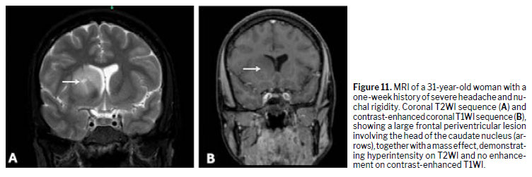
Tumor-like lesions in NBD are very rare and may require brain biopsy to differentiate from those of neoplastic or infectious etiology. Such lesions are most often found in the thalamus and basal ganglia, commonly presenting with surrounding edema and a mass effect
(23), as depicted in Figure 11.
Ocular involvementOptic neuropathy is a rare manifestation of parenchymal NBD. In the acute phase, optic neuritis presents as swelling of the intraorbital portion of the optic nerve, which appears hyperintense on contrast-enhanced T2WI or FLAIR sequences
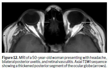
(18). Other MRI findings, such as thickening of the posterior ocular globe, may also be observed and can support the diagnosis of NBD when occurring in conjunction with retinal vasculitis (Figure 12).
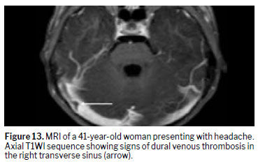
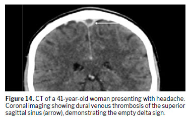
NON-PARENCHYMAL NBDCVTThe most common manifestation of non-parenchymal NBD is CVT. The superior sagittal sinus, transverse sinuses, and basal vein of Rosenthal are the sites most often affected
(6). On MRI, subacute thrombi typically appear hypointense on T2WI sequences
(24). In the chronic stage, particularly when recanalization is incomplete, lesions may show isointense signals on T1WI and isointense to hyperintense signals on T2WI sequences. An occasional complication of CVT is the development of a dural arteriovenous fistula (Figure 13). Follow-up imaging can show enlargement of the cortical arteries and veins adjacent to the affected area, changes attributed to compensatory arterial recruitment and venous hypertension
(24).
As shown in Figures 13 and 14, MRI venography in NBD can demonstrate direct and indirect signs of CVT
(17,24), the direct signs including loss of the normal flow void and irregular sinus contours after partial recanalization, whereas the indirect signs include collateral venous pathways and evidence of elevated intracranial pressure.
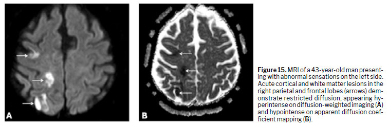
Intracranial aneurysm and cervical extracranial aneurysm/dissectionThe involvement of large arteries leading to ischemic infarcts is rare in NBD. A stroke-like presentation is not typical, and signs of arterial involvement should be interpreted with caution
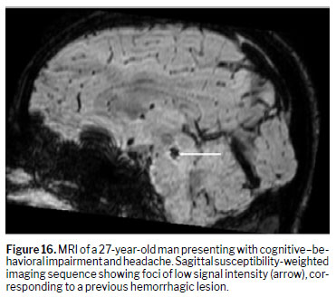
(6,25). These vascular complications may occur due to stenosis or dissection of the cervicocephalic arteries and present hyperintense signals on diffusion-weighted imaging, with a low apparent diffusion coefficient, indicating restricted diffusion (Figure 15). Foci of parenchymal cerebral hemorrhage and arteriovenous fistula may occur (Figure 16), although they are rare
(20,24,26).
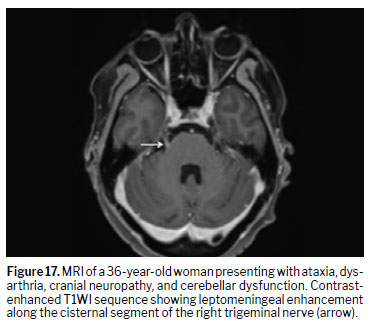
Acute meningeal syndromeAcute meningeal syndrome is an uncommon manifestation of NBD, often occurring concomitantly with parenchymal NBD. Isolated aseptic meningitis is very rare
(8,27). The MRI findings include meningeal thickening with contrast enhancement, with or without dural sinus thrombosis
(27), as depicted in Figure 17.
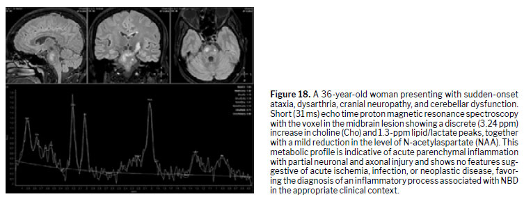
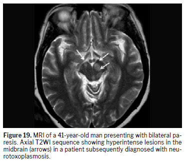
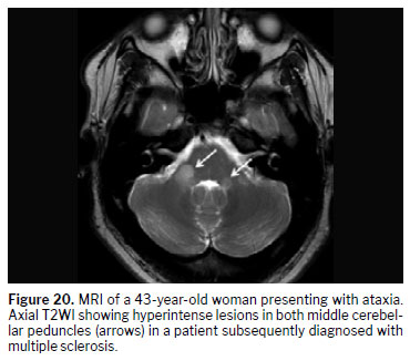
DIFFERENTIAL DIAGNOSISThe differential diagnoses of BD include inflammatory brain lesions, especially those involving the brainstem and midbrain–diencephalic region, such as multiple sclerosis, gliomas, and neurotoxoplasmosis
(4,6,28). In addition to imaging findings, clinical characteristics are important for a correct diagnosis. Proton magnetic resonance spectroscopy may reveal a low N-acetylaspartate peak, indicating neuronal and axonal loss or dysfunction. Although elevations in lipid/lactate peaks and in the choline/creatine ratio are nonspecific (Figure 18), they may indicate ongoing myelin breakdown
(29,30).
Similar to NBD, neurotoxoplasmosis may present with hyperintense lesions on T2W images and ring-enhancing lesions on gadolinium contrast-enhanced T1WI sequences. Clinical history and previous conditions such as immunosuppression should be assessed to assure a consistent diagnosis. Multiple sclerosis may present with hyperintensity on T2WI. In such cases, the diagnosis should be guided by the time–space dissemination criteria, including the presence of supratentorial lesions along with the clinical presentation
(4,28), as illustrated in Figures 19 and 20.
KEY MESSAGES• The gold standard for imaging assessment in NBD is MRI.
• In parenchymal NBD, the brainstem is the most commonly affected region, and involvement of the midbrain or pons, often extending to the thalamus, basal ganglia, or both, is a key radiologic indicator of this form of the disease.
• Spinal cord involvement in NBD may manifest as longitudinally extensive transverse myelitis. An MRI scan may also show two characteristic patterns: the bagel sign; and a motor neuron involvement pattern.
• The most common manifestation of non-parenchymal NBD is CVT.
• Because NBD is rare, it is essential to exclude potential mimics, such as CNS infections, neoplasms, and treatment-related toxicity, before confirming the diagnosis.
Conflicts of interest The authors report no conflicts of interest.
Funding This research did not receive any specific grants from funding agencies in the public, commercial, or not-for-profit sectors.
REFERENCES1. Emmi G, Bettiol A, Hatemi G, Prisco D. Behçet’s syndrome. The Lancet. 2024 Mar 16;403(10431):1093–108.
2. Al-Araji A, Kidd DP. Neuro-Behçet’s disease: epidemiology, clinical characteristics, and management. Lancet Neurol. 2009;8(2):192–204.
3. Uluduz D, Kürtüncü M, Yapici Z, Seyahi E, Kasapçopur O, Özdo-an H, et al. Clinical characteristics of pediatric-onset neuro-Behçet disease. Neurology;77(21):1900–5.
4. Kalra S, Silman A, Akman-Demir G, Bohlega S, Borhani-Haghighi A, Constantinescu CS, et al. Diagnosis and management of Neuro-Behçet’s disease: international consensus recommendations. J Neurol. 2014;261(9):1662–76.
5. Zhan H, Cheng L, Li Y. Neuro-Behçet’s disease: An update of clinical diagnosis, biomarkers, and immunopathogenesis. Clin Exp Immunol. 2025;219(1).
6. Belfeki N, Ghriss N, Fourati M, Leclercq D, Saadoun D. Neuro-Behçet’s disease: A review. Rev Med Interne. 2024;45(10):624–33.
7. Jawad O, Jawad H, Mohamed A, Mohamed C, Ahmed B. MRI findings in neuro-Behçet’s disease. About 104 moroccan patients and literature review. Mult Scler Relat Disord. 2025;102:106638.
8. Al-omoush O, AlBarakat MM, Alasmar D, Al-Khalaileh A, Alzoubi A, Tarakhan H, et al. Clinical and imaging features of parenchymal and non-parenchymal neuro-Behçet’s disease: a systematic review of case reports and series. BMC Neurol. 2025;25(1).
9. Hirohata S, Kikuchi H, Sawada T, Nagafuchi H, Kuwana M, Takeno M, et al. Clinical characteristics of neuro-Behcet’s disease in Japan: a multicenter retrospective analysis. Mod Rheumatol. 2012 Jun;22(3):405–13.
10. Hirohata S, Kikuchi H, Sawada T, Okada M, Takeno M, Kuwana M, et al. Recommendations for the Management of Neuro-Behçet’s Disease by the Japanese National Research Committee for Behçet’s Disease. Internal Medicine. 2020;59(19):2359.
11. Albayram S, Saip S, Hasiloglu ZI, Teke M, Ceyhan E, Tutuncu M, et al. Evaluation of parenchymal neuro-Behçet disease by using susceptibility-weighted imaging. AJNR Am J Neuroradiol. 2011;32(6):1050–5.
12. Akman-Demir G, Bahar S, Coban O, Tasci B, Serdaroglu P. Cranial MRI in Behçet’s disease: 134 examinations of 98 patients. Neuroradiology. 2003 Dec;45(12):851–9.
13. Kim SW, Kim TG, Oh J, Kim DY, Choi YC, Kim SM, et al. Clinical and Radiographic Characteristics of Neuro-Behçet’s Disease in South Korea. Journal of Clinical Neurology. 2019;15(4):429–37.
14. Bolek EC, Sari A, Kilic L, Kalyoncu U, Kurne A, Oguz KK, et al. Clinical features and disease course of neurological involvement in Behcet’s disease: HUVAC experience. Mult Scler Relat Disord. 2020;38:101512.
15. Akman-Demir G, Serdaroglu P, Tasçi B. Clinical patterns of neurological involvement in Behçet’s disease: evaluation of 200 patients. The Neuro-Behçet Study Group. Brain. 1999;122 ( Pt 11)(11):2171–81.
16. De Sousa DA, Mestre T, Ferro JM. Cerebral venous thrombosis in Behçet’s disease: A systematic review. J Neurol. 2011;258(5):719–27.
17. Borhani-Haghighi A, Kardeh B, Banerjee S, Yadollahikhales G, Safari A, Sahraian MA, et al. Neuro-Behcet’s disease: An update on diagnosis, differential diagnoses, and treatment. Mult Scler Relat Disord. 2020;39:101906.
18. Koçer N, Islak C, Siva A, Saip S, Akman C, Kantarci O, et al. CNS involvement in Neuro-Behcet syndrome: An MR study. American Journal of Neuroradiology. 1999;20(6):1015–24.
19. Cleaver J, Morrison H, Renowden SA, Atan D, Cossburn M, Rice CM. An important diagnostic clue for neuro-Behçet’s disease: the “cascade sign.” Rheumatology (Oxford). 2022;61(5):E130–1.
20. Uygunoğlu U, Siva A. Behçet’s Syndrome and Nervous System Involvement. Curr Neurol Neurosci Rep. 2018;18(7).
21. Tamanini JVG, Sabino JV, Cordeiro RA, Mizubuti V, Villarinho L de L, Duarte JÁ, et al. The Role of MRI in Differentiating Demyelinating and Inflammatory (not Infectious) Myelopathies. Semin Ultrasound CT MR. 2023;44(5):469–88.
21. Haghighi AB, Pourmand R, Nikseresht AR. Neuro-Behçet disease: A review. Vol. 11, Neurologist. 2005.
23. Fuentes ÓP, Ballester LL, Troncoso JÁ, Abánades CIS, Mozo AN, Marhuenda ÁR, et al. Inflammatory cerebral pseudotumour as a rare manifestation of neuro-Behçet’s disease: A case report and literature review. J Neuroimmunol. 2025;405.
24. Oliveira IM, Duarte JÁ, Dalaqua M, Jarry VM, Pereira FV, Reis F. Cerebral venous thrombosis: imaging patterns. Radiol Bras. 2022;55(1):54–61.
25. Borhani-Haghighi A, Kardeh B, Banerjee S, Yadollahikhales G, Safari A, Sahraian MA, et al. Neuro-Behcet’s disease: An update on diagnosis, differential diagnoses, and treatment. Mult Scler Relat Disord. 2020;39.
26. Nakaya Y, Hayashi K, Hashimoto N, Suzuki A, Mitsuhashi S, Sato M, et al. Dural Arteriovenous Fistula in Neuro-Behçet’s Disease: Association or Chance? Cureus [Internet]. 2024 Feb 27;16(2).
27. Gumà A, Aguilera C, Acebes J, Arruga J, Pons L. Meningeal involvement in Behçet’s disease: MRI. Neuroradiology. 1998;40(8):512–5.
28. Çoban O, Bahar S, Akman-Demir G, Taşcı B, Yurdakul S, Yazıcı H, et al. Masked assessment of MRI findings: is it possible to differentiate neuro-Behçet’s disease from other central nervous system diseases? [corrected]. Neuroradiology. 1999;41(4):255–60.
29. Faria A V., Reis F, Zanardi VA, Menezes JR, Cendes F. The pattern of proton magnetic resonance spectroscopy in non-neoplastic encephalic lesions. Arq Neuropsiquiatr. 2004;62(2B):429–36.
30. Scully SE, Stebner FC, Yoest SM. Magnetic resonance spectroscopic findings in neuro-Behçet disease. Neurologist. 2004;10(6):323–6.
1. Department of Orthopedics, Rheumatology, and Traumatology, (São Paulo) State University of Campinas, Campinas, SP, Brazil
2. Department of Radiology and Oncology, (São Paulo) State University of Campinas, Campinas, SP, Brazil
a.
https://orcid.org/0000-0002-5340-6894b.
https://orcid.org/0000-0001-6764-612Xc.
https://orcid.org/0009-0009-3592-184Xd.
https://orcid.org/0009-0003-9949-1208e.
https://orcid.org/0000-0002-2874-0268f.
https://orcid.org/0000-0003-1216-9690g.
https://orcid.org/0000-0003-2256-4379Correspondence:Carlos Eduardo Garcez Teixeira
MD, Rheumatologist. Vasculitis Outpatient Clinic, University of Campinas (UNICAMP),
Rua Tessália Vieira de Camargo, 126, Cidade Universitária Zeferino Vaz, Campinas, SP, Brazil, 13083-887.
Email:
carlosgarcezt@gmail.comEditor in charge: Dr. Valdair Francisco Muglia.
Received in
December 26 2025.
Accepted em
January 26 2026.
Publish in
August 28 2026.

 |
|







 PDF English
 Print
Print
 Send this article by email
 How to cite this article
 Submit a comment
 Mendeley
 Pocket
 Share on Facebook
 Share on Twitter