Radiologia Brasileira - Publicação Científica Oficial do Colégio Brasileiro de Radiologia
AMB - Associação Médica Brasileira CNA - Comissão Nacional de Acreditação
 Vol. 43 nº 2 - Mar. / Apr. of 2010
|
ORIGINAL ARTICLE
|
|
Estimation of placental volume by three-dimensional ultrasonography with the XI VOCAL method in the first gestational trimester |
|
|
Autho(rs): Paulo Martin Nowak, Luciano Marcondes Machado Nardozza, Edward Araujo Júnior, Liliam Cristine Rolo, Hélio Antonio Guimarães Filho, Antonio Fernandes Moron |
|
|
Keywords: Early placental phase, First trimester of pregnancy, Gestational age, Organ volume, Three-dimensional imaging |
|
|
Abstract:
IMDs, Department of Obstetrics, Universidade Federal de São Paulo (Unifesp), São Paulo, SP, Brazil
INTRODUCTION Human placenta is a villous, hemochorionic organ essential for the nutrient uptake, mother/fetus gas exchange, and also for elimination of waste resulting from the fetal metabolism. There are evidences supporting an association between first trimester placentation and the subsequent gestational outcome(1). Three-dimensional ultrasonography has shown to be a method capable of evaluating the placental volume (PV) at the first gestational trimester, but till the present moment only the multiplanar method(2,3) and Virtual Organ Computer-aided Analysis (VOCAL) have been applied in vivo(4–6). Recently, a novel volumetric technique named eXtended Imaging Virtual Organ Computer-aided AnaLysis (XI VOCAL) has been available as a part of the software Three-dimensional eXtended Imaging (3D XI) (Medison; Seoul, Korea). Such technique consists in the delimitation of sequential adjacent planes displayed on the equipment screen (multislice view); at the end of the process, the equipment performs the summation of the areas and automatic volume calculation(7). A sole study has utilized this technique for evaluating in vivo the volume of fetuses between 11 and 14 gestational weeks(8), but a description of the PV is not observed. The present study is aimed at determining reference values for PV between 7 and 10 + 6 weeks of gestation by means of three-dimensional ultrasonography with the novel XI VOCAL method.
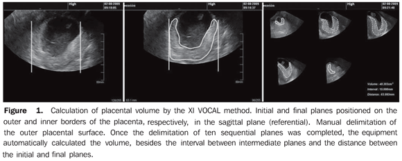
MATERIALS AND METHODS A cross-sectional study was developed in the period between October 2006 and March 2008 with 70 healthy pregnant women. This study was approved by the Committee for Ethics in Research of Universidade Federal de São Paulo (Unifesp), under No. 1491/06, and the women who agreed to voluntarily participate signed a term of free and informed consent. Inclusion criteria were the following: singleton gestation with a viable embryo, gestational age determined by the date of the last menstruation in women with regular menses, or by means of ultrasonography performed up to 10 + 6 weeks of gestation utilizing the crown-rump length (CRL) as a parameter; and absence of vaginal bleeding in the current gestation. Exclusion criteria were the following: pregnant women with chronic diseases, smoking habit or drug users during the current gestation; use of abortifacient drugs; presence of uterine malformations and pregnancy resulting from infertility treatments. The studies were performed by two observers at the Division of Three-Dimensional Ultrasonography, Department of Obstetrics of Unifesp, in an Accuvix XQ unit (Medison; Seoul, Korea) with a multifrequency endocavitary volumetric transducer (4–9 MHz). The three-dimensional placental volume is obtained by pressing the "3D" key, so a scanning window (BOX) is displayed and positioned to cover the whole placental extent (region of interest – ROI). A scanning angle of 75° was utilized with a slow velocity to achieve the best possible volume measurement quality. A clear separation between the basal layer of the placenta and the uterine wall was achieved with the harmonic mode and the focus adjusted for the placental depth. Once the automatic scanning was completed, the three-dimensional image was displayed on the screen perpendicular to each other in three orthogonal (axial, sagittal and coronal) planes. The placental volume was evaluated with the XI VOCAL method utilizing the sagittal plane as a reference, with the initial and final planes positioned respectively on the external and internal placental borders. Ten sequential planes were manually delimited and once the last plane delimitation was concluded, the equipment automatically calculated the structure volume as well as the interval between the intermediate planes and the distance between the initial and final planes (Figure 1). The volumetric analyses were performed off-line by a single observer with the SonoView Pro software version 1.03 (Medison; Seoul, Korea).
Data were stored on Excel 2003 worksheets (Microsoft; Redmond, WA, USA) and analyzed by the Statistical Package for Social Sciences (SPSS) for Windows version 11.0 (SPSS Inc.; Chicago, IL, USA). The correlation between the PV and the gestational age was evaluated by means of a scatter plot adjusted by the determination coefficient (R2). Means, medians, standard deviations and minimum/maximum values for PV, besides the 5th, 25th, 50th, 75th and 95th percentiles were calculated for each gestational age. The significance level was set at 0.05.
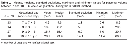
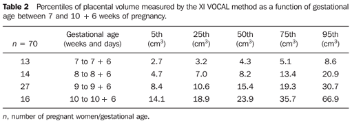
RESULTS The maternal aged ranged from 19 to 41 years with mean 29 ± 5.5 years (standard deviation). The number of pregnancies ranged from 1 to 9 (mean 2.5 ± 1.5), while the number of deliveries ranged from 0 to 8 (mean 1 ± 1.6). Mean PV ranged from 4.6 ± 1.8 cm3 (2.6–8.6 cm3) to 28.9 ± 14.1 cm3 (11.4–66.9 cm3) between 7 and 10 weeks + 6 days of gestation. Table 1 presents means, medians, standard deviations, and minimum/maximum values for PV at the evaluated gestational ages. Table 2 presents the 5th, 25th, 50th, 75th and 95th percentiles for PV as a function of the gestational age.
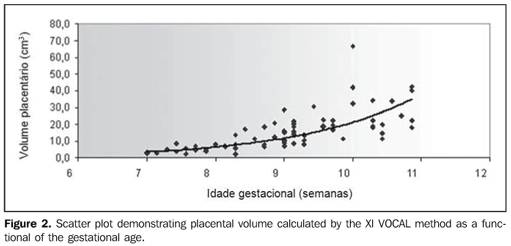
A strong correlation was observed between PV and gestational age (GA), with the best fit exponential regression model being represented by the following equation: PV = exp(0.582 × GA + 0.063); R2 = 0.82 (Figure 2).
DISCUSSION Three-dimensional ultrasonography has shown to be an appropriate technique for evaluating the PV at the first gestational trimester(2–6), as well as other structures such as the yolk sac, as recently evidenced by the authors' group(9). The multiplanar(2,3) and VOCAL(4–6) techniques are currently utilized for such purpose. The multiplanar technique involves the delimitation of areas of a determined object on a reference plane, while a cursor moves on another plane from a border to another; and once this course is completed, the equipment performs the summation of the areas and automatic volume calculation. The limitation of such technique is the long time required for the volumetric measurement(3). In the VOCAL technique, the structure is rotated around its axis and an area is determined at each rotational movement; at the end of the rotational process, the volume is calculated by the software and a reconstructed three-dimensional image of the structure is generated. The advantage of this technique is the relatively shorter time required for volumetric measurement, and the capacity of changing the margins of the delimited areas after the volumetric measurement is completed(10). A previous study developed by the authors has proved that the multiplanar (1.0 mm interval), VOCAL 30° (6 planes) and VOCAL 12° (15 planes) methods agreed in the evaluation of the PV between 6 and 10 + 6 weeks of gestation(5). The XI VOCAL method is an extension of the 3D XI software(7). By this technique, areas of sequential planes are delimited and displayed on the monitor screen (multislice view), and once the last plane delimitation is concluded, the equipment automatically calculates the structure volume as well as the interval between the intermediate planes and the distance between the initial and final planes. This software allows the delimitation of 5, 10 and 15 sequential planes. The sole study evaluating the application in vivo of such technique was recently published(8). In this study, the authors observe that the XI VOCAL method (10, 15 and 20 planes) can be permutably utilized with the multiplanar method (1.0 mm intervals); XI VOCAL (10 planes) and VOCAL 18°, besides XI VOCAL (15 planes) and VOCAL 12°, likewise can be permutably utilized in the evaluation of fetal volume between 11 and 14 gestational weeks. The authors have observed a strong correlation between PV measured with the XI VOCAL method (10 planes) and gestational age, being best fitted with the exponential regression model. A previous study developed by the authors group has demonstrated a strong correlation between the PV measured by the VOCAL 30º and the CRL, also with a best fit exponential regression model(6). Mean PV by the XI VOCAL method ranged from 4.6 to 28.9 cm3 between 7 and 10 + 6 weeks of gestation. In a previous study(5) with VOCAL 30° in the same gestational period, the mean PV ranged from 3.4 to 36.3 cm3 for a CRL between 9 and 40 mm of CRL. Another study(2) with the multiplanar method found a mean PV of 41.3 cm3 for a CRL between 35 and 44 mm2. Such results demonstrate a high intermethod agreement between XI VOCAL (10 planes) and VOCAL 30° in the evaluation of PV in the early pregnancy. However, the VOCAL 30° shows to be more advantageous because of the shorter time required for PV measurement and easier accessibility.
CONCLUSION In summary, the relevance of the present study is related to the introduction of a new applicability of the method in vivo, besides determining reference values for PV by the novel XI VOCAL method in the first trimester of pregnancy. Further studies with populations at risk for early gestational loss are required for proving the actual applicability of such reference curve.
REFERENCES 1. Smith GC. First-trimester origins of fetal growth impairment. Semin Perinatol. 2004;28:41–50. [ ] 2. Metzenbauer M, Hafner E, Hoefinger D, et al. Three-dimensional ultrasound measurement of the placental volume in early pregnancy: method and correlation with biochemical placenta parameters. Placenta. 2001;22:602–5. [ ] 3. Chen M, Leung KY, Lee CP, et al. Placental volume measured by three-dimensional ultrasound in the prediction of fetal alpha-thalassemia: a preliminary report. Ultrasound Obstet Gynecol. 2006;28:166–72. [ ] 4. Wegrzyn P, Faro C, Falcon O, et al. Placental volume measured by three-dimensional ultrasound at 11 to 13 + 6 weeks of gestation: relation to chromosomal defects. Ultrasound Obstet Gynecol. 2005;26:28–32. [ ] 5. Nowak PM, Nardozza LM, Araujo Júnior E, et al. Comparison of placental volume in early pregnancy using multiplanar and VOCAL methods. Placenta. 2008;29:241–5. [ ] 6. Nardozza LM, Nowak PM, Araujo Júnior E, et al. Evaluation of placental volume at 7–10+6 weeks of pregnancy by 3D-sonography. Placenta. 2009; 30:585–9. [ ] 7. Guimarães Filho HA, da Costa LL, Araujo Júnior E, et al. XI VOCAL (eXtended Imaging VOCAL): a new modality for three-dimensional sonographic volume measurement. Arch Gynecol Obstet. 2007;276:95–7. [ ] 8. Cheong KB, Leung KY, Chan HY, et al. Comparison of inter- and intraobserver agreement between three types of fetal volume measurement technique (XI VOCAL, VOCAL and multiplanar). Ultrasound Obstet Gynecol. 2009;33:287–94. [ ] 9. Rolo LC, Nardozza LMM, Araujo Júnior E, et al. Correlação do volume da vesícula vitelínica obtida por meio da ultrassonografia tridimensional com a idade gestacional entre a 7ª e a 10ª semanas usando o método multiplanar. Radiol Bras. 2009;42:359–62. [ ] 10. Peralta CF, Cavoretto P, Csapo B, et al. Lung and heart volumes by three-dimensional ultrasound in normal fetuses at 12-32 weeks' gestation. Ultrasound Obstet Gynecol. 2006;27:128–33. [ ] Received December 25, 2009. * Study developed at the Division of Three-Dimensional Ultrasonography, Department of Obstetrics, Universidade Federal de São Paulo (Unifesp), São Paulo, SP, Brazil. |
|
Av. Paulista, 37 - 7° andar - Conj. 71 - CEP 01311-902 - São Paulo - SP - Brazil - Phone: (11) 3372-4544 - Fax: (11) 3372-4554


