Radiologia Brasileira - Publicação Científica Oficial do Colégio Brasileiro de Radiologia
AMB - Associação Médica Brasileira CNA - Comissão Nacional de Acreditação
 Vol. 43 nº 2 - Mar. / Apr. of 2010
|
ORIGINAL ARTICLE
|
|
Magnetic resonance imaging of knee osteonecrosis: a study of 19 cases |
|
|
Autho(rs): Daniel Leme da Cunha, Antonio Carlos Pires Carvalho, Elísio José Salgado Ribeiro, Romeu Côrtes Domingues |
|
|
Keywords: Osteonecrosis, Magnetic resonance imaging, Subchondral abnormalities |
|
|
Abstract:
IMaster Fellow degree, Program of Post-Graduation, Faculdade de Medicina da Universidade Federal do Rio de Janeiro (UFRJ), MD, Radiologist at Clínicas de Diagnóstico Por Imagem (CDPI) and Multi-Imagem, Rio de Janeiro, RJ, Brazil
INTRODUCTION Spontaneous osteonecrosis of the knee is typically found in white women in their sixth and seventh decades of life. This condition is characterized by sudden onset of significant gonalgia not related to local trauma, meniscal surgery or even previous corticoid therapy. Yamamoto & Bullough(1) indicate that, additionally to the supposed vascular origin associated with increase in the intraosseous pressure, the mechanism of insufficiency fracture would play a role of relevant etiopathogenic factor besides osteoarthrosis, so the term "spontaneous" is questioned, considering that causal factors might by implied. The diagnosis is based on images that confirm the clinical suspicion. At the early phases of the disease, conventional radiography presents normal findings or minimal changes(2). In cases where conventional radiological signs are normal or dubious, magnetic resonance imaging is particularly useful for confirming the diagnostic suspicion since it demonstrates suggestive or even typical abnormalities in signal intensity, besides the advantage of noninvasiveness and high-definition multiplanar images(3). Ramnath & Kattapuram(4) have related the presence of a "linear" subchondral signal abnormality in cases supposedly originated in insufficiency fracture, such factor being absent in cases of osteoarthrosis (which also presents similar abnormalities related to osteonecrosis in the subchondral bone). The present study was aimed at describing epidemiological, clinical and mainly magnetic resonance imaging findings of osteonecrosis of the knee.
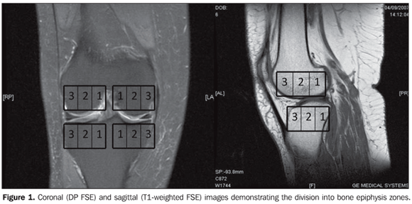
MATERIALS AND METHODS Nineteen magnetic resonance imaging studies of knees of 12 women and 7 men with no previous history of causal factors, with acute symptoms and signs compatible with osteonecrosis at examination were evaluated in the period from March to December/2007. Patients with previous history of trauma, local surgery of corticoid therapy were excluded. All the patients included in the present study were white, with ages ranging between 45 and 77 years (mean, 61 years) presented gonalgia. The following characteristics were taken into consideration in the clinical analysis: pain pattern (sudden or insidious; focal or diffuse) and association with joint edema, blockage and instability. Magnetic resonance imaging studies were performed in a GE 1.5 T Signa system (General Electric Medical Systems, Milwaukee, WI, USA), with acquisition of sagittal, T1-weighted sequence (repetition time [TR] = 405; time of excitation [TE] = 13.9; number of excitations [NEX] = 1; echo train length [ETL] = 2) and with proton density, fat-suppression in the coronal plane (TR = 1775; TE = 14; NEX = 2; ETL = 4), axial plane (TR = 1975; TE = 21; NEX = 2; ETL = 4) and sagittal plane (TR = 2250; TE = 14; NEX = 2; ETL = 4). Subchondral bone abnormalities were evaluated by two radiologists with experience in musculoskeletal system. Such abnormalities are defined as focal changes in signal intensity, with hypointense signal on T1-weighted images and variable signal intensity (hyper or hypointense) on T2-weighted images. The lesions location was defined as follows: distal femoral extremity (medial or lateral condyle) or tibial (internal or external plateau). Subsequently, each of the above described locations was arbitrarily divided into three zones, both in the coronal and sagittal planes to define the lesion epicenter. On the coronal plane (Figure 1), zone 1 would correspond to the internal third; zone 2 to the middle third, and zone 3, to the external third of the femoral condyle/tibial plateau. On the sagittal plane, 1, 2 and 3 would correspond, respectively, the posterior, middle and anterior thirds of the evaluated regions. Additionally, the presence or not signal intensity linear subchondral abnormality was evaluated. The presence of ipsilateral associated bone marrow edema, articular cartilage abnormality and meniscal lesion (whether unstable or not). Bone marrow edema was classified into grades I to III (respectively mild, moderate or significant). Subchondral lesions were measured (in millimeters) on the three planes (longitudinal × anteroposterior × cross-sectional planes).
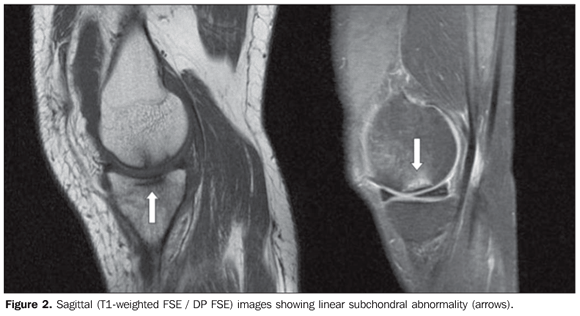
RESULTS As regards clinical presentation, 14 patients (73.6%) had a sudden pain onset, and in 5 patients the gonalgia onset was insidious. Eight patients reported joint blockage, two reported increased joint volume (edema), and three reported joint instability. All the patients presented lesions with hypointense signal on T1-weighted sequence and predominantly hyperintense signal (89.4%) on proton density weighted images with fat suppression. In eight cases, lesions were located in the external tibial plateau, in nine cases, in the medial femoral condyle, and in the internal tibial plateau in only two patients. Involvement of the lateral femoral condyle was observed in none of the cases. Sagittal images demonstrated a higher number of lesions with epicenter in the zone 2 (17 cases; 89.4%), two cases in the zone 3, and none in the zone 1. On coronal images, most of findings were observed in the zone 3 (11 cases; 57.8%), followed by the zone 2 (7 cases) and the zone 1 in a sole case. The presence of linear subchondral abnormality (Figure 2) was observed in 15 among the 19 cases (78.9%). Such abnormality was absent in the four remainder cases.
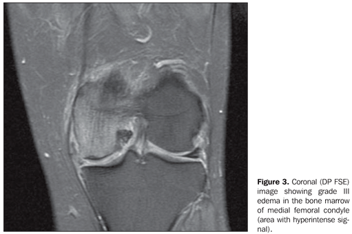
Association with grade III bone marrow edema was observed in 16 cases, and grade II in two cases (Figure 3).
Lesions size ranged from 1.5 to 8 mm on the longitudinal axis, from 2 to 30 mm on the anteroposterior axis, and from 6 to 26 mm on the cross-sectional axis [mean: 4.5 mm (lateral axis); 16.8 mm (ântero-posterior axis); and 11.8 mm (cross-sectional axis) – mean estimated volume, 466 mm3]. Chondral abnormalities were found in 63.1% of the patients, while associated ipsilateral meniscal lesion was observed in 14 cases (73.6%), with instability criteria (radial lesion, root ligament lesion) in 6 (31.5%) of these cases.
DISCUSSION Spontaneous osteonecrosis of the knee is a condition that affects predominantly female individuals, particularly in their seventh decade of life(5), with no association with systemic disorders, alcohol abuse, previous corticoid therapy, meniscal surgery or local trauma. In the first known description, Albäck et al.(6) have studied 40 patients (6 men and 34 women) with mean age of 70 years. Eschard et al.(2) have reported a clear female predominance (23 female × 11 male patients). In the present 19-patient sample, 12 were women (63.1%) and 7 (36.9%) were men, with mean age of 61 years. Lotke & Ecker(7) have defined sudden acute gonalgia onset in patients aged above 60 years without associated report of trauma as clinical criteria for the diagnosis of osteonecrosis. In the study developed by Eschard et al.(2), acute pain onset was observed in only 25% of the expected incidence, differently from the general literature. In the present study, at the time of the magnetic resonance imaging study, the greatest majority of the evaluated patients reported sudden focal pain onset. In the initial description presented by Albäck et al.(6), that was corroborated by the studies developed by Lotke & Ecker(7), the most frequently found site of osteochondral abnormalities was the medial femoral condyle. In the series developed by Björkengren et al.(8), this same location was reported in all of the 16 cases. Similarly, in the present study, there was a subtle predominance of involvement of the medial femoral condyle, followed by the internal tibial plateau. Previous studies had already emphasized the association of spontaneous osteonecrosis with meniscal lesion and, provided the diagnosis of the mentioned disease is not made when the patient is submitted to a meniscectomy, the acceleration of the clinical conditions and consequential articular involvement are evident. According to Muscolo et al.(9), in five cases with meniscal lesion there was a later onset of ipsilateral osteochondral abnormalities. Corroborating the literature, there was a considerable predominance of cases with homolateral meniscal lesion in association with osteochondral lesions, most frequently observed when instability was present. Because of the low sensitivity and the high negative likelihood ratio in chondral lesions diagnosis by magnetic resonance imaging(10), the absence of findings at this imaging study does not exclude the presence of such lesions. In the present study, more than half of cases (63.1%) presented associated chondral abnormalities. Such rate may be lower as compared with arthroscopy that is considered as the gold standard diagnostic method. Some studies(4,11) have already related the presence of insufficiency fracture in previously weakened bones as a trigger of the osteonecrosis process, based on correlation with similar findings described for femoral head osteonecrosis. Yamamoto & Bullough(1) have reported histological findings, postulating that one of the primary events that would lead to the development of osteonecrosis would be a subchondral insufficiency fracture in patients with bones mechanically weakened by non-neoplastic diseases, specifically osteoporosis, in spite of any confirmation by means of bone mass measurements in the evaluated patients. Ramnath & Kattapuram(4) have proposed that the presence of a linear subchondral signal at magnetic resonance imaging in association with the already described osteochondral abnormalities would affirm such relation, whereas the absence of this signal would suggest a stronger association with osteoarthrosis findings, casting a doubt in relation to the validity of the term "spontaneous" that is currently utilized. Additionally, in the present study, sudden pain onset was observed preferentially in weight-sustaining zones, besides a higher degree of associated bone marrow edema. However, in patients without such characteristics, the pain onset would be insidious and osteochondral abnormalities would be more evident, differently from the subchondral edema that is more subtle in such cases. In the greatest majority of patients in the present study the linear subchondral signal was observed, as well as a higher rate os association between the presence of this signal and edema as compared with cases without such alteration.
CONCLUSION Magnetic resonance imaging has demonstrated to be a noninvasive method with good sensitivity in the diagnosis of osteonecrosis of the knee, as well as associated lesions, besides the association of linear subchondral abnormality with sudden onset of gonalgia and high degree of edema in the adjacent bone marrow. Such condition is most frequently found in women (63% of cases).
REFERENCES 1. Yamamoto T, Bullough PG. Spontaneous osteonecrosis of the knee: the result of subchondral insufficiency fracture. J Bone Joint Surg Am. 2000; 82:858–66. [ ] 2. Eschard JP, Brochot P, Etienne JC. Osteonecrosis of the femoral condyles: a retrospective study of 34 cases. Eur J Orthop Surg Traumatol. 1997;7: 267–70. [ ] 3. Muglia VF, Simão MN, Elias Júnior J, et al. Erros comuns de interpretação de ressonância magnética de joelho: como reconhecê-los e evitá-los. Radiol Bras. 2001;34:161–6. [ ] 4. Ramnath RR, Kattapuram SV. MR appearance of SONK-like subchondral abnormalities in the adult knee: SONK redefined. Skeletal Radiol. 2004;33:575–81. [ ] 5. Lotke PA, Ecker ML, Alavi A. Painful knees in older patients: radionuclide diagnosis of possible osteonecrosis with spontaneous resolution. J Bone Joint Surg Am. 1997;59:617–21. [ ] 6. Ahlbäck S, Bauer GCH, Bohne WH. Spontaneous osteonecrosis of the knee. Arthritis Rheum. 1968;11:705–33. [ ] 7. Lotke PA, Ecker ML. Current concepts review. Osteonecrosis of the knee. J Bone Joint Surg Am. 1988;70:470–3. [ ] 8. Björkengren AG, AlRowaih A, Lindstrand A, et al. Spontaneous osteonecrosis of the knee: value of MR imaging in determining prognosis. AJR Am J Roentgenol. 1990;154:331–6. [ ] 9. Muscolo DL, Costa-Paz M, Ayerza M, et al. Medial meniscal tears and spontaneous osteonecrosis of the knee. Arthroscopy. 2006;22:457–60. [ ] 10. Karam FC, Silva JLB, Fridman MW, et al. A ressonância magnética para o diagnóstico das lesões condrais, meniscais e dos ligamentos cruzados do joelho. Radiol Bras. 2007;40:179–82. [ ] 11. Kattapuram TM, Kattapuram SV. Spontaneous osteonecrosis of the knee. Eur J Radiol. 2008;67: 42–8. [ ] Received September 27, 2009. * Study developed at Faculdade de Medicina da Universidade Federal do Rio de Janeiro (UFRJ) and Clínicas de Diagnóstico Por Imagem (CDPI) and Multi-Imagem, Rio de Janeiro, RJ, Brazil. |
|

Av. Paulista, 37 - 7° andar - Conj. 71 - CEP 01311-902 - São Paulo - SP - Brazil - Phone: (11) 3372-4544 - Fax: (11) 3372-4554