Radiologia Brasileira - Publicação Científica Oficial do Colégio Brasileiro de Radiologia
AMB - Associação Médica Brasileira CNA - Comissão Nacional de Acreditação
 Vol. 40 nº 1 - Jan. /Feb. of 2007
|
ORIGINAL ARTICLE
|
|
Radiological and histological correlation of non-palpable breast lesions in patients submitted to preoperative marking according to BI-RADS classification |
|
|
Autho(rs): Vaneska de Carvalho Melhado, Beatriz Regina Alvares, Orlando José de Almeida |
|
|
Keywords: Breast cancer, Mammography, BI-RADS, Histological diagnosis |
|
|
Abstract:
IMD, Resident in Gynecology and Obstetrics at Faculdade de Ciências Médicas da Universidade Estadual de Campinas
INTRODUCTION Mammography is the most specific and sensitive method fordiagnosis of breast cancer at its earliestpresentation(1). Annual mammographic screeningin women above 40 years of age identifies 100 to 200 new cases ofsuspect lesions for each 20,000 mammograms presenting likenon-palpable lesions and requiring histological study, thepreoperative localization being one amongst the availableoptions(2). So, notwithstanding the goodperformance of mammography in the identification of early stagesof breast neoplasms, only 15% to 30% of non-palpable lesionssubmitted to surgical biopsy are malignant(3).This has resulted in the elaboration of a proposal forclassifying mammographic findings aiming at improving theperformance of the method and reducing the frequency of biopsieswith benign diagnosis(4). The American College of Radiology has developed the BreastImaging Reporting and Data System (BI-RADS®) tostandardize the terminology employed for mammographic reportselaboration and for recommendations to be adopted. The fourthBI-RADS edition, of November/2003, proposed seven categories formammographic findings: negative for malignancy (1), benign (2),probably benign (3), suspect for malignancy (4), highly suspectfor malignancy (5), with proved malignancy (6) and requiringadditional evaluation (0). The category 4 was subdivided into A,B and C(5,6). The present study had the objective to evaluate the positive predictive value for BI-RADS (fourth edition) for categories 3, 4A, 4B, 4C and 5, correlating mammographic and histological diagnosis in non-palpable breast lesions, and verifying which are the findings of more relevance for breast cancer diagnosis in each category.
MATERIALS AND METHODS Experienced radiologists performed mammographic analysis of169 non-palpable breast lesions of patients submitted to biopsyin the period between September/2003 to April/2004. The findingswere classified according to BI-RADS, and categories 3, 4A, 4B,4C and 5 were evaluated. The mammograms evaluated in the presentstudy were performed in a Mammomat 3000 Nova (Siemens) equipment,in craniocaudal and medial-lateral oblique views, besidessupplementary views with magnification and focal compression. Thepatients were submitted to preoperative marking of lesions bymeans of stereotactic mammography in 90.53% (153) of cases, orultrasound in 9.46% (16). The breast tissue specimens obtained by surgical biopsies wereprocessed for sections in paraffin blocks and hematoxilin-eosinstaining, and the diagnoses were elaborated by pathologistsspecialized in breast pathology. This was a descriptive-analytical type study to evaluate the agreement between the updated BI-RADS classification and the histological diagnoses in non-palpable breast lesions, by calculating the positive predictive value (PPV). Also, a correlation was made between the most relevant radiological findings and malignant neoplasms for each category.
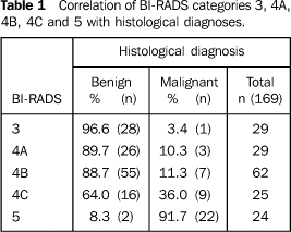
RESULTS Amongst the patients included in the present study, 36.1% wereless than 50 years old, 36.7% were 50–59 years old, and 27.2%were 60 or more years old. Of the total 169 cases evaluated, the percentual distributionof mammographic diagnoses according to BI-RADS was the following:17.2% (29) for category 3, 68.6% (116) for category 4, and 14.2%(24) for category 5. Focusing only on category 4, itssubcategories had the following percentual distribution: 25%(29/116) for the subcategory A, 53% (62/116) for the subcategoryB, and 22% (25/116) for subcategory C. Forty-two (24.8%) cases were diagnosed with breast cancer — one in category 3, three in the category 4A, seven in category 4B, nine in category 4C, and 22 in category 5. Therefore, PPV were: 3.4% (1/29) for BI-RADS 3, 10.3% (3/29) for BI-RADS 4A, 11.3% (7/62) for BI-RADS 4B, 36% (9/25) for BI-RADS 4C and 91.7% (22/24) for BI-RADS 5 (Table 1).
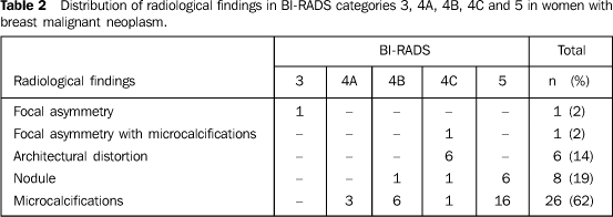
Amongst lesions classified as BI-RADS 3 there was only one case of breast cancer, whose radiological finding was focal asymmetry. In the other categories, the mammographic findings most frequently associated with breast cancer were those included in category 4A, punctate microcalcifications with segmental distribution in 66.7% (2/3) of cases; in category 4B, amorphous, heterogeneous punctate microcalcifications, in 57.1% (4/7) of cases; in category 4C, spiculated architectural distortion, in 66.7% (6/9) of cases; for lesions classified as BI-RADS 5, pleomorphic microcalcifications in a branching pattern were present in 72.7% (16/22) of breast cancers. Overall, the radiological findings most frequently associated with malignant disease were microcalcifications present in 61.5% of total cases (Table 2).
The most frequent malignant breast neoplasm was ductalcarcinoma in situ in 59.5% (25/42), followed by invasiveductal carcinoma in 33.3% (14/42), lobular carcinoma insitu in 4.8% (2/42) and invasive lobular carcinoma in onecase. The diagnosis of atypical ductal hyperplasia occurred in 7.1% (12/169) of total cases, all of them in the category 4. In 66.7% (8/12) of total cases, the findings were microcalcifications, and in 33.3% (4/12), architectural distortion. The two cases classified as BI-RADS 4A presented with clustered, round, linear and punctate microcalcifications, tending to coalescence. On the other hand, category 4B had four cases of amorphous, heterogeneous and punctate microcalcifications, and one case of clustered microcalcifications with linear distribution. In category 4C, one case of clustered, pleomorphic calcifications with linear distribution, and four cases of architectural distortion.
DISCUSSION The BI-RADS classification has represented the first attemptto standardize mammographic findings in descriptive terms,constituting an important ancillary tool in both in cases ofsuspect malignancy and definition of conduct to beadopted(5,7–9). Studies correlatingmammographic and histological findings in non-palpable breastlesions employing the BI-RADS classification have found PPV forbreast cancer between 12.3% and47.8%(4,7,8,10–12). In the present study, 24.8%from the total of biopsied non-palpable breast lesions had ahistological diagnosis of malignant disease. The percentage of probably benign mammographic findingssubmitted to biopsy was high (17.2%), above the values found byother studies which have ranged between 2% and11%(4,7,8,10). As 96.5% of lesions classifiedas category 3 were benign, this has contributed for decreasingthe PPV in the global evaluation of categories by the presentstudy. Analyzing the PPV exclusively in relation to category 3,the value was 3.4% — compatible with mean values reported byother studies(4,7,8,11). The high number ofsurgical biopsies in probably benign lesions can be explained bythe difficulty to perform a semiannual mammographic follow-up inmany patients originating from other locations. Correlating histological and radiological findings in category4 breast lesions, in our study, the PPV was 16.4%, while otherauthors have found PPV ranging between 4% and45%(4,7,8,10–12). In the present study, we have found 12 cases of breast lesionsclassified as BI-RADS category 4 with diagnosis of atypicalductal hyperplasia which have not been included in the PPVcalculation. According to Heywang-Köbrunner et al., the"atypical ductal hyperplasia represents a borderline lesion,whose malignancy risk in comparison with the normal population isincreased in four to five times"(13). If thecases of atypical ductal hyperplasia were included in thecategory 4 PPV calculation, we would found an increase to26.7%. Analyzing the findings in subcategories 4A, 4B e 4C, we havefound, respectively, PPV of 10.3%, 11.3% and 36%. If we includedatypical ductal hyperplasia in this category, the values wouldchange to 17.2%, 19.4% and 56%. Therefore, these findings havedemonstrated an increasing sensitivity of BI-RADS subcategories4A, 4B and 4C for detecting suspect breast lesions and thoseconsidered with risk for malignancy. More recent studies on subcategories 4A, 4B and 4C have notbeen found in the literature, so it has not been possible toestablish a correlation with the values obtained in the presentstudy(9–12). As regards category 5, we have found a PPV of 91.7% for malignancy, a value compatible with data of the literature(4,7,8, 10,11). In lesions classified as BI-RADS 5, we have observed one case of sclerosing adenosis, and another of radial scar, situations where a differential diagnosis is not feasible by means of mammography, so this is a mandatory indication for an excisional biopsy(6,14).
CONCLUSION The present study has demonstrated that the BI-RADS classification allows a safe prediction of high suspicion for malignancy in lesions classified as category 5, and minimal suspicion in lesions classified as category 3. As regards category 4, a progressive increase in PPV was observed in subcategories A, B and C, demonstrating that this subdivision contributes in a more detailed and accurate way for indicating lesions suspect for malignancy.
REFERENCES 1. Bellantone R, Rossi S, Lombardi CP, et al. Nonpalpable lesions of the breast. Diagnostic and therapeutic considerations. Minerva Chir 1994; 49:327–333. [ ] 2. Frasson A, Farante G, Sacchini V, et al. Localização pré-operatória de lesões mamárias não-palpáveis. Rev Bras Ginecol Obstet 1993;3:35–44. [ ] 3. Hall FM, Storella JM, Silverstone DM, Wyshak G. Nonpalpable breast lesions: recommendations for biopsy based on suspicion of carcinoma at mammography. Radiology 1988;167:353–358. [ ] 4. Orel SG, Kay N, Reynolds C, Sullivan DC. BI-RADS categorization as a predictor of malignancy. Radiology 1999;211:845–850. [ ] 5. D'Orsi CJ. Illustrated Breast Imaging Reporting and Data System. 4th ed. Reston: American College of Radiology, 2003. [ ] 6. Rocha DC, Bauab SP. Atlas de imagem da mama. Correlação mamografia/ultra-sonografia, incluindo ressonância magnética e BI-RADS. 2ª ed. Rio de Janeiro: Revinter, 2004. [ ] 7. Liberman L, Abramson AF, Squires FB, Glassman JR, Morris EA, Dershaw DD.The Breast Imaging Reporting and Data System: positive predictive value of mammographic features and final assessment categories. AJR Am J Roentgenol 1998;171: 35–40. [ ] 8. Bérubé M, Curpen B, Ugolini P, et al. Level of suspicion of mammographic lesion: use of features defined by BI-RADS lexicon and correlation with large-core breast biopsy. Can Assoc Radiol J 1998;49:223–228. [ ] 9. Vieira AV, Toigo FT. Classificação BI-RADS: categorização de 4.968 mamografias. Radiol Bras 2002;35:205–208. [ ] 10. Kestelman FP, Canella EO, Arvellos NA, et al. Classificação radiológica nas lesões não-palpáveis da mama. Análise de resultados do Hospital do Câncer III – INCA-MS. Radiol Bras 2001;34 (Supl 1):20. [ ] 11. Giannotti IA, Giannotti Filho O, Scalzaretto AP, Visentainer M, Elias S. Correlação entre diagnóstico por imagem e histologia de lesões não-palpáveis de mama. Rev Bras Cancer 2003;49:87–90. [ ] 12. Roveda Jr D, Campos MSDA, Ferezin PC, Ferreira ACP, Valle MJR. Valor preditivo das anormalidades mamográficas pelo sistema BI-RADS na ausência de lesões palpáveis das mamas. Radiol Bras 2001;34(Supl 1):21. [ ] 13. Heywang-Köbrunner SH, Schreer I, Dershaw DD, et al. Mama: diagnóstico por imagem. Correlação entre mamografia, ultra-sonografia, ressonância magnética, tomografia computadorizada e procedimentos intervencionistas. 1ª ed. Rio de Janeiro: Revinter, 1999;141–155. [ ] 14. Stefenon CC, Carvalho AA, Djahjah MCR, Koch HA. Cicatriz radial/lesão esclerosante complexa: aspectos radiológicos com correlação clínica, ultra-sonográfica e anatomopatológica. Radiol Bras 2003;36:95–103. [ ]
Received October 7, 2005.
* Study developed at Centro de Atenção Integral à Saúde da Mulher (CAISM) Department of Radiology – Universidade Estadual de Campinas, Campinas, SP, Brazil, with financial support from Fundação de Amparo à Pesquisa do Estado de São Paulo (Fapesp). |
|
Av. Paulista, 37 - 7° andar - Conj. 71 - CEP 01311-902 - São Paulo - SP - Brazil - Phone: (11) 3372-4544 - Fax: (11) 3372-4554


