Radiologia Brasileira - Publicação Científica Oficial do Colégio Brasileiro de Radiologia
AMB - Associação Médica Brasileira CNA - Comissão Nacional de Acreditação
 Vol. 40 nº 1 - Jan. /Feb. of 2007
|
ORIGINAL ARTICLE
|
|
Breast ultrasound: evaluation of echographic criteria for differentiation of breast lesions |
|
|
Autho(rs): Maria Julia Gregorio Calas, Hilton Augusto Koch, Maria Virginia Peixoto Dutra |
|
|
Keywords: Ultrasound, Breast lesions, Diagnosis |
|
|
Abstract:
IMaster's Degree in Medicine by Faculdade de Medicina da Universidade Federal do Rio de Janeiro
INTRODUCTION Breast cancer is one of the most important causes of deathamongst women(1). Mammography is the onlydiagnostic technique recognized for its proved contribution tothe early detection and decrease in breast cancer mortality.However, the accuracy of this method is highly dependent on thebreast parenchyma composition and tissularcharacteristics(1–4). The association of diagnostic methods has been successfullyemployed in the search of a more and more early diagnosis of thispathological entity(1–4). Breast ultrasound,both for diagnostic and interventional purposes, plays asignificant role as a method supplementary to the clinicalmammography, and has become a well established and invaluablemethod for diagnosis of breastdiseases(1–4). The first reference to breast ultrasound in the literatureappears in 1951, with a study by Wild and Neal, describing invivo sonographic features of two breast tumors, one malignantand another benign(5). With the introduction ofthe gray scale in the seventies by Kossof, Jellins et al. and,along the last decades, with the utilization of dynamic study,high-frequency linear transducers (7.5 MHz to 13 MHz) andelectronic focus, the breast ultrasound has been established as amethod for diagnostic evaluation in the mammarypropedeutics(5). This method is well tolerated and accepted by patients for notrequiring ionizing radiation nor compression, besides being fastand easy to perform(1–4). Additionally, todayultrasound is the only real-time imaging method widely available,representing an excellent modality for guidance of interventionalinvasive procedures, allowing the choice of the shortest routebetween the skin and the area of interest, with higher swiftnessand minimum discomfort for the patient(6). The echographic interpretation is based on the knowledge ofthe normal mammary structure, its variants and multiple aspectsof breast diseases. Abnormal images have been evaluated anddefined according to morphological characteristics. The capacityfor evaluating morphological criteria of echographic images anddifferentiating between benign and malignant alterations has beensubject for several studies, but there are controversies in theliterature regarding the predictive capacity of each echographiccharacteristic(7–11). Many authors consider that the combination of echographiccriteria results in higher sensitivity and specificity of thefindings when compared with the evaluation of an isolatecharacteristic(7,8). For any isolateechographic characteristic to have practical applicability in thedifferentiation between malignant and benign lesions, thischaracteristic must be a finding with a high rate ofinterobserver agreement(7,8). Considering thatultrasound is an operator-dependent method, the professionalqualification, besides the operator experience, is clearlymandatory, allowing not only the images visualization, but alsothe utilization of morphological characteristics fordifferentiation between malignant and benignlesions(7–11). A quantitative approach issuggested by other studies, aiming at analyzing tumors marginsand texture on the basis of several mathematical techniques.These techniques have been employed as powerful ancillarydiagnostic tools (computer aided diagnostics – CAD-type systems)for assessing different types of medical images, considering thatquantification does not depend on the operatorexperience(12). The standardization of terms employed for describing breastlesions is important since it allows a more objective analysis ofimages, creating an uniform nomenclature capable of indicatingthe degree of malignancy suspicion according to the morphology ofthe echographic findings and, consequently, allowing a moreaccurate orientation for the conduct to beadopted(13,14). The present study aims at identifying those echographic criteria which are most closely related to lesions benignancy and malignancy, besides evaluating echographic characteristics to determine their predictive value for malignancy.
MATERIALS AND METHODS Our casuistic included 637 cases of patients who had beenreferred by request of their doctors for undergoingultrasound-guided fine needle aspiration (206 cases), andultrasound-guided core-biopsy (431 cases), in the period betweenJanuary and December of 2003. All examinations were performed with a GE Logic MD 400equipment with a multi frequency transducer, with orthogonalphotographic recording of each image, besides appropriatematerial for each procedure. The photographic records review, aswell as the evaluation for the presence of echographic criteriaon images, was performed by one of the authors of the presentstudy who has ten-year experience in breast imaginology andinvasive procedures. Of the 637 cases, 187 were excluded because the material wasconsidered as unsatisfactory for the purposes of the presentstudy. The remaining 450 cases are distributed as follows: 9cases of simple cysts, 42 of complicated cysts, 2 of complexcysts and 397 cases of solid breast lesions. This study was developed in a private service of diagnosticimaging to which the patients were referred by request of theirdoctors. It was up to the patients' doctors to make a decisionabout their treatment on the basis of information originated bythe examinations (results of the invasive procedures),independently from the present study. For the 450 selected cases, the standard test consisted of the histopathological results in the surgical cases (provided by the patients' doctors), and of the echographic follow-up of the lesions in later examinations performed during a six-to twenty four-month period, in the non-surgical cases. Information on the follow-up was obtained in the service data-base or was provided by the patients' doctors. With the experience acquired through the daily practice, results from a previous study, and a literature review, we utilized the characteristics described by the majority of authors in this rating methodology as follows: the shape may be well-defined (rounded, oval-shaped, elongated) or ill-defined; limits may be precise (clear), imprecise (unclear) or partially precise. The contour or margins may be regular, partially regular (macrolobular aspect) or irregular (angular, microlobular, spiculated or indistinct aspect). The echogenicity is defined by comparison between images and the surrounding fibroadipous tissue, and may be anechoic, hypoechoic, isoechoic, hyperechoic and mixed (for example, anechoic and hypoechoic, or hypoechoic and hyperechoic). The echotexture is defined as homogeneous or heterogeneous. Echo transmission may be absent, present acoustic enhancement or posterior shadow. The orientation is defined as horizontal or vertical. Besides the aforementioned characteristics, the following secondary signs are taken into consideration: thickening and/or skin retraction; increase in echogenicity of the subcutaneous, peritumoral or parenchymal cellular tissues; thickening of Cooper ligaments; intratumoral calcifications; parenchymal architectural distortion; muscular and lymph nodes involvement (Figures 1–9).
In order to perform a binary evaluation of the properties of adiagnostic test, that is to say, sensitivity and specificity ofmorphological characteristic and their respective positive andnegative predictive values, it was necessary to arrange certaincharacteristics into groups: Regular margins (regular andmacrolobular); and irregular margins (indistinct, spiculated,angular and microlobular). Images with partially precise andimprecise limits were grouped as lesions with imprecise limits.As regards echo transmission, the characteristics were grouped aspresent echo transmission in the presence of acoustic shadow orposterior enhancement, and absent echo transmission, in theabsence of retrotumoral acoustic phenomenon. The software Epiinfo6.04b CDC-USA, World Health Organization (Oct.1997) was employedfor such evaluation. The project was submitted to the Committee on Ethics in Research of Hospital Universitário Clementino Fraga Filho at Universidade Federal do Rio de Janeiro, and was approved with no restriction.
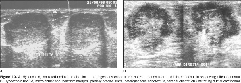
RESULTS On ultrasound, the size of the 450 lesions ranged between 4 mmand 55 mm. In seven cases, the lesions could not be measuredbecause of their large volumes. The results of the procedures (fine-needle aspiration and corebiopsy) were compared by means of the standard test (surgery andfollow-up), resulting in 82 (18.2%) cases of cancer, and 368(81.8%) cases of benign lesions. The most frequent benign lesions were fibroadenomas (43.1%), followed by non-proliferative lesions (35.4%). Amongst the malignant findings, the infiltrating ductal carcinoma was prevalent, corresponding to 72.7% of cases (Figures 10A and 10B). In the studied casuistic, the patients' ages ranged between 16and 88 years (mean age 52 years). Amongst patients who presentedwith carcinomas, the ages ranged between 29 and 88 years (meanage 60 years). On the other hand, the ages of patients withbenign lesions ranged between 16 and 81 years (mean age 45years). A list of the breast lesions echographic characteristics is on Table 1, where the column "Benign" includes lesions in follow-up or those histopathologically diagnosed as benign, and the column "Malignant" includes those lesions considered as malignant at surgery.
Amongst the lesions presenting a defined shape, 94.5%(362/383) were benign, and amongst those with ill-defined shape,91.0% (61/67) were malignant. Amongst lesions with regular margins, 98.2% (336/342) werebenign. Amongst lesions with irregular margins, 70.4% (76/108)were malignant, the microlobular type being the most frequentlyfound — 30.5% of cases (33/108). Amongst lesions with precise limits, 97.1% (338/348) werebenign, and amongst those with imprecise limits, 70.6% (72/102)were malignant. Amongst anechoic and hyperechoic images, 100% of lesions werebenign. As regards hypoechoic images, 69.7% were benign, and30.3% were malignant. Amongst isoechoic images, 98.7% werebenign, and 1.3% malignant. Of those images presenting mixedechogenicity, 69.8% were benign, and 30.2% malignant. Amongst the images with homogeneous echotexture, 91.3%(314/344) were benign, and 49.0% (52/106) of those withheterogeneous echotexture were malignant, and 51% (54/106 werebenign. Echo transmission was absent in 72.4% (326/450), and thepresence of acoustic enhancement or shadow was found in 27.6% ofcases (124/450). Absent echo transmission was described in 83.7%(273/326) of benign cases; acoustic enhancement was reported in95.5%, and posterior acoustic shadow, in 66.2% of benignlesions. Amongst images with horizontal orientation, 91.8% (348/379)were benign. Vertical orientation was present in 57.6% (34/59) ofthe malignant lesions. In 12 cases, images orientation could notbe analyzed because lesion were on a deep plane and presented amarked acoustic shadow, complicating a correct evaluation of thelesion size, and, consequently, a precise evaluation of thelesion orientation. Secondary signs were found just in 14 (17%) cases ofmalignancy, and were absent in 100% of benign cases. The evaluation of characteristics more closely related to lesions malignancy was considered for analyzing the diagnostic properties, as per Table 2. The most sensitive characteristic was the irregular margins. Two characteristics presented a very high specificity: vertical orientation (99.3%) and ill-defined shape (98.4%). The negative predictive values were high, in contrast to the positive predictive values in the presence of echo-transmission (23.4%), and heterogeneous echotexture (49.1%).
DISCUSSION The quality of a procedure is highly dependent on theoperator's knowledge of the equipment, the appropriate technique,the results interpretation, the patient's history, besides themalignant, benign and functional alterations of thebreast(2,4,15). Notwithstanding all the studies reviewed have utilizedhigh-resolution equipment and trained professionals, thedivergences observed in the literature are due to differentmethodologies applied, either utilizing different morphologicalcharacteristics or different criteria for distinguishingmalignant from benign lesions(7–9,16–19). Thisdiversity reflects the fact that not always all the morphologicalcharacteristics of an image with all their variables can beutilized(19). Some features are unique to ultrasound, such as orientationand echogenicity, and some are fundamental to interpretingimages, such us shape and margins(20). Amongst the studies selected for the purpose of comparativeanalysis, all of them utilized the description of the contour ormargin, followed by echo transmission, echogenicity, echotextureand orientation. The features less utilized by the authors were:limits, compressibility, branching and size of thelesion(7–17,19,21–24). Some authors(10,15,19,20,24,25) describe notonly morphological signs of the lesions but also the alterationsin surrounding tissues, the parenchyma architectural distortionbeing the most frequently described secondary sign. An universal standardization of terms for description ofechographic features is still pending, however, with thepublication of the Illustrated Breast Imaging and Reporting DataSystem (BI-RADS®): Ultrasound by the AmericanCollege of Radiology(26), most probably thisstandardization and consequent uniformization will beaccepted. Although studies involving interpretation and classificationof sonographic images recommend an analysis of the concordancebetween observers, and limitation of the generalization based onstudies by only one observer, Skaane andEngedal(17), Zonderland etal.(19), Buchberger etal.(27), and Chen et al.(28)have considered that studies interpreted by only one observermight be consistent in the application of criteria for a lesiondefinition. The study developed by Chen et al.(28)suggests that sonographic characteristics considered in thelesions differentiation might vary in tumors, depending on theirsize. However, they consider that the most significant feature inlesions differentiation, independently from their size, is thecontour or margins, according to the results reported by Skaaneand Engedal(17). Paulinelli et al.(9) evaluated the influenceof the age and size of the tumor on the interpretation of thesonographic characteristics of solid breast nodules. Theseauthors have concluded that the presence of irregular margins,internal heterogeneous echoes and posterior shadowing in benigntumors is directly proportional to the patient's age. Thepresence of internal heterogeneous echoes, anterior halo andthickening of Cooper ligaments in malignant tumors is directlyproportional to the size of the tumor. Additional studies are necessary to reach a consensus on whichechographic characteristics would be more significant, lesssubjective, and more reproducible, and, besides, it is necessaryto analyze other factors which could affect the sonographicimages interpretation and the risk ofmalignancy(9,11,14,20). For most of the authors(16,17,19,21,22,25, 27–29), regular or macrolobular margins represented themain criterion for defining a lesion as benign. The shapedescribed as defined, oval, rounded or ellipsoid was the secondmost important feature, in agreement with the results of thepresent study, where the regular and macrolobular margins wereidentified in 98.2% of the benign cases, and the defined shape,in 94.5%. The malignancy characteristics most frequently described inthe literature(9,10, 16–19,21,22,25,27–29) were irregular margins, presenceof posterior acoustic shadowing, vertical orientation andirregular shape. In the present study, the more characteristic signs ofmalignancy were: ill-defined shape, irregular margins, partiallyprecise limits and hypoechoic lesions. Secondary signs werepresent in only 14 of 82 cases of malignancy, and absent in the368 benign cases. The literature suggests a criterion forbenignity in the absence of suchsigns(11,15,25,29), and a criterion formalignancy in their presence.(15,17,29). In the 12 cases where orientation could not be identified duethe size and localization of the lesion, the term "indeterminate"orientation might be employed, according to Zonderland etal.(19). This pattern suggests a criterion formalignancy, since the 12 cases of indeterminate orientation hadtheir malignancy confirmed on the surgical piece. Main features considered by the severalauthors(11,15,16,22,24,25,27,29) asnon-significant for lesions classification were: echotexture,echo transmission, when absent or in the presence of posterioracoustic enhancement, and echogenicity. In the present study, the criteria non-significant forechographic differentiation were: echotexture and echotransmission. Amongst 106 images with heterogeneous echotexture,52 cases (49%) were malignant and 54 cases (51%) were benign. Asregards posterior acoustic findings, acoustic shadowing waspresent in 66.2% of the benign lesions and in 33.8% of themalignant lesions, in contrast to other authorsobservations(7,10,11,17,19,21–24,27–29) . A contradiction is observed in the literature. While thefeatures most utilized by the authors for echographicallydescribing a lesion were echo transmission, echogenicity andechotexture, such features were not significant fordifferentiating a malignant from a benign lesion. The definition of the echotexture of a lesion as homogeneousor heterogeneous depends on the diversity of the tissues in suchlesion. For example, a lesion might be heterogeneous forpresenting areas of necrosis (carcinomas) or due to fibroadenomas(benign) hyalinization(28). So, the utilizationof this single characteristic might lead to misinterpretation.The absence of a prognostic value of this variable was describedby Stavros et al.(11) and Skaane andEngedal(17). Also, it is important to note thatin the new standardization of the American College ofRadiology(26), this feature was not taken intoconsideration. Echogenicity might be defined as a shade of the gray scaledemonstrating a lesion and representing one of the main problemsin the description of such lesion(21,24). Inthe absence of standardization, the echogenicity becomes a sourceof subjectivity, since the gray scale parameters utilized forthis type of characterization are still to be known. In the study developed by Stavros etal.(11), the lesions echogenicity was comparedwith the normal adjacent adipous tissue, i.e., with a structureof echogenicity approximating the gray scale spectrum, that wasuniform and present in all of the patients. For Stavros etal.(11), the hyperechogenicity of a lesionrepresented the feature with higher negative predictive value(100%). In a different way, Soon et al.(24)have compared the lesions echogenicity with the adjacentglandular tissue in 393 cases of carcinoma, and have found twocases (0.5%) of carcinomas as hyperechoic lesions. For Zonderlandet al.(19), the gain adjustment in theequipment, as well as the breast thickness, might affect theechogenicity description. In the study developed by Chen et al.(28),echogenicity was the main feature in the differentiation betweeninfiltrating carcinomas and carcinomas in situ, the latestpresenting more isoechoic when compared with infiltratingcarcinomas. The echo transmission is a function of the equipment, thetransducer, the compression exerted during the procedure, thesize, localization and even the type of thelesion(21). The echo transmission has emerged in the beginnings of thebreast ultrasonography as an essential, almost pathognomonic signof malignancy, indispensable for diagnosis(5).As a matter of fact, the experience has proved that this is asign found in 20% to 60% of cases of breast cancer, especially inthose where the tumor is larger than 2 cm(21).This is the reason, mainly in cases of non-palpable tumors (with< 1 cm), for which the absence of echo transmission does notallow neither the exclusion of malignancy, nor assurance ofbenignity. An article published by Weinstein etal.(30), presents a group of benign lesionswhich might present posterior acoustic shadowing, likefibroadenomas, radiate scar, diabetic mastopathy, steatonecrosis,post-surgical scar, focal fibrosis, and sclerosing adenosis. As regards diagnostic properties found by several authors and in the present study (Table 3), the irregular margins (microlobular, spiculated or angular aspect) and orientation were the echographic characteristics with highest rates of sensitivity, specificity and predictive values. In summary, one may conclude that the main features fordifferentiating malignant from benign lesions are margins andshape of the lesion, with the first one presenting the highestsensitivity, and the second, the highest specificity. Therefore, the quantitative study of the margins of a lesionmight be a powerful tool for helping the observer todifferentiate malignant tumors from the benign ones. Alvarenga et al.(31), utilizing a methodbased on mathematical morphology for images segmentation, havefound 95.7% sensitivity and 96.7% specificity in thedifferentiation of tumors by means of margins analysis. The next step in this study will be to develop an interobserver study (with qualitative criteria), comparing it with the results obtained with quantitative methods applied to margins and echogenicity of lesions. Based on this comparison, we will be able to evaluate the contribution that the quantification may add to the final diagnosis of such lesions.
REFERENCES 1. Paulinelli RR, Moreira MAR, Freitas Júnior R. A importância do diagnóstico precoce do câncer de mama. Femina 2004;32:233–237. [ ] 2. Fine RE, Staren ED. Updates in breast ultrasound. Surg Clin North Am 2004;84:1001–1034. [ ] 3. Paulinelli RR, Moreira MAR, Freitas Júnior R. Ultra-sonografia no diagnóstico do câncer de mama: realidade atual e possibilidades para o futuro. Rev Bras Mastol 2003;13:168–174. [ ] 4. Fonseca ALA. Ultra-sonografia da mama. In: Pasqualette HA, Koch HA, Soares-Pereira PM, Kemp C, editores. Mamografia atual. 1ª ed. Rio de Janeiro: Revinter, 1998:205–215. [ ] 5. Dempsey PJ. The history of breast ultrasound. J Ultrasound Med 2004;23:887–894. [ ] 6. Lucena CEM. Procedimentos intervencionistas mamários guiados por ultra-som. Femina 2002; 30:537–541. [ ] 7. Arger PH, Sehgal CM, Conant EF, Zuckerman J, Rowling SE, Patton JA. Interreader variability and predictive value of US descriptions of solid breast masses: pilot study. Acad Radiol 2001;8: 335–342. [ ] 8. Baker JA, Kornguth PJ, Soo MS, Walsh R, Mengoni P. Sonography of solid breast lesions: observer variability of lesion description and assessment. AJR Am J Roentgenol 1999;172:1621–1625. [ ] 9. Paulinelli RR, Freitas-Júnior R, Moreira MAR, et al. Risk of malignancy in solid breast nodules according to their sonographic features. J Ultrasound Med 2005;24:635–641. [ ] 10. Paulinelli RR, Vidal CSR, Ruiz AN, Moraes VA, Bernardes Júnior JRM, Freitas Júnior R. Estudo prospectivo das características sonográficas no diagnóstico de nódulos sólidos da mama. Rev Bras Ginecol Obstet 2002;24:195–199. [ ] 11. Stavros AT, Thickman D, Rapp CL, Dennis MA, Parker SH, Sisney GA. Solid breast nodules: use of sonography to distinguish between benign and malignant lesions. Radiology 1995;196:123–134. [ ] 12. Azevedo CM, Alvarenga AV, Pereira WCA, Infantosi AFC. Análise computacional de imagens por ultra-som de lesões da mama em pacientes mastectomizadas e em pacientes com lesões sistêmicas. Rev Imagem 2004;26:279–286. [ ] 13. Calas MJG, Castro R, Manoel VR, Pasqualette HA, Soares-Pereira PM. Proposta de normatização dos laudos de ultra-sonografia mamária. Femina 2002;30:103–110. [ ] 14. Pasqualette HAP, Soares-Pereira PM, Calas MJG, et al. Revisão e validação de uma proposta de classificação de laudos de ultra-sonografia mamária. Rev Bras Mastol 2003;13:159–167. [ ] 15. Kossoff MB. Ultrasound of the breast. World J Surg 2000;24:143–157. [ ] 16. Rahbar G, Sie AC, Hansen GC, et al. Benign versus malignant solid breast masses: US differentiation. Radiology 1999;213:889–894. [ ] 17. Skaane P, Engedal K. Analysis of sonographic features in the differentiation of fibroadenoma and invasive ductal carcinoma. AJR Am J Roentgenol 1998;170:109–114. [ ] 18. Watson L. The role of ultrasound in breast imaging. Radiol Technol 2000;71:441–459. [ ] 19. Zonderland HM, Hermans J, Coerkamp EG. Ultrasound variables and their prognostic value in a population of 1103 patients with 272 breast cancers. Eur Radiol 2000;10:1562–1568. [ ] 20. Mendelson EB, Berg WA, Merritt CR. Toward a standardized breast ultrasound lexicon, BI-RADS: ultrasound. Semin Roentgenol 2001;36: 217–225. [ ] 21. Michelin J, Levy L. Ultra-sonografia da mama – diagnóstica e intervencionista. 1ª ed. Rio de Janeiro: Medsi, 2001. [ ] 22. Murad M, Bari V. Ultrasound differentiation of benign versus malignant solid breast masses. J Coll Physicians Surg Pak 2004;14:166–169. [ ] 23. Taylor KJ, Merritt C, Piccoli C, et al. Ultrasound as a complement to mammography and breast examination to characterize breast masses. Ultrasound Med Biol 2002;28:19–26. [ ] 24. Soon PSH, Vallentine J, Palmer A, Magarey CJ, Schwartz P, Morris DL. Echogenicity of breast cancer: is it of prognostic value? Breast 2004;13: 194–199. [ ] 25. Blohmer JU, Schmalisch G, Kürten A, Chaoui R, Lichtenegger W. Relevance of sonographic criteria for differential diagnosis of mammary tumours. Eur J Ultrasound 1997;6:35–41. [ ] 26. American College of Radiology. Illustrated Breast Imaging Reporting and Data System Atlas (BI-RADS®): Ultrasound. 4th ed. Reston: American College of Radiology, 2003. [ ] 27. Buchberger W, Dekoekkoek-Doll P, Springer P, Obrist P, Dünser M. Incidental findings on sonography of the breast: clinical significance and diagnostic workup. AJR Am J Roentgenol 1999; 173:921–927. [ ] 28. Chen SC, Cheung YC, Su CH, Chen MF, Hwang TL, Hsueh S. Analysis of sonographic features for the differentiation of benign and malignant breast tumors of different sizes. Ultrasound Obstet Gynecol 2004;23:188–193. [ ] 29. Chao TC, Lo YF, Chen SC, Chen MF. Prospective sonographic study of 3093 breast tumors. J Ultrasound Med 1999;18:363–370. [ ] 30. Weinstein SP, Conant EF, Mies C, Acs G, Lee S, Sehgal C. Posterior acoustic shadowing in benign breast lesions: sonographic-pathologic correlation. J Ultrasound Med 2004;23:73–83. [ ] 31. Alvarenga AV, Infantosi AFC, Azevedo CM, Pereira WCA. Aplicação de operadores morfológicos na segmentação e determinação do contorno de tumores de mama em imagens por ultra-som. Rev Bras Eng Bioméd 2003;19:91–101. [ ]
Received January 11, 2006.
* Study developed, as a part of a Master's Degree Dissertation, in the Sector of Radiology at Faculdade de Medicina da Universidade Federal do Rio de Janeiro, Rio de Janeiro, RJ, Brazil. |
|


Av. Paulista, 37 - 7° andar - Conj. 71 - CEP 01311-902 - São Paulo - SP - Brazil - Phone: (11) 3372-4544 - Fax: (11) 3372-4554