Radiologia Brasileira - Publicação Científica Oficial do Colégio Brasileiro de Radiologia
AMB - Associação Médica Brasileira CNA - Comissão Nacional de Acreditação
 Vol. 41 nº 3 - May / June of 2008
|
ORIGINAL ARTICLE
|
|
Infra-inguinal angioplasty in patients with critical limb ischemia Rutherford grade III, category 5 |
|
|
Autho(rs): Abdo Farret Neto, Eduardo Baptista Faria, Alessander Laurentino |
|
|
Keywords: Balloon angioplasty, Percutaneous transluminal angioplasty, Popliteal artery, Leg arteries, Limb salvage, Lower extremity |
|
|
Abstract:
ITitular Member of Colégio Brasileiro de Cirurgiões, Member of Sociedade Brasileira de Angiologia e Cirurgia Vascular, MD, Angioradiologist at Hospital Universitário Onofre Lopes da Universidade Federal do Rio Grande do Norte (UFRN), Natal, RN, Brazil
INTRODUCTION Developments in the manufacturing technology for materials utilized in endovascular techniques have allowed angioplasty in more and more distal arterial segments(1). Percutaneous transluminal angioplasty (PTA) has proved its usefulness in the management of iliac atherosclerotic disease, with short- and long-term results comparable to surgical treatment(2). PTA is considered as an acceptable therapeutic procedure for the management of lesions in the femoropopliteal and infrapopliteal segments, with an increasing number of reports in the literature(3). Recent series demonstrate that PTA in this segment, with or without stent implantation, presents a reasonable rate of limbs salvage, although controversies still remain about indications, patients selection and long-term results(4,5). Considering this undefined scenery, the present study was primarily aimed at demonstrating the PTA effectiveness in this controversial arterial segment. For this purpose, the authors have adopted an exclusively clinical parameter in the follow-up of angioplasties patency and results. Such parameter corresponded to achieving the healing of the trophic lesion without major amputation, exclusively by means of endovascular correction of the baseline vascular disease, with clinical follow-up of 36 patients with critical lower limb ischemias submitted to 84 infra-inguinal angioplasties without stenting.
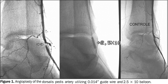
MATERIALS AND METHODS The present study was approved by the Committee for Ethics in Research of the involved institutions. Thirty-six patients submitted to infra-inguinal angioplasty were evaluated in the period between April 1, 2004 and August 3, 2005. All of them had presented with Rutherford grade III, category 5 critical limb ischemia(6). Twenty patients were men and 16, women, in the age range between 43 and 83 years (mean, 65.17 years). Main risk factors associated were diabetes (61%) and arterial hypertension (22%). The technical objective to be achieved was the obtention of at least one patent artery to the foot, in the case of tibial arteries, or to the ankle, with collateral vessels to the foot, in the case of fibular artery; this would guarantee a good blood supply to allow the feasibility of cicatrization. The patients were previously given anti-platelet drugs — AAS 100 mg/day, and had undergone diagnostic angiography. After the procedure, all of them remained under continuous, combined anti-platelet therapy (AAS 100 mg/day plus clopidogrel 75 mg/day) during four weeks. For lesions located below the middle third of the superficial femoral artery, the approach of choice was anterograde, with 4F introducer and without therapeutic catheter. In cases of associated lesions in the iliac, common femoral, deep femoral arteries and proximal segment of the superficial femoral artery, the contralateral approach was adopted, with a 6F contralateral introducer (Balkin). Arterial access was obtained with 0.035² guide wires, and 0.014² Galeo M guide wires (Biotronik GmbH&Co. KG; Berlin, Germany) to cross the lesions and sustain the angioplasties. Following the catheter implantation, 5000 UI heparin/500 ml saline solution was injected through this device at a rate of 40 drops/min, along the procedure. The following balloon-catheters were utilized: Gazelle® Monoraill (Boston Scientific Ireland Ltd.; Galway, Ireland) for diameters = 4 mm with < 2 cm in length, Pheron® for more extensive lesions, with > 4 mm in diameter, and Pleon® for lesions with < 4 mm in diameter, both from Biotronik (Biotronik GmbH&Co. KG; Berlin, Germany). It is important to note that long balloons were not utilized in the present casuistic. Balloons were selected according to the diameter of the vessels to be treated, without oversizing, and based on calibrated measurements performed during the procedure. Only 0.014² guide wires were utilized for crossing the lesions, under road-mapping guidance in a Philips Integris® V5000 digital angiography equipment (Philips Medical Systems; Best, Netherlands). In cases of leg arteries angioplasty, 5 ml (5 mg) isosorbide mononitrate (Monocordil® - Baldacci S.A.; São Paulo, Brazil) previously diluted in 9 ml saline solution was injected through the catheter. Immediately after each procedure, a follow-up angiography was performed to confirm the effectiveness of the angioplasty and the necessity or not to reinflate the balloon. In case of dissection, a longer balloon inflation was performed. If a residual stenosis of > 30% was suspected, the stenosis was measured by a software installed in the digital angiography equipment. Then, the maximum balloon inflation pressure was applied, or a larger balloon was utilized. After the satisfactory angioplasty result, a final angiography was performed to demonstrate the good distal perfusion and absence of embolization (Figure 1). The catheter was removed at the end of the procedure, since full heparinization was not required. The lesions dressings management could not be standardized because the patients came from and went back to several health institutions in different cities; however, general principles of hygienization were adopted, with removal of necrosed tissues. The follow-up was performed with physical examination and clinical evaluation of the lesion progression. In cases where the cicatricial progression was not satisfactory, the patients were referred again to the author's institution. The procedure was considered as successful in cases where cicatrization of the trophic lesion was achieved, without major amputation. Failure was determined by non-cicatrization, necessity of surgical revascularization or major amputation.
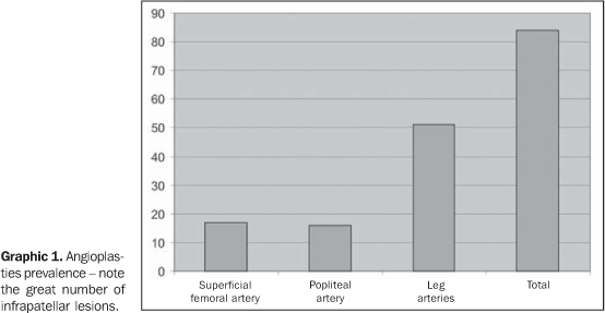
RESULTS Thirty-six patients were submitted to angioplasty for 84 lesions, 17 (20.2%) of them in the superficial femoral artery, 16 (19%) in the popliteal artery, and 51 (60.8%) in leg arteries (Graphic 1).
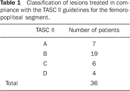
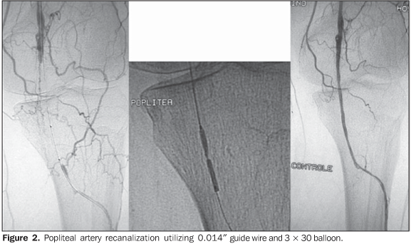
Only eight patients presented with lesions restricted to the femoropopliteal segment, without affecting the leg arteries. In 18 patients, the lesions involved the legs arteries, with no alteration in the femoropopliteal segment. Twenty-six patients (72.2%) presented with more than one lesion, independently from the segment involved, and 12 (33.3%), more than two lesions. Nine (10.7%) of the 84 treated lesions corresponded to occlusions. Eight of these lesions could be successfully recanalized (Figure 2). Most of these lesions had < 3 cm in length, tabulated according to the TASC II Inter-Society Consensus for the Management of PAD(7), as shown on Table 1.
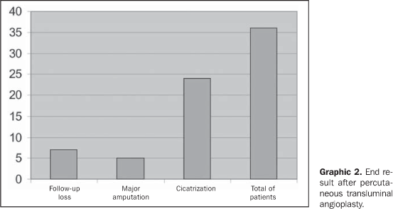
No residual stenosis > 30% was observed at the end of the procedures. Measurements were performed with an appropriate software installed in the digital angiography equipment. Also, no acute flow-limiting dissection was observed therefore, no case required stenting. No arterial rupture, acute arterial occlusion or death was observed during the procedures. Seven of the 36 patients missed the follow-up, so the clinical progression and, consequently, the angioplasties effectiveness, was evaluated in 29 patients. Twenty-four (82.76%) of these 29 patients could achieve the end-objective of the present study, that is to say that cicatrization of their trophic lesions was achieved, without major amputation. Four of these 24 patients underwent minor amputations, one surgical debridement, and the remainder 19 patients, ambulatory management of wound dressings. Four (13.79%) of the five (17.24%) patients who could not achieve the end-objectives of the present study underwent amputation at the level of the leg, and one (3.45%), at the level of the thigh, the latest as a result of Fournier gangrene, four months after the procedure, progressing to death. However, before the amputation, the patient presented with palpable dorsalis pedis pulse.
DISCUSSION In the present study, there was a high prevalence of diabetic patients. This fact may determine a lower long term patency of the angioplasties, considering that diabetes is associated to a worse prognosis(8). Because of the diabetes prevalence, most o lesions were found in leg arteries and, frequently, the patients presented with multiple lesions (Table 1). Despite the higher rate of limbs salvage in patients submitted to stenting reported by some studies in the literature(9), in the present study stenting was not required, considering the absence of immediate residual stenosis > 30% or flow-limiting dissection. The authors attribute this fact to the cautious selection of low profile balloon, without oversizing, utilizing only 0.014² guide wires to cross the lesions, to the careful, slow balloon deinflation avoiding the risk for implosion of the treated area, and to the longer angioplasty procedures in cases of dissection or recoil. Despite the high rate of missed follow-ups (19.44%), the number of patients who achieved the end-objective, i.e., the trophic lesion cicatrization with or without minor amputations, was relatively high (Graphic 2).
It is known that both traditional and endovascular procedures for arterial flow restoration have a limited duration, but the objective of lesion healing should be achieved through the less morbid approach. Likewise in the conventional surgery (bypass), the clinical success is the primary end-objective to be achieved, with improvement or healing of symptoms, which is corroborated by numberless authors(10,11). The authors believe that the ideal follow-up of patients submitted to femoropopliteal or infrapopliteal angioplasty should include echo-Doppler studies regularly repeated at scheduled intervals, but all of the patients in the present casuistic come from the SUS - Sistema Único de Saúde (the Brazilian universal public health system), where the echo-Doppler method is not available; additionally, the patients cannot afford the costs of such studies. However, if the study is aimed at evaluating the clinical progression of the lesions, there will be room to allow that the follow-up of this patients is performed by a non-specialist in more remote regions of the country. Therefore, taking the cicatrization of trophic lesion cicatrization as an end-objective, femoropopliteal and infra popliteal angioplasties have shown to be a highly successful technical procedure, with low morbidity and mortality rates, and high effectiveness in the healing of patients with critical lower limb ischemia.
REFERENCES 1. Klonaris C, Katsargyris A, Giannopoulos A, et al. Advances in endovascular treatment of femoropopliteal arterial occlusive disease. Perspect Vasc Surg Endovasc Ther. 2006;18:329-41. [ ] 2. Kudo T, Chandra FA, Ahn SS. Long-term outcomes and predictors of iliac angioplasty with selective stenting. J Vasc Surg. 2005;42:466-75. [ ] 3. Rastogi S, Stavropoulos SW. Infrapopliteal angioplasty. Tech Vasc Interv Radiol. 2004;7:33-9. [ ] 4. Gray BH, Laird JR, Ansel GM, et al. Complex endovascular treatment for critical limb ischemia in poor surgical candidates: a pilot study. J Endovasc Ther. 2002;9:599-604. [ ] 5. Feiring AJ, Wesolowski AA, Lade S. Primary stent-supported angioplasty for treatment of below-knee critical limb ischemia and severe claudication: early and one-year outcomes. J Am Coll Cardiol. 2004;44:2307-14. [ ] 6. Rutherford RB, Becker GJ. Standards for evaluating and reporting the results of surgical and percutaneous therapy for peripheral arterial disease. Radiology. 1991;181:277-81. [ ] 7. Norgren L, Hiatt WR, Dormandy JA, et al. Inter-Society Consensus on Peripheral Arterial Disease (TASC II). Eur J Vasc Endovasc Surg. 2007;33 Suppl 1:S1-70. [ ] 8. Faglia E, Dalla Paola L, Clerici G, et al. Peripheral angioplasty as the first-choice revascularization procedure in diabetic patients with critical limb ischemia: prospective study of 993 consecutive patients hospitalized and followed between 1999 and 2003. Eur J Vasc Endovasc Surg. 2005;29:620-7. [ ] 9. Gray BH, Laird JR, Ansel GM, et al. Complex endovascular treatment for critical limb ischemia in poor surgical candidates: a pilot study. J Endovasc Ther. 2002;9:599-604. [ ] 10. Matsagas MI, Rivera MA, Tran T, et al. Clinical outcome following infra-inguinal percutaneous transluminal angioplasty for critical limb ischemia. Cardiovasc Intervent Radiol. 2003;26: 251-5. [ ] 11. Hoffmann U, Schulte KL, Heidrich SH, et al. Complete ulcer healing as primary endpoint in studies on critical limb ischemia? A critical reappraisal. Eur J Vasc Endovasc Surg. 2007;33:311-6. [ ] Received July 11, 2007. Accepted after revision September 12, 2007. * Study developed at Hospital Universitário Onofre Lopes da Universidade Federal do Rio Grande do Norte (UFRN) and Hospital do Coração de Natal, Natal, RN, Brazil. |
|

Av. Paulista, 37 - 7° andar - Conj. 71 - CEP 01311-902 - São Paulo - SP - Brazil - Phone: (11) 3372-4544 - Fax: (11) 3372-4554