Radiologia Brasileira - Publicação Científica Oficial do Colégio Brasileiro de Radiologia
AMB - Associação Médica Brasileira CNA - Comissão Nacional de Acreditação
 Vol. 41 nº 3 - May / June of 2008
|
ORIGINAL ARTICLE
|
|
Correlation between ultrasonographic and magnetic resonance imaging findings in fetal sacrococcygeal teratoma |
|
|
Autho(rs): Erika Antunes, Heron Werner Jr., Pedro Augusto Daltro, Leise Rodrigues, Bruno Amim, Fernando Guerra, Romeu Côrtes Domingues, Emerson Leandro Gasparetto |
|
|
Keywords: Fetus, Sacrococcygeal teratoma, Ultrasonography, Magnetic resonance imaging |
|
|
Abstract:
IMD, Trainee at Clínica de Diagnóstico Por Imagem (CDPI), Rio de Janeiro, RJ, Brazil
INTRODUCTION Sacrococcygeal teratoma is a benign tumor containing components arising from the three germ layers, and derived from the pluripotential cell line originating in the Hensen's node. Although rare, this is the most frequent congenital tumor, with an incidence of 1:40000 live births(1), with 75% of female prevalence(2). The earliest sign of a fetal sacrococcygeal teratoma may be a greater-than-expected uterine increase, however, most of times the lesion is asymptomatic, being incidentally found on routine ultrasound (US)(3). Although the majority of tumors occur sporadically, heritable forms of the disease have been reported. Perinatal morbimortality is high, as a result of complications secondary to this tumor such as high output cardiac failure, preterm delivery, anemia, dystocia, and tumor rupture(1). Prognosis seems to be related not to the size of the mass but rather to its extent and content(1); for this reason, magnetic resonance imaging (MRI) has becoming essential in the evaluation of these patients. US still remains as the method of choice for fetal evaluation, considering the fact that this is a real time, non-invasive(4) and low-cost examination(5,6). The prenatal diagnosis of sacrococcygeal teratoma can be performed by US where the majority of tumors can be visualized as a solid mass, a mix of cystic and solid elements or, occasionally, as a completely cystic variant(7,8). Sacrococcygeal teratomas generally are extremely vascular, which can be easily demonstrated by the color Doppler technique(3,9). However, US presents some limitations such as a restricted field of view, accoustic shadowing resulting from fetal pelvic bones, oligohydramnios, and maternal obesity(5). Besides providing higher anatomical detail, MRI presents a larger field of view as compared with US. The development of faster MRI sequences has increased the utilization of this imaging modality in the fetal evaluation(1). These faster sequences can be acquired during a single breath-hold of the mother, practically eliminating the necessity of sedation(10). Therefore, MRI allows a better evaluation of the content and intrapelvic extent of fetal sacrococcygeal teratoma, which represent significant factors in the definition of the prognosis and treatment of these fetuses. The present study was aimed at describing three cases of fetal sacrococcygeal teratoma, establishing a correlation between US and MRI findings, and demonstrating the relevance of MRI for an accurate characterization of the tumor.
MATERIALS AND METHODS The present study was developed at Clínica de Diagnóstico por Imagem (CDPI) do Rio de Janeiro, in the period between January and December/2006. The pregnant women (n = 3) in the age range between 25 and 26 years (mean age, 25.6 years), and between the 30th and 35th weeks of gestation (mean, 32.6 weeks), had been referred by Instituto Fernandes Figueiras and a private clinic, after undergoing obstetric ultrasound between the 25th and 34th gestational weeks (mean, 29.0 weeks) whose findings suggested the diagnosis of fetal sacrococcygeal teratoma. The three patients were submitted to fetal MRI, and subsequently to US for imaging findings correlation. All of them signed a term of free and informed consent. The MRI studies were performed between the 30th and 35th weeks of gestation (mean, 32.6 weeks). The three patients were given information and instructions about the procedures. A Magnetom Avanto with a 1.5 tesla magnet (Siemens Medical Systems; Erlangen, Germany) was utilized for MR images acquisition, with the patients in dorsal or left lateral decubitus, with their head or feet facing the open end of the scanner (at the patients' discretion), and with the surface coil positioned on their abdomens. The fetal images acquisition took approximately 20 minutes, with T1-weighted sequences (repetition time [TR]: 201 ms; echo time [TE]: 4,72 ms; field-of-view [FoV]: 250-400 mm; matrix: 256 × 90-256) and T2-weighted HASTE (TR: 1000 ms; TE: 85-87 ms; FoV: 250-380 mm; matrix: 256 × 112-256) in the axial, coronal and sagittal planes, and with 3.07.0 mm-thick slices. US studies were performed in Logic 500 and Voluson 730 (GE Healthcare; Wisconsin, USA) equipment coupled with 3.5 MHz, 5.0 MHz and volumetric (3D and 4D) transducers. Tumors location, size and content were evaluated. The study was complemented by color Doppler ultrasound for evaluating the tumors vascularization. The images were evaluated by two radiologists experienced in fetal medicine, aiming at establishing a correlation between the imaging findings by both methods. Tumors size, location, extent and content were evaluated. Additionally, based on the MRI findings, the sacrococcygeal teratomas were classified into four types, according to the system developed by the Surgical Section of the American Academy of Pediatrics(11): Type I - predominantly external tumors, with minimum presacral involvement. Type II - tumors with significant external and intrapelvic components. Type III - apparently external tumors, but with the majority of the lesion extending into intrapelvic and intrabdominal spaces. Type IV - tumors located entirely within the pelvis and abdomen. The three patients were followed-up for both the gestational and postnatal progression. All the pregnancies were interrupted by Cesarean section between the 37th and 38th weeks (mean, 37.3 weeks). The three neonates (all of them female) were submitted to surgery for tumor resection between the second and seventh postnatal days (mean, 4 days), and the diagnosis was histopathologically confirmed.
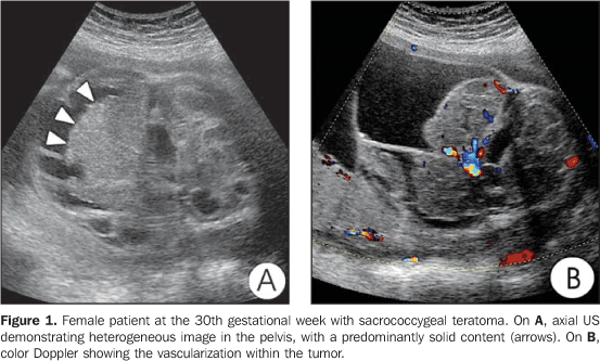
RESULTS All the tumors (n = 3) identified at obstetric US could be demonstrated by MRI. Findings regarding tumors location, size and content were similar for both methods. All the lesions were found in the sacrococcygeal region, with sizes varying between 5.0 cm × 5.7 cm and 8.9 cm × 13.0 cm. As regards the tumors content, two of them were mixed solid-cystic, and one, entirely cystic (Figure 1). The solid component corresponded to about 20% of the tumor content in one case, and 75% in the other. As regards the tumors extent, US could not accurately determine the degree of involvement of the tumor. On the other hand, MRI demonstrated intrapelvic extension in approximately 25% and 30% in two of the cases evaluated.
Based on the MRI findings (Figure 2), the tumors were classified according to the system developed by the Surgical Section of the American Academy of Pediatrics. One case was classified as type I (subtle pelvic involvement), and two as type II (significant intrapelvic extension).
Calcifications or intratumoral complications such as hemorrhage or necrosis were not found.
DISCUSSION Fetal tumors present unique histological characteristics, anatomical distribution and physiology, and their biological behavior may be different as compared with a same tumor diagnosed later in life(3). Teratomas constitute the most frequent and significant group of fetal tumors and the sacrococcygeal region is the most frequent site of involvement(1,3,4). Sacrococcygeal teratomas are derived from the pluripotential cells line originating in the Hensen's node located anteriorly to the coccyx. Ectodermal components, especially neural tissues, are prevalent in the fetal teratoma. Mesodermal tissues, including fat, bone, smooth muscle and cartilage are frequently found(3). The prognosis seems to be related not only to the size but also to the tumor content(12). The size of the solid component of the tumor is the most significant factor in the prognosis of these patients(3). Benign teratomas are formed only by mature tissue, including fluid, fat and calcification(13). On the other hand, malignant teratomas present predominantly solid content and frequent hemorrhage and necrosis(13). The accurate diagnosis plays a critical role in the treatment of the fetus, mother and neonate(3). The most frequent anomalies associated with sacrococcygeal teratomas occur in the genitourinary tract and include hydronephrosis, renal dysplasia, urethral atresia, urinary ascites and hydrocolpos. Prenatal complications include polyhydramnios, oligohydramnios, preterm delivery, HELLP syndrome and hyperemesis(3). Cesarean section is indicated in cases of fetuses with large-sized tumors to avoid dystocia, tumor hemorrhage and avulsion. Usually, the coccyx is involved, even in benign cases, and must be resected with the tumor(13). Developments in obstetric US have increased the possibility of early detection of fetal malformations(1). However, in cases of sacrococcygeal teratomas, factors such as mass size, hemorrhagic transformation and intrapelvic or intraspinal extension may be incompletely evaluated by ultrasonography(1). Imaging findings in cases of sacrococcygeal teratomas depend on the tumor content(13). On US images, these tumors may appear as cystic, solid, or mixed cystic-solid lesions(12). Additionally, characteristic echogenic patterns may be observed because of tumor necrosis, cystic degeneration, internal hemorrhage and calcification(12). In the present study, ultrasonography has appropriately evaluated the three cases of sacrococcygeal teratomas in relation to their location, size and content. All of them were localized in the sacrococcygeal region and presented a mean size of 6.0 cm × 9.0 cm. The tumors were mixed solid-cystic in two cases and cystic in one. However, the degree of intrapelvic extension of the lesions was inadequately characterized by US, considering the limitations of this method: operator dependency, restricted field of view, and difficult visualization of the fetus in cases of oligohydramnios and maternal obesity. As regards the MRI evaluation of patients with sacrococcygeal teratomas, T2-weighted images clearly show the fetal anatomy, and play a significant role in the evaluation of tumor extent and content. Both the tumor extent and content constitute relevant factors in the definition of the prognosis and treatment of these fetuses. Cystic areas present hyposignal on T1-weighted images, and hypersignal on T2-weighted images. Areas with fat tissue present hypersignal on T1-weighted images, while calcifications and bone tissues do not generate any signal(13). MRI can be considered as a safe method for fetal evaluation after the 12th gestational week, but its utilization should be limited to the cases where ultrasonographic results are incomplete or dubious(5). In the present study, MRI was considered as a method complementary to US, providing appropriate information about tumors size, location and content. MRI was more accurate than US in the evaluation of the intrapelvic extension, allowing the correct classification of the tumors (one case - type I, and two cases - type II), with an accurate definition of the tumors content (two mixed cystic/solid, and one entirely cystic) and measurement of their size, allowing a more appropriate surgical planning for each case. This is a critical factor in the patients´ survival, considering that the treatment of choice is based on an extensive tumor resection(14).
CONCLUSION After evaluating the US and MRI images of three patients with fetal sacrococcygeal teratoma, it may be concluded that fetal MRI plays a significant role in the definition of the prognosis, pre- and perinatal management of the patient, considering the high accuracy of this method in the evaluation of the tumor content and extent. US, despite its limitations as compared with MRI, remains as the method of choice in the fetal evaluation, playing the primary role in the prenatal screening for malformations, and should be complemented by MRI in case of an inconclusive diagnosis or for a better evaluation of lesions extent. Some fetal tumors, although histologically benign, may be fatal, depending on the region involved and the size of the lesion. Therefore, an accurate evaluation of the size, content and extent of these tumors is essential for an improved therapeutic planning for these patients. The association between fetal MRI and US results in a better characterization of fetal sacrococcygeal teratomas.
REFERENCES 1. Avni FE, Guibaud L, Robert Y, et al. MR imaging of fetal sacrococcygeal teratoma: diagnosis and assessment. AJR Am J Roentgenol. 2002; 178:179-83. [ ] 2. Lees RF, Williamson BR, Brenbridge NA, et al. Sonography of benign sacral teratoma in utero. Radiology. 1980;134:717-8. [ ] 3. Woodward PJ, Sohaey R, Kennedy A, et al. From the archives of the AFIP: a comprehensive review of fetal tumors with pathologic correlation. Radiographics. 2005;25:215-42. [ ] 4. Shinmoto H, Kashima K, Yuasa Y, et al. MR imaging of non-CNS fetal abnormalities: a pictorial essay. Radiographics. 2000;20:1227-43. [ ] 5. Frates MC, Kumar AJ, Benson CB, et al. Fetal anomalies: comparison of MR imaging and US for diagnosis. Radiology. 2004;232:398-404. [ ] 6. Coakley FV, Glenn OA, Qayyum A, et al. Fetal MRI: a developing technique for the developing patient. AJR Am J Roentgenol. 2004;182:243-52. [ ] 7. Weinstein BJ, Lenkey JL, Williams S. Ultrasound and CT demonstration of a benign cystic teratoma arising from the retroperitoneum. AJR Am J Roentgenol. 1979;133:936-8. [ ] 8. Sheth S, Nussbaum AR, Sanders RC, et al. Prenatal diagnosis of sacrococcygeal teratoma: sonographic-pathologic correlation. Radiology. 1988;169:131-6. [ ] 9. Hata K, Hata T, Kitao M. Antenatal diagnosis of sacrococcygeal teratoma facilitated by combined use of Doppler sonography and MR imaging. AJR Am J Roentgenol. 1991;156:1115-6. [ ] 10. Amin RS, Nikolaidis P, Kawashima A, et al. Normal anatomy of the fetus at MR imaging. Radiographics. 1999;19:201-14. [ ] 11. Wells RG, Sty JR. Imaging of sacrococcygeal germ cell tumors. Radiographics. 1990;10: 701-13. [ ] 12. Danzer E, Hubbard AM, Hedrick HL, et al. Diagnosis and characterization of fetal sacrococcygeal teratoma with prenatal MRI. AJR Am J Roentgenol. 2006;187:350-6. [ ] 13. Kocaoglu M, Frush DP. Pediatric presacral masses. Radiographics. 2006;26:833-57. [ ] 14. Rypens FF, Avni EF, Abehsera MM, et al. Areas of increased echogenicity in the fetal abdomen: diagnosis and significance. Radiographics. 1995; 15:1329-44. [ ] Received June 5, 2007. Accepted after revision July 17, 2007. * Study developed at Multi-Imagem and Clínica de Diagnóstico Por Imagem (CDPI), Rio de Janeiro, RJ, Brazil. |
|
Av. Paulista, 37 - 7° andar - Conj. 71 - CEP 01311-902 - São Paulo - SP - Brazil - Phone: (11) 3372-4544 - Fax: (11) 3372-4554


