Radiologia Brasileira - Publicação Científica Oficial do Colégio Brasileiro de Radiologia
AMB - Associação Médica Brasileira CNA - Comissão Nacional de Acreditação
 Vol. 41 nº 3 - May / June of 2008
|
WHICH IS YOUR DIAGNOSIS?
|
|
Which is your diagnosis? |
|
|
Autho(rs): Sérgio Daher, Murilo Tavares Daher, Wilson Eloy Pimenta Júnior, Márcio Martins Machado, Renato Tavares Daher, Ricardo Tavares Daher |
|
|
IMD, Orthopedist, Surgeon-in-Chief, Unit of Surgery of Spine - Department of Orthopedics and Traumatology at Faculdade de Medicina da Universidade Federal de Goiás (DOT-FM/UFG), Goiânia, GO, Brazil
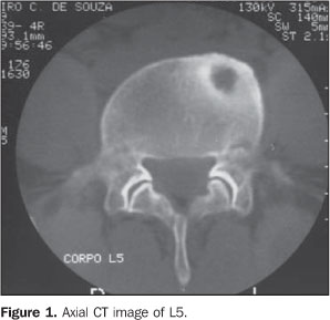
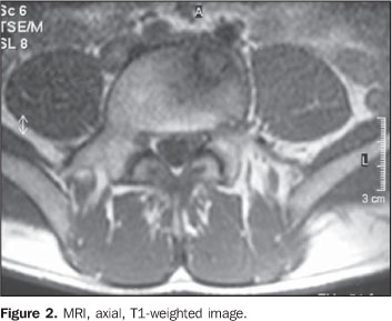
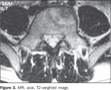
A 30-year-old, male patient, driver, has presented with lumbar pain irradiating especially to his lower limbs, and worsening with deambulation, for six months. At clinical examination, the patient presented pain and significantly limited flexion of the lumbar spine. No apparent deformity was observed. No alteration was found at neurological examination. Imaging findings A radiograph of the lumbar spine was requested and did not demonstrate any bone alteration. Technetium-99 bone scintigraphy was subsequently performed, demonstrating increased uptake in the fifth lumbar vertebra, compatible with a mild osteogenic process. At computed tomography (CT), a lytic lesion was observed in the anterior and lateral portions of L5, with an adjacent sclerotic area (Figure 1), which at magnetic resonance imaging (MRI) presented low-intensity signal on T1-weighted, and a slightly high-intensity signal on T2-weighted sequences (Figures 2 and 3).
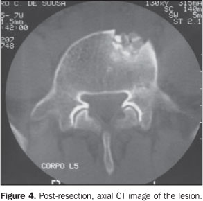
The patient underwent surgery for complete excision of the lesion by anterior, retroperitoneal approach, with insertion of an iliac bone autograft. Histopathologic analysis of the excised material demonstrated a tumor nidus constituted by thick vascular bars of osteoblastic tissue surrounded by vascular fibrous tissue and presence of adjacent sclerotic cortical bone. The patient progressed with pain relief, and a new CT study confirmed the complete excision of the lesion (Figure 4).
Diagnosis: Osteoid osteoma of the L5 vertebral body.
COMMENTS Osteoid osteoma was firstly described in 1935 by Jaffe(1), and represents about 10% of all benign bone tumors, affecting mainly the metaphysis and diaphysis of lower limbs long bone. In 10-20% of cases, osteoid osteomas affect the spine, particularly in the lumbar and thoracic regions(2-4). The prevalence is higher in the male population, especially in the age range between five and 25 years, the differential diagnosis being important in dorsodynia, particularly in children and adolescents(5). Microscopically, this lesion consists of a well delimited nidus with a bone growth pattern, containing numerous osteoblasts producing osteoid and bone tissue, very similar to osteoblastoma, although this later is a typically more aggressive lesion, without sclerotic ring and with a nidus > 1.5 cm in its largest axis(4,6). Other differential diagnoses are osteosarcoma and osteomyelitis. In cases where the axial skeleton is involved, the posterior spinal elements are affected. Presentation in the vertebral body, like in the present case, is unusual, (2-4,7). Clinically, the main symptom is pain which classically worsens at night and is well-relieved with non-steroidal anti-inflammatory and salicylate drugs. Radicular symptoms may be or not be present, and neurological deficit is rarely detected(2-4). Frequently, scoliosis is associated, with non-rotational curves, typically with the lesion localized on the apex of the deformity, in the concavity of the curve(2,6). Usually, in cases where the lumbosacral transition is involved, the deformity apex is cranial to the lesion, with presence of pelvic obliquity(3). In the present case, such deformities were not found, despite the localization of the lesion in the L5. Radiographically, the lesion shows up like a sclerotic area, and a radiolucent nidus may be or not be observed(6). Because of the complex anatomy of the vertebrae, this lesion is hardly or even not visualized(4). Bone scintigraphy shows hyperuptake, and is highly sensitive for early assessment of patients with back pain and suspicion of osteoid osteoma(3). Computed tomography shows a lytic lesion surrounded by sclerosis, with or without central calcification. Classically, this is considered as the best method for diagnosis and localization of this type of lesion. At MRI, a heterogeneous lesion can be observed, with hyperintense signal on T2-weighted images, and calcification within the nidus (if present) and reactive sclerosis with hypointense signal on all sequences. Bone marrow and soft tissues edema may be present, tending to simulate more aggressive lesions such as osteomyelitis and malignant neoplasms(4,8). The treatment for osteoid osteoma is surgical; nidus ablation must be complete to avoid the lesion recidivation(9-11). Open resection(9-11) or CT-guided percutaneous procedures may be utilized in the lesion ablation(12-14). In the present case, because of the anterior localization of the lesion in the vertebral body, the anterior retroperitoneal approach was adopted, with curettage and implantation of iliac bone autograft. Arthrodesis must be performed in cases where the lesion is localized in the posterior spinal elements and the spinal stability may be affected as a result of the lesion excision(11). Several methods are available for an accurate intraoperative localization of the lesion: preoperative radioactive isotope injection, specimen CT, preoperative identification with Kirschner wire or simply intraoperative radiography(3). None of these methods were utilized in the present case, however, during the clinical evaluation, a complete curettage of the nidus was observed, and later confirmed by CT at the postoperative follow-up. The vertebral involvement is relatively unusual in cases of osteoid osteomas typically localized in posterior spinal elements. Localization in the vertebral body is rare. This lesion is a relevant cause of back pain, particularly in children and adolescents and especially in the presence of secondary scoliosis.
REFERENCES 1. Jaffe HL. Osteoid osteoma: a benign osteoblastic tumor composed of osteoid and atypical bone. Arch Surg. 1935;31:709-28. [ ] 2. Domans JP, Moroz L. Infection and tumors of the spine in children. J Bone Joint Surg Am. 2007;89 Suppl 1:S79-97. [ ] 3. Crist BD, Lenke LG, Lewis S. Osteoid osteoma of the lumbar spine: a case report highlighting a novel reconstruction technique. J Bone Joint Surg Am. 2005;87:414-5. [ ] 4. Defino HLA, Pereira CU, Barbosa CSP. Tumores benignos e lesões pseudotumorais da coluna vertebral. Rio de Janeiro: Revinter; 2002. [ ] 5. Basile Jr R, Barros Filho TEP, Bonetti VL, et al. Dor nas costas em crianças e adolescentes. Rev Bras Ortop. 1994;29:144-8. [ ] 6. Ozaki T, Liljenqvist U, Hillmann A, et al. Osteoid osteoma and osteoblastoma of the spine: experiences with 22 patients. Clin Orthop Relat Res. 2002;(397):394-402. [ ] 7. MacLellan DI, Wilson FC Jr. Osteoid osteoma of the spine. A review of the literature and report of six new cases. J Bone Joint Surg Am. 1967;49: 111-21. [ ] 8. Assoun J, Richardi G, Railhac JJ, et al. Osteoid osteoma: MR imaging versus CT. Radiology. 1994;191:217-23. [ ] 9. Maiuri F, Signorelli C, Lavano A, et al. Osteoid osteomas of the spine. Surg Neurol. 1986;25: 375-80. [ ] 10. Pettine KA, Klassen RA. Osteoid osteoma and osteoblastoma of the spine. J Bone Joint Surg Am. 1986;68:354-61. [ ] 11. Avanzi O, Joilda FG, Dezen EL, et al. Tumores benignos e lesões pseudotumorais da coluna vertebral. Análise de 60 pacientes. Rev Bras Ortop. 1996;31:131-42. [ ] 12. Labbé JL, Clement JL, Duparc B, et al. Percutaneous extraction of vertebral osteoid osteoma under computed tomography guidance. Eur Spine J. 1995;4:368-71. [ ] 13. Mazoyer JF, Kohler R, Bossard D. Osteoid osteoma: CT-guided percutaneous treatment. Radiology. 1991;181:269-71. [ ] 14. Vanderschueren GM, Taminiau AHM, Obermann WR, et al. Osteoid osteoma: clinical results with thermocoagulation. Radiology. 2002;224:82-6. [ ] Study developed in the Unit of Surgery of the Spine - Department of Orthopedics and Traumatology at Faculdade de Medicina da Universidade Federal de Goiás (DOT-FM/UFG), Goiânia, GO, Brazil. |
|
Av. Paulista, 37 - 7° andar - Conj. 71 - CEP 01311-902 - São Paulo - SP - Brazil - Phone: (11) 3372-4544 - Fax: (11) 3372-4554


