Radiologia Brasileira - Publicação Científica Oficial do Colégio Brasileiro de Radiologia
AMB - Associação Médica Brasileira CNA - Comissão Nacional de Acreditação
 Vol. 41 nº 1 - Jan. /Feb. of 2008
|
ORIGINAL ARTICLE
|
|
Effects of low X-radiation doses on the central nervous system development: an experimental study in rats |
|
|
Autho(rs): Liliane Lins, Laís Gomes, Lis Gomes, Marcele Trindade, Leonardo Dias, Ricardo Bragança, Rodrigo Pimentel |
|
|
Keywords: Irradiation, Malformations, Hemorrhage, Central nervous system |
|
|
Abstract:
IPhD, Titular Professor of Embriology at Escola Bahiana de Medicina e Saúde Pública, Salvador, BA, Brazil
INTRODUCTION Neuroblasts, probably the most abundant type of cell in mammals embryo-fetuses, are extremely radiosensitive and represent an intermediate stage between neuroepithelial cells and neurons. In mice, neuroblasts appear at the 7th day of gestation, while in the human species they are formed 18 days after fecundation. Both in the human species and in mice, these cells are present from the gestation to the neonatal period, interconnecting the developing tissues and organs(1). So, the use of radiation during the embryonal period where there is the highest concentration of neuroblasts, frequently results in congenital anomalies of the central nervous system and associated structures, such as microphthalmos, anophtalmia, micrencephaly and anencephaly. Studies demonstrate that an X-ray dose of some hundreds Gy may induce a significant number of encephalic hemorrhages, the quantity of lesion being exponentially proportional to the dose increase(2). However, there is still a necessity for analysis of the effect of low X-radiation doses on the development of the nervous system of rat fetuses. The effect of radiation on the embryo in the pre-implantation phase may be best described as "everything or nothing", dichotomically represented either by an early embryo death or the normal development of the embryo. It is considered that the chromosomal damage caused by Xirradiation is preponderant in determining the embryo death because of the resulting primitive cells degeneration. The primary effect of the radiation exposure on the organogenesis is the development of malformations(3). Such abnormalities are related to the structures involved in the organogenesis at the moment of irradiation, the differentiation stage and the dose administered(4). Severe and persistent deficits in the adult brain, disorganized cortical architecture, decrease in the cortical size and cerebral weight, micrencephaly and motor dysfunction have been observed in the study developed by Miki et al.(5), who have successively applied X-radiation at doses ranging between 1.0 Gy and 2.0 Gy in the period between the 13th and 18th day of gestation. Takai et al.(6) have exposed adult rats to an X-radiation 1.5 Gy dose on the hippocampus, resulting in cognitive dysfunction related to cell ectopia. Still, as regards hippocampal alterations, Schmitz et al.(7), studying gamma radiation, have reported Purkinje cells decrease, and increase in the cerebral volume as a result of a cumulative dose of 3.0 Gy applied in the period between the 13th and 16th days of gestation. Considering the need for additional histomorphological studies on the effects of Xirradiation on the embryonal development, the present study is aimed at evaluating the effects of low Xradiation doses on the development of the nervous system of rat fetuses.
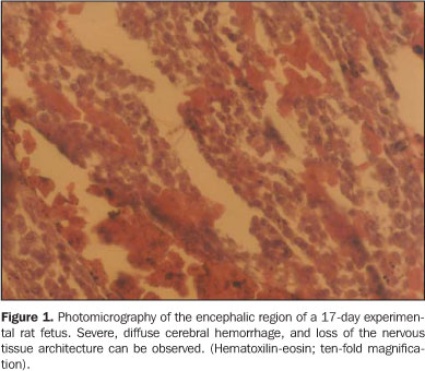
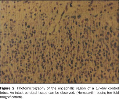
MATERIALS AND METHODS The sample studied included 15 (10 female and 5 male) eight-week-old Rattus norvegicus albinus, Wistar, weighting on average 275 g. Both male and female rats were kept under a 12-hour light-dark cycle, with unrestricted access to food and water. The female animal models were fertilized according to an adaptation from the method of Chahoud & Kwasigroch(8). An otoscope was utilized for detecting the vaginal plug to determine the day of conception(9). Five female animal models at the 8th gestational day, with 37 embryos, were kept under standard survival conditions, constituting the control group. Other five female animal models, also at the 8th gestational day, with 39 embryos, had their abdominal region exposed to a single 0.3 Gy Xradiation dose for 30 seconds. A Gnatus, 70 kV, 10 mA odontological X-ray equipment. All of the rats were kept in separate cages, anesthetized with ether and submitted to the irradiation and perfusion procedures. On the 17th day of gestation, both groups were submitted to hysterectomy and sacrificed with an ether injection into the abdominal cavity under 0.2 ml/100 g ketamine anesthesia. The animals' uteri were fixed in a 10% formaldehyde solution, dehydrated and embedded in paraffin. A microtome was utilized to obtain several 4 µm-tissue sections. Selected sections were hematoxilin-eosin stained and evaluated for comparatively analyzing the fetuses brains in both groups. The macroscopic analysis was based on the closure of the rostral neuropore, while the microscopic analysis consisted in a morphological evaluation of the brains, utilizing a ten-fold magnification. The analysis covered the hippocampus and cerebellum, besides neuronal ectopia, loss of the tissue architecture and hemorrhage, rated as mild, moderate and severe according to the vessels congestion intensity and blood cells flow out. The Epi-Info 2004 software (CDC. 3.3 version) was utilized for statistical analysis, according to the chi-square test for hemorrhage variables and alterations in the nervous tissue architecture, comparing both the experimental and control groups. A p value < 0.05 was considered as statistically significant.
RESULTS Thirty-seven and 39 embryos, respectively in the control and experimental groups, were evaluated, with no follow-up loss being observed. Clinical examinations demonstrated absence of macroscopic differences between groups, considering that 100% of the animal models presented a normal closure of the rostral neuropore, and none (0%) presented signs of anophtalmia, micrencephaly and anencephaly. Microscopic analyses demonstrated cerebral cortex hemorrhage ranging between moderate and severe. Twenty-seven per cent of the animal models in the experimental group presented moderate cerebral hemorrhage, and 77% presented severe hemorrhage (p < 0.05; Figure 1). All the cases of severe cerebral hemorrhage were associated with loss of the nervous tissue architecture (p = 0.05). None of the animals in the control group presented cerebral hemorrhage or loss of nervous tissue architecture (Figure 2).
DISCUSSION All of the gestations developed normally up to the analyses period, without any behavioral alteration being observed in the experimental group as compared with the control group. An otoscope was effectively utilized for detecting the vaginal plug, allowing the accurate determination of the exact day of conception, and increasing the predictability of the gestation, as demonstrated by Voipio & Nevalainen(9). This method did not cause any stress or pain to the animals, besides being rapid and easy to perform. Therefore the otoscope insertion did not present any morphological effect on the gestation development, and the rats were not affected by any external interference during the study. Considering that the nervous system cells are extremely sensitive to radiation, nervous tissue lesions and hemorrhages were the main findings in the present study. However, other effects such as motor and cognitive dysfunctions, Purkinje cells atrophy, micrencephaly and hippocampal ectopia have been observed and reported by other authors(5,6), nevertheless they were not investigated in the present study. Hemorrhages detected in the brain of rats exposed to radiation constitute evidences compatible with the findings of Yang & Tobias(2), who have demonstrated that a low X-radiation dose may cause cerebral hemorrhage and, consequently, nervous tissue lesion. According to the findings of Friedberg et al.(4), none of the animal models presented any abnormality such as anophtalmia, micrencephaly and anencephaly, probably because of the low radiation dose applied. Despite the high rate of cerebral hemorrhage in the experimental group, there was no morphological difference between the experimental and control groups. These findings are compatible with those reported by Wang et al.(10), who have found few alterations resulting from X-irradiation. The description of the effect of X-irradiation during the pre-implantation period as "everything or nothing"(3) was confirmed in the present study, considering that the embryos developed normally up to the 17th gestational day, and no follow-up loss was recorded. Vos(3) also reports that the primary effect of X-ray exposure during the organogenesis phase is the development of malformations. Nevertheless, in the present study, the control group was irradiated during the organogenesis phase and no malformation could be evidenced. However, loss of nervous tissue architecture and hemorrhage probably would have resulted in later developmental disorders. According to Schmitz et al.(7), the prenatal irradiation results not only in neuronal loss, but also in a massive volumetric decrease in the cerebral regions investigated. Probably because of the low radiation dose applied, this could not be observed. It is known that progenitor cells may be injured by irradiation and cannot divide during radiation exposure, but, maybe, a single X-radiation dose is not enough to cause damages during the cells development stages. These authors have utilized different levels of irradiation, and this may have affected the growth factors. Finally, based on their findings, the authors support the assertion that X-radiation in the form and conditions described in the present study may determine the occurrence of cerebral hemorrhages and nervous tissues lesions in rat fetuses. Acknowledgement The present study was partially supported by Fundação Bahiana para Desenvolvimento das Ciências.
REFERENCES 1. Rugh R. X-ray-induced teratogenesis in the mouse and its possible significance to man. Radiology. 1971;99:433–43. [ ] 2. Yang T, Tobias C. Effects of heavy ion irradiation on the brain vascular system and embryonic development. Adv Space Res. 1984;4:239–45. [ ] 3. Vos O. Effects and consequences of prenatal irradiation. Boll Soc Ital Biol Sper. 1989;65:481–500. [ ] 4. Friedberg W, Faulkner DN, Neas BR, et al. Dose-incidence relationships for exencephalia, anophthalmia and prenatal mortality in mouse embryos irradiated with fission neutrons or 250 kVX-rays. Int J Radiat Biol Relat Stud Phys Chem Med. 1987;52:223–36. [ ] 5. Miki T, Fukui Y, Takeuchi Y, et al. A quantitative study of the effects of prenatal X-irradiation on the development of cerebral cortex in rats. Neurosci Res. 1995;23:241–7. [ ] 6. Takai N, Sun XZ, Ando K, et al. Ectopic neurons in the hippocampus may be a cause of learning disability after prenatal exposure to X-rays in rats. J Radiat Res. 2004;45:563–9. [ ] 7. Schmitz C, Born M, Dolezel P, et al. Prenatal protracted irradiation at very low dose rate induces severe neuronal loss in rat hippocampus and cerebellum. Neuroscience. 2005;130:935–48. [ ] 8. Chahoud I, Kwasigroch TE. Controlled breeding of laboratory animals In: Neubert D, Merker HJ, Kwasigroch TE, editors. Methods in prenatal toxicology. Stuttgart: Georg Thieme; 1977. p.78–91. [ ] 9. Voipio HM, Nevalainen T. Improved method for vaginal plug detection in rats. Scand J Lab Anim Sci. 1998;25:5–9. [ ] 10. Wang H, Chen D, Gao C, et al. Effects of low level prenatal 60Co gamma-irradiation on postnatal growth and behavior in mice. Teratology. 1993; 48:451–7. [ ]
Received May 15, 2006. Accepted after revision August 16, 2007.
* Study developed at Escola Bahiana de Medicina e Saúde Pública, Salvador, BA, Brazil. |
|

Av. Paulista, 37 - 7° andar - Conj. 71 - CEP 01311-902 - São Paulo - SP - Brazil - Phone: (11) 3372-4544 - Fax: (11) 3372-4554