Radiologia Brasileira - Publicação Científica Oficial do Colégio Brasileiro de Radiologia
AMB - Associação Médica Brasileira CNA - Comissão Nacional de Acreditação
 Vol. 41 nº 1 - Jan. /Feb. of 2008
|
ORIGINAL ARTICLE
|
|
Doppler and maternal serum screening in the prediction of pregnancy complications |
|
|
Autho(rs): Fabrício da Silva Costa, Rebeca Silveira Rocha, Sérgio Pereira da Cunha, Francisco Cândido dos Reis, Aderson Tadeu Berezowski, José Antunes-Rodrigues |
|
|
Keywords: Doppler, Serum screening, Preeclampsia, Complications, Pregnancy |
|
|
Abstract:
IPhD, Associate Professor of Gynecology and Obstetrics in the Course of Medicine, Universidade Estadual do Ceará (UECE), Fortaleza, CE, Brazil
INTRODUCTION During the last three decades, numerous clinical, biophysical and biochemical tests have been suggested for early detection of several gestational disorders, especially preeclampsia. Some of these tests are simple, other, invasive; some of them have been extensively evaluated, others still remain under clinical investigation. The literature review demonstrates a high level of disagreement as far as the sensitivity and predictive value of several of these tests are concerned, and an ideal test for screening the main gestational and perinatal pathologies is still to be found(1). The greatest majority of current studies about markers for gestational and perinatal complications associated with alterations in the trophoblastic invasion, show a trend towards the identification of a sole marker(2). Preeclampsia and intrauterine growth restriction (IUGR), however, are multisystemic diseases, with placentation abnormalities probably due to genetic and immunological predisposition(3), and a series of humoral and cardiovascular alterations probably related to endothelial dysfunction(4), so the discovery of a good sole marker is practically unachievable. Some studies have demonstrated that the association between serum markers and an abnormal uterine artery Doppler may potentialize the identification of pregnant women at risk, preventing the development of gestational complications(5,6). The present study is aimed at comparing the effectiveness of uterine arteries Doppler and the maternal sera screening — alpha-fetoprotein (AFP), human chorionic gonadotropin (hCG), atrial natriuretic peptide (ANP), and nitric oxide (NO), in the prediction of pregnancy complications.
MATERIALS AND METHODS The present casuistic included 49 primigravidae, with no chronic, gestational or gynecologic disease, assisted at the prenatal assistance clinic of Department of Gynecology and Obstetrics at Hospital das Clínicas da Faculdade de Medicina de Ribeirão Preto da Universidade de São Paulo (HCFMRP-USP) or at the public health service network of Ribeirão Preto, SP. This study was approved by the HCFMRP-USP Committee for Ethics on Research, and the tests and examinations were performed only after the patients had been given an explanation and signed a term of informed consent. The determination of gestational age was based on the first day of last menstrual period, and on transvaginal ultrasound study performed during the first trimester of gestation. The pregnant women were included in the study at their 18th gestational week, when a blood sample was collected for serum screening. Uterine artery Doppler was performed between the 24th and 26th gestational weeks. Additionally, the patients were given guidance about parturition in the HCFMRP-USP Obstetric Center. The women's prenatal follow-up was kept at their original health units up to the end of gestation. Extra-protocol tests and studies were offered in cases of intercurrences or by request of the prenatal doctor. In the setting of complications, the patients were referred for follow-up in the High-Risk Pregnancy Clinic of the HCFMRP-USP Department of Gynecology. Exclusion criteria of the present study were: presence of chronic, gestational or gynecologic diseases, chronic use of medications, smoking, multiple gestation, presence of fetal malformation, miscarriage, failure do appear at scheduled returns and childbirth in other hospitals. Failure to appear at scheduled returns (two cases), and childbirth in other hospitals (two cases) were the reasons for exclusion of four women during the development of the present study. The following data were obtained: women's age, color and familial history of chronic diseases (chronic arterial hypertension and diabetes mellitus). Upon their inclusion in the study at the 18th gestational week, the women had a 15 ml blood sample collected, after 30-minute seating rest, by peripheral venous puncture with a 20 ml syringe (without heparin). AFP and hCG screening was performed by chemiluminescence, with Immulite AFP (2000) and Immulite hCG (2000) kits (Diagnostic Products Corporation; Los Angeles, CA, USA). The essays were performed at the Laboratory of Tocogynecological Physiology and Pharmacology, Department of Gynecology and Obstetrics of HCFMRP-USP. The measurements were performed with an Immulite chemiluminescence automatic analyzer (DPC Cirrus Inc., a DPC subsidiary). Values found for AFP and hCG were converted into multiples of median. AFP and hCG medians were determined by means of a normality curve corresponding to the Brazilian population. Values > 2.0 MoM for AFP and hCG screening were considered as abnormal according to the literature(1). ANP screening was performed by radioimmunoessay, with Euro-Diagnostica (2000) kits (ALPCO – American Laboratory Products Company; Windham, NH, USA). The essays were performed at Laboratory of Neurophysiology, Department of Physiology of FMRP-USP. In the present study, this essay sensitivity for ANP was 3.5 pg/ml. The intra-essay coefficient of variation was 8.6%, with mean and standard deviation of 12.6 ± 1.1 pg/ml. All of the measurements were performed in a single essay, so eliminating the inter-essay variation. Values > 237.4 pg/ml for ANP were considered as abnormality, corresponding to the 95% of the present sample. NO screening was performed by comparison with a standard colorimetric curve with the following parameters or characteristics: slope = 2.70; intercept = –26.00; R = 0.99992 and standard deviation = 6.38 µmol/l. All of the measurements were performed in a single essay, so eliminating the inter-essay variation. Values > 17.8 µmol/l for NO were considered as abnormality, corresponding to the 95th percentile of the present sample. Uterine artery Doppler examinations were always performed by a same observer, between the 24th and 26th gestational weeks, with the patient in dorsal decubitus, the table tilted at 45°, after spontaneous urination. Mean examination duration was 30 minutes. The equipment utilized was an ATL HDI 3000 model (Advanced Technologies Laboratories; Bothell, WA), with a wide band 2–5 MHz convex transducer, pulse Doppler, color-Doppler and power-Doppler, and 100 Hz filter setting. The vessels evaluated by the Doppler method were the right and left uterine arteries and respective ascending branches. For the purpose of examination, the transducer was placed on the abdomen inferior lateral quadrant, in a medially angulated position. The color Doppler was used to identify the uterine artery homolateral to the transducer, on the intersection with the external iliac artery, and the volume sample was placed approximately 1 cm above the crossing point. The presence of bilateral notch in the Doppler study performed between the 24th and 26th gestational weeks was considered as an abnormality in the flow velocity waveform. The childbirths occurred in the HCFMRP-USP Obstetric Center. The neonates were received and followed-up by the team of the Pediatrics and Puericulture Department in the mentioned hospital. One has considered as gestational complications: development of preeclampsia, premature separation of normally implanted placenta (PSNIP) and preterm delivery (PTD). The diagnosis of preeclampsia was based on increase in the arterial pressure > 140/90 mmHg, unchanged after six-hour rest, associated with proteinuria (> 0.3 g in 24 hours)(6). One has considered as perinatal complications: centralization of fetal blood flow, presence of thick meconium in the amniotic fluid at the time of birth, and small infants for the gestational age (SGA). SGA neonates were those who, at the birth, presented a weight below the 10th percentile on the growth curve established for our population. For statistical analysis, the unpaired t-test was utilized for quantitative continuous variables of normal distribution; the Mann-Whitney test for non-parametric quantitative samples; the exact Fisher test for qualitative parameters; and the Pearson's for correlations evaluation. Sensitivity, specificity, positive predictive value (PPV), and negative predictive value (NPV) were calculated by the cut-off established in the literature for AFP and hCG(1), and values of ANP and NO 95th percentile. The multivariate analysis by means of multiple logistic regression was utilized for evaluation of all the markers as a whole.
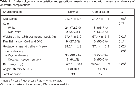
RESULTS The present study included 45 pregnant women whose epidemiological characteristics and gestational results are described on Table 1.
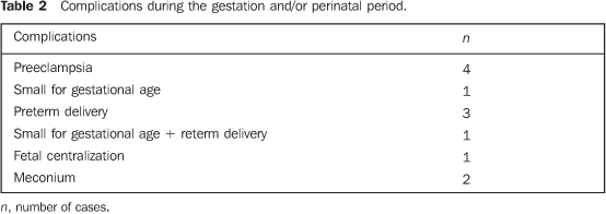
The patients were divided into two groups: normal – with no obstetric and/or perinatal complications; complicated – with obstetric and/or perinatal complications. Table 2 is a list of the 12 cases of gestational and/or perinatal complications found in the present study. All of these complications are included in the spectrum of alterations related to abnormality in the placental adaptation. No case of PSNIP was observed.
Table 3 shows mean and standard deviation for AFP, hCG, ANP and NO serum screening both in normal and complicated gestations.
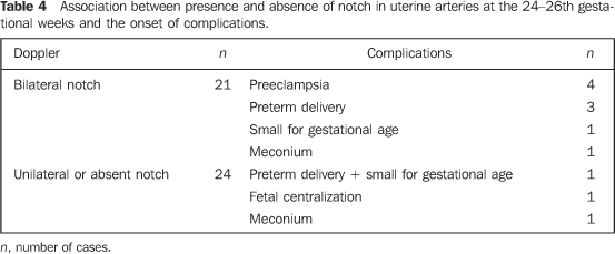
The presence of bilateral notch in the uterine arteries was found in the studies performed between the 24th and 26th gestational weeks, in 21 cases (46.7%), unilateral notch in three cases (6.6%), and absent in 21 cases (46.7%). The presence of bilateral notch in the uterine arteries between the 24th and 26th weeks was strongly associated with the onset of complications during pregnancy. Nine pregnant women presented intercurrences, representing 75% of the total of complications. All the cases of preeclampsia (four), one case of SAG neonate, three cases of PTD, and one case of thick meconium at the time of delivery, presented alteration of the flow velocity waveform (FVW). The presence of unilateral notch was observed only in three cases. One pregnancy progressed normally, while one case resulted in PTD with SAG neonate, and another in "centralization of fetal circulation" resulting in a term neonate with no neonatal complication. The FVW normality has not been strongly associated with the onset of diseases during gestation. Of 21 patients who have not presented notch in the uterine arteries, only one progressed with complication in the perinatal period. Table 4 demonstrates the association between the presence of gestational and/or perinatal complications and the qualitative evaluation of the uterine arteries FVW.
The presence of any gestational and/or perinatal complication has demonstrated to be more frequently associated with the presence of bilateral notch in uterine arteries at Doppler, according to the exact Fisher test (p < 0.04). Table 5 shows the analysis by multiple logistic regression utilized for evaluating the simultaneous or multivariate effect from AFP, hCG, ANP, NO serum screening and uterine artery Doppler on the prediction of pregnancy complications.
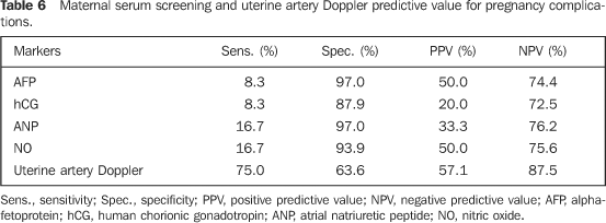
The presence of bilateral notch is the only variable presenting correlation with the onset of complications in the multivariate analysis (odds ratio of 5.2). Table 6 shows the sensitivity, specificity, PPV and NPV of the different markers in the prediction of gestational complications.
DISCUSSION More than 100 tests have already been evaluated for the prediction of disorders associated with abnormality in the placental adaptation, particularly preeclampsia, but the ideal predictive test has not been described yet(7). In 1988, investigators obtained, overall, 26% of adverse gestational results after inexplicable high serum level of AFP: 6% of fetal or neonatal deaths, 15% of low weight at birth, and 6.2% of congenital abnormalities(8). On the other hand, some authors have contested the value of the AFP serum screening in the prediction of preeclampsia(9). Researchers have studied 72 patients with inexplicable increase in the AFP serum level during the gestation, and compared their findings with those from normal control groups. Adverse perinatal result has occurred in 38.9% of cases with high serum levels of AFP, and in 31.9% of controls (p = 0.50), allowing the authors to conclude that AFP offers only a small, additional predictive value in a high-risk population(9). In the present study, the AFP values have not demonstrated a statistically significant difference between the group with gestational and/or perinatal complications (mean 0.98 ± 0.48 MoM) and the control group (mean 0.91 ± 0.36 MoM), with p = 0.89. With a cut-off value for abnormality > 2.0 MoM, the AFP sensitivity was only 8.3%, specificity 97.0%, the PPV, 50.0%, and NPV, 74.4% for prediction of pregnancy complications. Some studies in the literature have shown that the hCG and/or plasmatic b-hCG serum screening between 14–20 amenorrhea weeks seems to be an excellent test for prediction of vascular complications in primigravidae(10). A study has demonstrated that b-hCG levels twofold above the mean level were associated with an increase in the relative risk for hypertension induced by gestation and preeclampsia (RR = 1.7 and 5.1, respectively)(10). Similarly to AFP, some authors have contested the value of hCG serum screening in the prediction of gestational complications. In a study developed in 1998, the b-hCG levels resulted in a ROC curve to evaluate its potential in the screening for preeclampsia. Nineteen patients (4.4%) developed preeclampsia and the mean free b-hCG level in the second trimester of pregnancy was significantly high in comparison with the control group (1.52 versus 1.10; p = 0.03). The ROC curve demonstrated that, for a sensitivity of 79%, the specificity was only 54%, allowing the author to conclude that the isolate measurement of b-hCG free fraction during the second gestational trimester is not clinically useful as a maternal serum screening for preeclampsia in primigravidae.(11). In the present study, hCG values have not demonstrated a statistically significant difference between the group with gestational and/or perinatal complications (mean = 1.19 ± 0.78 MoM) and the control group (mean = 1.08 ± 0.75 MoM). With a cut-off value for abnormality above de 2.0 MoM, the hCG sensitivity in the prediction of complications was only 8.3%, specificity 87.9%, PPV 20.0% and NPV 72.5%. ANP seems to play a significant role as a predictive marker for preeclampsia and other related diseases, considering that ANP secretion may be a response to hemodynamic changes possibly preceding the clinical onset of the disease(1). Ultrasonography has allowed the identification of na increase in the left atrium dimensions and a frequent occurrence of pericardial effusion in women with preeclampsia and with high ANP serum levels in the third gestational trimester and early in the puerperium, suggesting that the preeclampsia increases the intracardiac pressure, stimulating the ANP secretion(12). Data in the present casuistic have not shown a statistically significant difference in the ANP serum concentration, considering the group with gestational and/or perinatal complications (mean 139.3 ± 77.1 pg/ml) and the control group (mean 119.6 ± 47.0 pg/ml). With a cut-off value above the 95th percentile for abnormality (272.7 pg/ml), the ANP sensitivity for complication prediction was only 16.7%, and specificity 97.0%, PPV 33.3% and NPV 76.2%. Aiming at demonstrating that the decrease in the production of NO may play a role in the preeclampsia physiopathology, investigators have measured the serum nitrate levels in 20 women with preeclampsia, 20 healthy pregnant women and 12 non-pregnant women in childbearing age. The serum nitrate concentration was significantly higher in the patients with preeclampsia, in comparison with the healthy pregnant and the non-pregnant women(13). These results do not support the hypothesis that a decrease in the endothelial NO production plays a significant role in the preeclampsia physiopathology; on the contrary, increased serum nitrate levels may represent an increase in the NO production or decrease in its renal clearance as a response to the disease. The role of the maternal serum NO in the prediction of complications early in the first half of the pregnancy, is still to be established. This has encouraged its investigation as a probable marker in the prediction of gestational complications. No statistically significant difference was found in the serum NO concentration, between the group with gestational and/or perinatal complications (mean 11.1 ± 4.6 µmol/l) and the control group (mean 10.0 ± 3.4 µmol/l). With a cutoff value above the 95th percentile for abnormality (18.9 µmol/l), the NO sensitivity for gestational complications prediction was only 16.7%, specificity 93.9%, PPV 50.0% and NPV 75.6%. The placenta, in the process of implantation and development, changes the uterine circulation from low flow with high resistance to high flow with low resistance(6). As result, the uterine arteries protodiastolic notch disappears around the 24–26th gestational. In the present group of pregnant women, bilateral notches were present in uterine arteries in 21 cases (46.7%) at the 24–26th gestational weeks, with a statistically significant difference as compared with normal pregnancies (p < 0.04). In 1998, in a study with uterine arteries Doppler between the 19th and 21st gestational weeks, investigators observed the presence of bilateral notch in 12.4% of cases, and the Doppler study was considered as altered in 22.8% of cases(14). In the same year, in a study of non-selected patients in the 24th gestational week, the author observed the notch in 27.7% of cases(15). On the other hand, another study has shown a high incidence of notches between the 18th and 26th gestational weeks, with the presence of bilateral notches in 40.7% of cases(16). Considering the easiness, safety and non-invasiveness of uterine artery Doppler, the sensitivity and specificity of this method in the prediction of gestational complications have already been widely evaluated(17,18). In 1998, in a study on the diagnosis of the presence of bilateral notches with Doppler, the sensitivity was 61.9%, the specificity 88.7%, PPV 11.1%, and NPV 99.0%. On the other hand, for the diagnosis of IUCR, the sensitivity was 36.8%, specificity 89.2%, PPV 17.9%, and NPV 95.7%. With the high resistance as a criterion for Doppler abnormality, the authors reported 71.4% sensitivity, 78.2% specificity, 6.9% PPV, and 99.2% NPV for the diagnosis of preeclampsia. For the diagnosis of IUCR, the sensitivity was 47.4%, specificity 78.7%, PPV 12.5%, and NPV 95.9%(14). In 1998, also with the presence of bilateral notch or high RI as criteria for Doppler abnormality, the investigators demonstrated a good sensitivity, but low PPV. Bilateral notch as a predictor of toxemia presented 100.0% sensitivity, 76.3% specificity, 19.0% PPV, and 100.0% NPV; high RI showed 83.3% sensitivity, 84.7% specificity, 23.3% PPV, and 98.9% NPV(15). In 2000, another study demonstrated 91.0% sensitivity, 42.0% specificity, and 37.0% PPV for the prediction of preeclampsia(16). For the prediction of SAP neonates, the sensitivity was 84.0%, specificity 39.0%, and PPV 33.0%. A study developed in 2005 prospectively evaluated the level of amniotic fluid in low-risk pregnant women, concluding that this is not a good predictor of gestational and perinatal complications, whereas the presence of bilateral notch in the uterine arteries confirmed by Doppler in the period between the 24th and 26th gestational weeks is a good predictor of such complications, with 90.0% sensitivity, and 62.5% specificity(19). Another study has shown that the presence of a high RI between the 24th and 26th gestational weeks presented sensitivity and specificity similar to the presence of bilateral notch in uterine arteries, with a slight improvement in the PPV(20). In 1998, investigators evaluated simultaneously the N-terminal proatrial natriuretic peptide, the free b-hCG and AFP in the prediction of preeclampsia by means of maternal serum screening between the 15th and 19th gestational weeks in a population of 637 primigravidae. The sensitivity, specificity and predictive values were calculated considering a cut-off value of 2.0 MoM for AFP and b-hCG. An inexplicable increase in AFP levels above 2.0 MoM was observed in 2% of the pregnant women, whereas the b-hCG was high in 16%. AFP sensitivity and specificity for prediction of preeclampsia were, respectively, 3% and 98%, and the b-hCG sensitivity and specificity, respectively 3% and 95%(1). These low sensitivities in the detection of gestational complications were similar to those found in the present study. The N-terminal proatrial natriuretic peptide serum levels were not high in the patients with gestational hypertension (330 pmol/l) and in cases of preeclampsia. These data are similar to the ones of the present study, considering that significant differences in the ANP levels between complicated and normal gestations were not found. With a gradual logistic regression model, the same authors have evaluated the simultaneous effect of the levels of N-terminal ANP, AFP, b-hCG, maternal age and mean arterial pressure (MAP) between the 19–24th gestational weeks on the chances of preeclampsia development. Serum levels and maternal age cannot predict preeclampsia, the MAP being the only factor identified as a risk predictor (p = 0.01). In the present study, a gradual logistic reversion model was utilized for simultaneously evaluating AFP, hCG, ANP, NO and uterine arteries Doppler as gestational complications predictors. In the multivariate analysis, the presence of bilateral notch on uterine arteries Doppler is the only variable presenting correlation with the onset of gestational and/or perinatal complications, with odds ratio of 5.2.
CONCLUSION The present casuistic demonstrates the relevance of uterine artery Doppler, particularly between the 24th and 26th gestational weeks, even for low-risk pregnant women, allowing the selection of patients with uteroplacental flow alterations for follow-up in reference centers and adoption of prophylactic measures.
REFERENCES 1. Pouta AM, Hartikainen AL, Vuolteenaho OJ, et al. Midtrimester N-terminal proatrial natriuretic peptide, free beta-hCG, and alpha-fetoprotein in predicting preeclampsia. Obstet Gynecol. 1998; 91:940–4. [ ] 2. Broughton Pipkin F, Sharif J, Lal S. Predicting high blood pressure in pregnancy: a multivariate approach. J Hypertens. 1998;16:221–9. [ ] 3. Robillard PY, Hulsey TC, Périanin J, et al. Association of pregnancy-induced hypertension with duration of sexual cohabitation before conception. Lancet. 1994;344:973–5. [ ] 4. Roberts JM, Taylor RN, Musci TJ, et al. Preeclampsia: an endothelial cell disorder. Am J Obstet Gynecol. 1989;161:1200–4. [ ] 5. Elsandabesee D, Srinivas M, Kodakkattil S. The clinical value of combining maternal serum screening and uterine artery Doppler in prediction of adverse pregnancy outcome. J Obstet Gynaecol. 2006;26:115–7. [ ] 6. Cnossen JS, van der Post JAM, Mol BWJ, et al. Prediction of pre-eclampsia: a protocol for systematic reviews of test accuracy. BMC Pregnancy and Childbirth. 2006;6:29. [ ] 7. Dekker GA, Sibai BM. Early detection of preeclampsia. Am J Obstet Gynecol. 1991;165:160–72. [ ] 8. Burton BK. Outcome of pregnancy in patients with unexplained elevated or low levels of maternal serum alpha-fetoprotein. Obstet Gynecol. 1988;72:709–13. [ ] 9. Phillips OP, Simpson JL, Morgan CD, et al. Unexplained elevated maternal serum alpha-fetoprotein is not predictive of adverse perinatal outcome in an indigent urban population. Am J Obstet Gynecol. 1992;166:978–82. [ ] 10. Sorensen TK, Williams MA, Zinghein RW, et al. Elevated second-trimester human chorionic gonadotropin and subsequent pregnancy-induced hypertension. Am J Obstet Gynecol. 1993;169: 834–8. [ ] 11. Luckas M, Hawe J, Meekins J, et al. Second trimester serum free beta human chorionic gonadotrophin levels as a predictor of pre-eclampsia. Acta Obstet Gynecol Scand. 1998;77:381–4. [ ] 12. Pouta AM, Räsänen JP, Airaksinen KEJ, et al. Changes in maternal heart dimensions and plasma atrial natriuretic peptide levels in the early puerperium of normal and pre-eclamptic pregnancies. Br J Obstet Gynaecol. 1996;103:988–92. [ ] 13. Smárason AK, Allman KG, Young D, et al. Elevated levels of serum nitrate, a stable end product of nitric oxide, in women with pre-eclampsia. Br J Obstet Gynaecol. 1997;104:538–43. [ ] 14. Kurdi W, Campbell S, Aquilina J, et al. The role of color Doppler imaging of the uterine arteries at 20 weeks' gestation in stratifying antenatal care. Ultrasound Obstet Gynecol. 1998;12:339–45. [ ] 15. Montenegro CAB, Chaves E, Pessoa LG, et al. Valor preditivo para a toxemia do Doppler das artérias uterinas. Prog Diagn Prenat. 1998;10:16–9. [ ] 16. Coleman MA, McCowan LM, North RA. Mid-trimester uterine artery Doppler screening as a predictor of adverse pregnancy outcome in high-risk women. Ultrasound Obstet Gynecol. 2000; 15:7–12. [ ] 17. Nardozza LMM, Camano L, Moron AF, et al. Alterações ultra-sonográficas na gravidez Rh negativo sensibilizada avaliada pela espectrofometria do líquido amniótico e pela dopplervelocimetria da artéria cerebral média. Radiol Bras. 2006;39: 11–3. [ ] 18. Pastore AR. Dopplervelocimetria da artéria cerebral média fetal: o divisor de águas no diagnóstico da anemia fetal [editorial]. Radiol Bras. 2006;39(1):iii. [ ] 19. Costa FS, Cunha SP, Berezowski AT. Avaliação prospectiva do índice de líquido amniótico em gestações normais e complicadas. Radiol Bras. 2005;38:337–41. [ ] 20. Costa FS, Cunha SP, Berezowski AT. Qual o melhor período para a realização do Doppler das artérias uterinas na predição de complicações da gestação? Radiol Bras. 2006;39:97–102. [ ]
Received March 14, 2007. Accepted after revision May 24, 2007.
* Study developed at Hospital das Clínicas da Faculdade de Medicina de Ribeirão Preto da Universidade de São Paulo (HCFMRP-USP), Ribeirão Preto, SP, Brazil. |
|
Av. Paulista, 37 - 7° andar - Conj. 71 - CEP 01311-902 - São Paulo - SP - Brazil - Phone: (11) 3372-4544 - Fax: (11) 3372-4554


