Radiologia Brasileira - Publicação Científica Oficial do Colégio Brasileiro de Radiologia
AMB - Associação Médica Brasileira CNA - Comissão Nacional de Acreditação
 Vol. 42 nº 2 - Mar. / Apr. of 2009
|
ORIGINAL ARTICLE
|
|
Comparative analysis of dose-volume histograms between 3D conformal and conventional non-conformal radiotherapy plannings for prostate cancer |
|
|
Autho(rs): Sílvia Moreira Feitosa, Adelmo José Giordani, Rodrigo Sousa Dias, Helena Regina Comodo Segreto, Roberto Araujo Segreto |
|
|
Keywords: Prostate cancer, Radiotherapy, Conformal plan, Non-conformal plan |
|
|
Abstract:
IMasters, MDs, Universidade Federal de São Paulo/Escola Paulista de Medicina (Unifesp/EPM), São Paulo, SP, Brazil
INTRODUCTION With the introduction of megavolt equipment (cobalt-60 and linear accelerators), external beam radiotherapy has become universally accepted as an appropriate therapeutic modality for localized prostate cancer in patients above the age of 50 years, because of the lower skin dose and homogeneity of radiation in depth(1). The conventional or non-conformal technique utilizes the four-field beam arrangement (antero-posterior, postero-anterior, right latero-lateral and left latero-lateral), also known as four-field or box technique that may be performed with isocentric fields or fields with a fixed source-surface distance, technique of beams in two rotational arcs (120°), a combination of both techniques or three-field technique (one anterior field, two parallel, lateral fields and opposed left and right fields). The treatment with the rotational technique (360°) can also be utilized as a dose boost (in a second phase), however, with this technique the dose delivered to the anterior wall of the rectum is higher(2,3). One of the difficulties observed in these non-conformal techniques for treatment of localized prostate cancer is the dose delivered to the adjacent healthy tissues. In these techniques, the maximum achievable doses range between 65 and 70 Gy. Many times such doses are not sufficient to eradicate the malignant clone cells, which make the management of the disease more difficult. Studies developed by the Patterns of Care Study, in 1984, demonstrated that the incidence of grade 3 and 4 rectal and bladder toxicity have doubled (from 3.5% to 6.9%) in cases where doses > 70 Gy were prescribed with the non-conformal planning(1). In the nineties, technological development allowed the conformal radiotherapy planning. The 3D planning system varies in some aspects, but is based on a common principle: the utilization of images (computed tomography or magnetic resonance imaging) for delimitating the prostate, organs at risk and generating high-resolution 3D reconstructions. This technique provides a precise anatomical definition of target volumes (prostate and seminal vesicles) and adjacent healthy tissues (rectum, bladder and coxofemoral joint), allowing an individual radiation beam conformation to the target-volumes(4,5). Because of the volumetric appreciation of the target volume, the treatment planning is based on the ability to anatomically define each sub-volume within the total irradiated volume and accurately calculating the dose delivery to each point, allowing for radiation dose escalation, i.e., high doses to the target volumes and limited doses and volumes to the adjacent healthy tissues, within the recommended tolerance parameters(6). In Brazil, many radiotherapy centers still lack a system for conformal radiotherapy planning. Under the practical point of view it would be interesting to compare conformal isocentric and non-conformal plannings. The present study is aimed at comparatively analyzing radiation doses in target volume and organs at risk in 3D conformal, non-conformal isocentric and non-conformal plannings utilizing the source-surface distance for patients with localized prostate cancer submitted to radiotherapy.
MATERIALS AND METHODS The present study evaluated radiotherapy plannings for 40 patients with localized prostate cancer assisted in the Unit of Radiotherapy at Universidade Federal de São Paulo/Escola Paulista de Medicina (Unifesp/EPM), in the period between 2004 and 2005. Conformal, conformal isocentric and non-conformal plannings utilizing the source-surface distance were simulated for each of the patients for comparison of radiation dose in target volumes and organs at risk. The sample of the present study included patients with prostate tumors clinically staged T1-T2c according to the American Joint Committee on Cancer (2002), without distant metastases and previous surgical treatment, submitted or not to neoadjuvant hormone therapy. Planning methods Pre-planning - The patients were previously submitted to a simulation in a simulator. They were positioned in dorsal decubitus with the hands on the anterior region of their chest, a pillow under their head and with a feet support. The pre-tomography isocenter was marked with the localization of a 10 × 10 cm field, with the inferior margin coinciding with the inferior branch of the ischium and with the field center between the pubic bones, on the anterior field and, rotating the gantry 90°, the field center was located at 1.5 cm posteriorly to the anterior pubic border on the lateral field. Planning tomography - The patient preparation included 300 ml hydric ingestion and fleet enema two hours before the planning tomography. The procedure was performed in the same position of the previous simulation with 5 and 5 mm sections of the pelvic region, from the transition between the 4th and 5th lumbar vertebrae to the upper third of the femur, including all the adjacent lateral soft tissues. Target volumes delineation - Target volumes were delineated through delimitation of the gross tumor volume (GTV), clinical target volume (CTV) and planning target volume (PTV) for each patient. Delineation of organs of interest - The bladder was completely delineated through its external wall; the rectum was delineated through the external wall from the rectosigmoid junction to the anal margin; and the femoral contour was delineated through the external wall of the bone to the minor trochanter. Planning techniques - With the aid of a computational Eclipse (Varian; Palo Alto, USA) planning system, two non-conformal and one conformal planning were performed for the 40 patients, in two phases for a 6 MeV linear accelerator. The first non-conformal planning with fixed source-surface distance utilized the four-field technique (antero-posterior, postero-anterior, right latero-lateral and left latero-lateral) in the first phase with a 100 cm source-surface distance up to the dose of 5040 cGy, with 1.5 cm margins around the whole the prostate and seminal vesicles for intermediate and high-risk patients, and 1.5 cm margins around the whole prostate and proximal third of seminal vesicles for low risk patients; the isodoses were normalized at maximum, a 95% isodose was selected, encompassing the target volume. In the second phase of the same planning, the technique of two isocentric rotational arcs up to the dose of 1980 Gy was utilized, with two 120° rotational arcs and with a central, anterior and posterior 60° interval (the first arc encompassed 30° to 150° and the second arc, 210° to 330°), with 1.5 cm margins encompassing the prostate and the proximal third of seminal vesicles. In both treatment phases, the total dose prescribed was 7020 cGy; the isodoses were normalized (in the isocenter), and a 95% dose was selected, encompassing the target volume. In the first phase of the second non-conformal isocentric planning the technique of four isocentric fields (antero-posterior, postero-anterior, right latero-lateral and left latero-lateral) was utilized up to the dose of 5040 cGy, with 1.5 cm margins encompassing the prostate and the proximal third of the seminal vesicles for intermediate and high-risk patients; the isodoses were normalized (in the isocenter) and a 95% dose was selected, encompassing the target volume. In the second phase of the same planning, the technique of two isocentric rotational arcs up to the dose of 1980 cGy was utilized, with two 120° rotational arcs with a central, anterior and posterior 60° interval (the first arc encompassed 30° to 150°, and the second arc, 210° to 330°), with 1.5 cm margins encompassing the prostate and the proximal third of the seminal vesicles; the isodoses were normalized (in the isocenter), and a 95% dose was selected, encompassing the target volume. In both phases of the treatment, the total prescribed dose was 7020 cGy. The center of the treatment field was placed on the center of the prostate, utilizing the planning tomography, but with symmetric collimators to open the treatment fields. Collimation blocks were not utilized. In the isocentric conformal planning, the treatment scheme was simulated for a 6 MeV accelerator in two phases. For low-risk patients, GTV and CTV corresponded to the prostate, and PTV corresponded to the prostate and the proximal third of the seminal vesicles besides a 1 cm margin around the whole region, except posteriorly where the added margin measured 0.8 cm. For intermediate and high-risk patients, GTV and CTV1 (CTV of the first treatment phase) corresponded to the prostate and seminal vesicles; CTV2 (CTV of the second treatment phase) corresponded to the prostate and the proximal third of the seminal vesicles. PTV1 (PTV of the first treatment phase) corresponded to the CTV1 besides a 1 cm margin around the whole region, except posteriorly where the added margin measured 0.8 cm. PTV2 (PTV of the second treatment phase) corresponded to the CTV2 besides a 1 cm margin around the whole region, except posteriorly where the added margin measured 0.8 cm. In all of the plannings a 0.5 cm block margin was added. In the first phase, the field arrangement ranged between four and six up to a dose of 5040 cGy, the selected prescription isodose encompassing the prostate and seminal vesicles (in cases of medium and high-risk patients) and prostate and proximal third of the seminal vesicles (for low-risk patients). In the second phase, the field arrangement ranged between four and six up to a dose of 2160 cGy, a prescription isodose encompassing the prostate and the proximal third of the seminal vesicles. The total dose for both phases corresponded to 7200 cGy prescribed for a 95% isodose curve (normalization at the isocenter). Evaluation methods The analysis covered dose-volume histograms for target volumes (CTV and PTV) and bladder, rectum and coxofemoral joint of the conformal plannings in comparison with non-conformal plannings. The dose-volume histograms (DVHs) are shown on Figure 1. In both plannings, maximum, mean and median doses were analyzed for the CTV and PTV. Regarding organs, the doses were evaluated in 10%, 15%, 25%, 40% and 60% of the rectal volume (percentages evaluated in the institution), besides the maximum, mean and median doses; in the bladder, the doses were evaluated in 30% and 60% of its volume (percentages evaluated in the institution), besides the maximum, mean and median doses; and in the coxofemoral joints, maximum, mean and median doses were evaluated in the whole volume. Statistical analysis The Friedman variance analysis was applied in the results evaluation, considering 0.05 or 5% (α < 0.05) as the level for null hypothesis rejection. Significant values were marked with an asterisk, and the non-significant ones with NS.
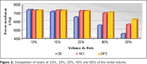
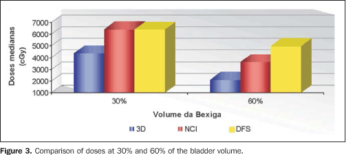
RESULTS The following paragraphs describe the results of the analysis of non-conformal plannings utilizing a fixed (100 cm) source-surface distance, non-conformal isocentric plannings and conformal plannings for the patients included in the present study. The median doses were significantly lower in the conformal planning for the following rectal volumes: 25%, 40% and 60% (Figure 2). The median doses were significantly lower in the conformal planning for the following bladder volumes: 30% and 60% (Figure 3).
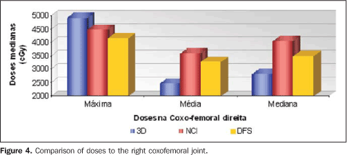
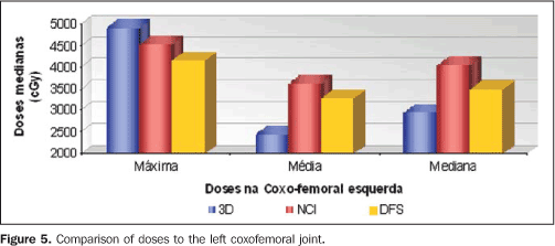
In the comparison of the maximum doses to the coxofemoral joints, the non-conformal plannings obtained higher median doses. The medians of the mean doses and medians to the coxofemoral joints were significantly lower in the conformal planning (Figures 4 and 5).
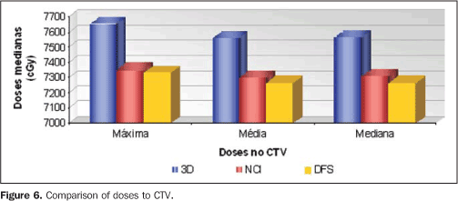
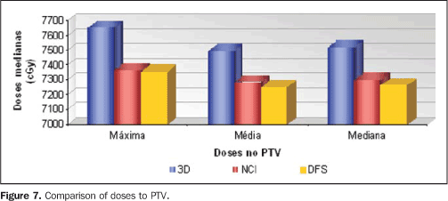
In the comparison of maximum, mean and median doses to CTV and PTV, the conformal planning obtained higher median doses (Figures 6 and 7).
DISCUSSION Regarding the volumes of the organs evaluated, the present study has found the following results: the median volume of the rectum was 63 cm3, the median volume of the bladder was 201 cm3, the median volumes of the right and left coxofemoral joints were respectively 158.4 cm3 and 153 cm3, the median GTV was 51 cm3. The comparative analysis of plannings for conventional, conformal, and intensity-modulated radiotherapy demonstrated median volumes of rectum, bladder, coxofemoral joint and GTV of 33 cm3, 79 cm3, 111 cm3 and 76 cm3, respectively, in the conventional and conformal plannings. The lower volumes reported by Oh et al may be explained by the fact that the organs at risk were delineated only 1.5 cm below and above the prostate and seminal vesicles(2). Another comparative study of doses and volumes for prostate cancer with conventional and conformal techniques have found a median GTV of 64.2 cm3, with the organs at risk delineated 2.0 cm above and below the prostate. The seminal vesicles have not been included in the GTV(7). The present study demonstrated higher values for volumes than the ones reported in the literature because of the different techniques utilized in the delineation of the targets and organs at risk. The mean and median doses observed on the dose-volume histograms for the rectum and bladder in the conformal planning were significantly lower as compared with the doses observed in the conventional planning (except for the mean and median dose to 10% of the rectal volume). Perez et al. have reported a higher mean dose in the bladder and rectum with the conventional technique, as compared with the conformal technique that utilizes oblique beams to reduce the radiation in these structures(8). Oh et al. have reported mean doses of 2132-2711 cGy in the bladder, and 2932-3620 cGy in the rectum in the conventional planning. On the other hand, with the conformal technique, they have found mean doses of 1241-1952 cGy in the bladder, and 2432-3502 cGy in the rectum, when the dose of 4500 cGy was prescribed in the isocenter(2). Another comparative study published in 2000 and updated in 2002, between the techniques studies, demonstrated the following percentages: with the conformal technique, 8.5 ± 11.8 for a dose > 70 Gy in the rectum, and with the conventional technique, 28.8 ± 28.9. In the bladder, the percentage of volume with a dose > 70 Gy was 6.3 ± 8.4 in the conformal technique, and 19.4 ± 24.4 in the conventional technique(8,9). In these studies developed by Perez et al., the conventional technique was performed with two 120° rotational arcs and prostate margins of 2 cm. On the other hand, the conformal technique was performed with seven fields and collimation with cerobend or multi-leaf collimator(8,9). In a randomized trial comparing toxicity between conventional and conformal radiotherapy for prostate cancer, Dearnaley et al. have reported median high-dose volume - defined as volume intersection in the space for therapy beams in the conformal planning - presented a 39% decrease with the utilization of the conformal technique. This study has reported 48% less in the volume of the rectum and 38% less in the vesical volume were treated with a 90% isodose in the conformal therapy as compared with the conventional therapy(10). Maximum doses observed in the present study on HDVs for rectum and bladder were significantly higher in the conformal planning. The authors believe that such fact is explained by the higher prescribed dose which was not compensated neither by collimation nor by the higher number of field entrances. In a study developed by Sale et al., approaching the two planning techniques, the conventional therapy utilized the area defined by the 95% isodose curve (encompassing the prostate) as target volume, and the conformal therapy utilized the area defined by the prostate with a 1.5 cm margin. Significantly higher maximum doses in the volume of the rectum and bladder were not found in the conformal planning. However, the doses in 100% of the vesical volume and in 100%, 90% and 50% of the rectal volume were significantly higher in the conformal planning. The authors argued that this has occurred because of the larger field size in the conformal planning, which has not been compensated by collimations(7). Variations in maximum doses were observed in the rectum and bladder, respectively, from 4807 to 4855 cGy and from 4661 to 4803 cGy utilizing the conventional technique, and from 5072 to 5304 cGy and 4705 to 5078 cGy in the conformal planning with a prescribed dose of 4500 cGy in the isocenter(2). The results of the present study demonstrate that the maximum doses in the coxofemoral joints were significantly higher in the conformal planning, while the mean and median doses were significantly lower with this technique. As regards doses in the coxofemoral joint, the literature does not show a sharp advantage between the conventional and conformal techniques. Maximum and mean doses of respectively 2718- 2891 cGy and 1614-2537 cGy have been reported for conventional planning. On the other hand, maximum and mean doses of respectively 3539-4901 cGy and 1850-2443 cGy have been reported for conformal planning(2). In the present study, maximum, mean and median doses found on the DVHs of CTV and PTV were significantly higher in the conformal planning. These data agree with the literature which reports that the conformal technique prescribes higher doses in PTV, as compared with the conventional technique. According to Oh et al., the mean dose in PTV ranged between 4455 and 4547 cGy with the conventional technique, and between 4671 and 4896 cGy with the conformal technique(2). Significantly higher doses at 100%, 90% and 50% of GTV and PTV in the conformal planning as compared with the conventional planning have also been reported(7). Other authors have reported mean and maximum doses in tumor of, respectively, 69.8 ± 2.6 Gy and 71.7 ± 2.4 Gy in the conformal technique, and 69.7 ± 2.8 Gy and 71.3 ± 2.8 Gy in the conventional technique(8). Pollack et al. have developed a study comparing two patients groups: one group that received a 68 Gy dose with the conventional technique, and another that received a 78 Gy dose with the conventional technique in the initial phase, with a dose boost in a second phase with conformal technique. This study has demonstrated that a significantly higher percentage of the bladder received doses > 60 Gy in the group submitted exclusively to conventional therapy. As regards the percentage of rectal volume treated with doses > 60 Gy, no statistically significant difference has been found between groups. The volume of prostate and seminal vesicles receiving 95% of the prescribed dose was significantly larger in the group that received a dose boost with conformal technique(11). In a study comparing the conventional technique (four-field) and two conformal techniques (four- and six-field), Magrini et al. have found lower mean doses in the rectum with conformal techniques (75% of the isocenter dose) than with the conventional technique (79% of the isocenter dose), although no statistical significance has been achieved. In the same study, the authors have observed that the mean dose in the bladder was statistically lower in the conformal techniques with four and six fields (respectively 57% and 50% of the isocenter dose) than in the conventional technique (79% of the isocenter dose). The percentage of CTV and PTV that received more than 95% of the isocenter dose was statistically higher in conformal techniques(12). The results of the present study demonstrate the benefits from the utilization of the 3D radiotherapy planning system in cases of prostate cancer. These results demonstrate that even with a higher prescription dose in the conformal technique, a higher target volume dose with lower doses in the organs at risk can be achieved.
CONCLUSION It may be concluded that the present study has evidenced that conformal radiotherapy planning for prostate cancer allows the delivery of higher doses to the target volume and lower doses to adjacent critical organs.
REFERENCES 1. Leibel SA, Fuks Z, Zelefsky MJ, et al. Technological advances in external-beam radiation therapy for the treatment of localized prostate cancer. Semin Oncol. 2003;30:596-615. [ ] 2. Oh CE, Antes K, Darby M, et al. Comparison of 2D conventional, 3D conformal, and intensity-modulated treatment planning techniques for patients with prostate cancer with regard to target-dose homogeneity and dose to critical, uninvolved structures. Med Dosim. 1999;24:255-63. [ ] 3. Fletcher GH. Textbook of radiotherapy. 3rd ed. Philadelphia: Lea & Febiger; 1980. [ ] 4. Perez CA, Halperin EC, Brady LW, et al. Principles and practice of radiation oncology. 4th ed. Philadelphia: Lippincott Williams & Wilkins; 2004. [ ] 5. Brundage M, Lukka H, Crook J, et al. The use of conformal radiotherapy and the selection of radiation dose in T1 or T2 low or intermediate risk prostate cancer - a systematic review. Radiother Oncol. 2002;64:239-50. [ ] 6. Mangar SA, Huddart RA, Parker CC, et al. Technological advances in radiotherapy for the treatment of localised prostate cancer. Eur J Cancer. 2005;41:908-21. [ ] 7. Sale CA, Yeoh EE, Scutter S, et al. 2D versus 3D radiation therapy for prostate carcinoma: a direct comparison of dose volume parameters. Acta Oncol. 2005;44:348-54. [ ] 8. Perez CA, Michalski JM, Purdy JA, et al. Three-dimensional conformal therapy or standard irradiation in localized carcinoma of prostate: preliminary results of a nonrandomized comparison. Int J Radiat Oncol Biol Phys. 2000;47:629-37. [ ] 9. Perez CA, Michalski JM, Mansur D, et al. Three-dimensional conformal therapy versus standard radiation therapy in localized carcinoma of prostate: an update. Clin Prostate Cancer. 2002;1:97-104. [ ] 10. Dearnaley DP, Khoo VS, Norman AR, et al. Comparison of radiation side-effects of conformal and conventional radiotherapy in prostate cancer: a randomised trial. Lancet. 1999;353:267-72. [ ] 11. Pollack A, Zagars GK, Starkschall G, et al. Conventional vs. conformal radiotherapy for prostate cancer: preliminary results of dosimetry and acute toxicity. Int J Radiat Oncol Biol Phys. 1996;34:555-64. [ ] 12. Magrini SM, Cellai E, Rossi F, et al. Comparison of the conventional 'box technique' with two different 'conformal' beam arrangements for prostate cancer treatment. Cancer Radiother. 1999;3: 215-20. [ ] Received June 3, 2008. * Study developed at Universidade Federal de São Paulo/Escola Paulista de Medicina (Unifesp/EPM), São Paulo, SP, Brazil. |
|
Av. Paulista, 37 - 7° andar - Conj. 71 - CEP 01311-902 - São Paulo - SP - Brazil - Phone: (11) 3372-4544 - Fax: (11) 3372-4554
