Sr. Editor,
Paciente do sexo masculino, 17 anos, com história de crises convulsivas tonicoclônicas generalizadas desde os cinco anos de idade, foi encaminhado ao nosso serviço para investigação. A ressonância magnética (RM) mostrou lesão expansiva no lobo frontal esquerdo, com hipossinal em T1 e hipersinal em T2, com halo de hipossinal nas duas sequências, sem realce pelo meio de contraste e com discreto edema circunjacente (Figuras 1A e 1B). A lesão foi totalmente ressecada (Figura 1C) e enviada para estudo histopatológico e imuno-histoquímico (Figura 1D), sendo concluído o diagnóstico de pseudoneoplasia calcificada do neuroeixo (
calcifying pseudoneoplasm of the neuraxis – CAPNON). O paciente está sendo acompanhado há dois anos, sem sinais clínicos ou radiológicos de recidiva.
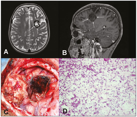 Figura 1.
 RM pré-operatória nas sequências T2 sem contraste (
A) e T1 pós-contraste (
B) mostrando lesão expansiva no lobo frontal esquerdo, com halo de hipossinal nas duas sequências, componente interno com hipersinal em T2 e hipossinal em T1, sem realce pelo meio de contraste e com discreto edema circunjacente.
C: Imagem intraoperatória da pseudoneoplasia calcificada após ressecção total.
D: Corte histológico do pseudotumor exibindo inúmeros focos de calcificação (microcalcificações), que correspondem às células da lesão. Raras células viáveis de permeio só podem ser demonstradas pelo estudo por imuno-histoquímica (hematoxilina e eosina, ampliação 20×).
CAPNON, também conhecida como lesão fibro-óssea do sistema nervoso central (SNC), é uma lesão benigna rara, primeiramente relatada por Rhodes et al. em 1978
(1). A CAPNON caracteriza-se por ser lesão calcificada, de crescimento lento, normalmente única e que pode ter localização em qualquer parte do SNC. A origem ainda não é totalmente conhecida, porém, com base na evolução clínica benigna e nos achados histopatológicos, acredita-se em um processo reacional a fatores como trauma, infecção, inflamação ou neoplasias
(2,3). Segundo García Duque et al.
(4), a maioria dos casos publicados na literatura ocorreu em homens (59,38%) e em sítio intracraniano (54,2%), principalmente supratentorial. Os principais sintomas relacionados à CAPNON são cefaleia, convulsões e sintomas focais neurológicos. Todavia, a apresentação clínica vai depender da localização, das dimensões e do consequente efeito de massa provocado pela lesão
(4,5).
Os exames de imagem são importantes ferramentas para o diagnóstico de lesões tumorais do SNC
(6–10), servindo também para a caracterização da CAPNON e para incluí-la na lista de diagnósticos diferenciais de lesão calcificada no SNC. A maior parte dos casos descritos na literatura apresenta-se na radiografia convencional e na tomografia computadorizada como lesão sólida e calcificada, ou com calcificações periféricas. Na RM, normalmente apresenta hipossinal em T1 e T2, com mínimo ou nenhum realce pelo meio de contraste e raramente apresenta edema
(11,12). Entretanto, alguns casos foram descritos com sinais variados em T1 e T2, ou ainda com presença de edema e realce importante pelo meio de contraste
(12).
O diagnóstico depende do estudo histopatológico, que se caracteriza pela presença de nódulos com matriz condromixoide, calcificações finas, corpos psamomatosos de permeio, quantidades variáveis de estroma fibroso e células epitelioides e fusiformes na periferia. A presença de cada componente é altamente variável entre os casos descritos. Os achados da imuno-histoquímica mais comuns são a presença de antígeno epitelial de membrana e vimentina, e a ausência de proteína acida glial fibrilar e da proteína S100
(3,12).
A ressecção cirúrgica é o tratamento de escolha e normalmente apresenta bom prognóstico, não havendo relato na literatura de recidiva após remoção completa da CAPNON
(11). Nos diagnósticos diferenciais por imagem incluem-se cavernoma, meningioma, lesões infecciosas (neurocisticercose, tuberculose), oligodendroglioma e astrocitoma com calcificações
(3,12,13). Em razão do aumento do número de casos descritos nos últimos anos, a CAPNON deve entrar no diagnóstico diferencial das lesões calcificadas no SNC.
REFERÊNCIAS1. Rhodes RH, Davis RL. An unusual fibro-osseous component in intracranial lesions. Hum Pathol. 1978;9:309–19.
2. Aiken AH, Akgun H, Tihan T, et al. Calcifying pseudoneoplasms of the neuraxis: CT, MR imaging, and histologic features. AJNR Am J Neuroradiol. 2009;30:1256–60.
3. Serrano-Pardo R, Valls AB, Benítez HS, et al. Pseudoneoplasia calcificante del sistema nervioso central. A propósito de un caso y revisión de la literatura. Rev Esp Patol. 2009;42;155–8.
4. García Duque S, Medina Lopez D, Ortiz de Méndivil A, et al. Calcifying pseudoneoplasms of the neuraxis: report on four cases and review of the literature. Clin Neurol Neurosurg. 2016;143:116–20.
5. Giardina F, Guerra G, Righi A, et al. Calcifying pseudotumor of the spine: a case report. Clin Surg. 2016;1:1042.
6. Queiroz RM, Abud LG, Abud TG, et al. Burkitt-like lymphoma of the brain mimicking an intraventricular colloid cyst. Radiol Bras. 2017;50: 413–4.
7. Langer FW, Santos D, Alves GRT, et al. Malignant peripheral nerve sheath tumor of the vagus nerve: an uncommon cause of progressive dyspnea. Radiol Bras. 2017;50:412–3.
8. Abreu PP, Muniz BC, Ventura N, et al. Intraventricular ganglioglioma with dissemination of cerebrospinal fluid. Radiol Bras. 2018;51:272–3.
9. Niemeyer B, Muniz BC, Ventura N, et al. Papillary tumor of the pineal region accompanied by Parinaud''s syndrome: magnetic resonance imaging findings. Radiol Bras. 2018;51:202–4.
10. Muniz BC, Niemeyer B, Ventura N, et al. Primary intraosseous meningioma: atypical presentation of a common tumor. Radiol Bras. 2018;51: 412–3.
11. Krejčí T, Buzrla P, Vecera Z, et al. Calcifying pseudoneoplasm of the neural axis – a case report. Cesk Slov Neurol N. 2015;78/111:568–75.
12. Stienen MN, Abdulazim A, Gautschi OP, et al. Calcifying pseudoneoplasms of the neuraxis (CAPNON): clinical features and therapeutic options. Acta Neurochir (Wien). 2013;155:9–17.
13. Lyapichev K, Bregy A, Shah AH, et al. Occipital calcified pseudoneoplasms of the neuraxis (CAPNON): understanding a rare pathology. BMJ Case Rep. 2014;2014.
1. Universidade Federal do Rio de Janeiro (UFRJ), Rio de Janeiro, RJ, Brasil;
https://orcid.org/0000-0002-8308-1920 2. Universidade Federal do Rio de Janeiro (UFRJ), Rio de Janeiro, RJ, Brasil;
https://orcid.org/0000-0002-4765-8526 3. Universidade Federal do Rio de Janeiro (UFRJ), Rio de Janeiro, RJ, Brasil;
https://orcid.org/0000-0003-2412-8239 4. Universidade Federal do Rio de Janeiro (UFRJ), Rio de Janeiro, RJ, Brasil;
https://orcid.org/0000-0002-9306-7145 5. Universidade Federal do Rio de Janeiro (UFRJ), Rio de Janeiro, RJ, Brasil;
https://orcid.org/0000-0001-8797-7380Correspondência: Dra. Rafaela Fonseca Albuquerque Pithon
UFRJ – Departamento de Radiologia
Rua Rodolpho Paulo Rocco, 255, Cidade Universitária, Ilha do Fundão
Rio de Janeiro, RJ, Brasil, 21941-913
E-mail:
rafaelafa@hotmail.com.brRecebido para publicação em 22/9/2017.
Aceito, após revisão, em 14/12/2017.

 |
|


 Read in English
 PDF Portuguese
 Print
Print
 Send this article by email
 How to cite this article
 Submit a comment
 Mendeley
 Pocket