Radiologia Brasileira - Publicação Científica Oficial do Colégio Brasileiro de Radiologia
AMB - Associação Médica Brasileira CNA - Comissão Nacional de Acreditação
 Vol. 51 nº 4 - July / Aug. of 2018
|
ORIGINAL ARTICLE
|
|
Facebook as a tool to promote radiology education: expanding from a local community of medical students to all of South America |
|
|
Autho(rs): Matheus Zanon1; Stephan Altmayer1; Gabriel Sartori Pacini1; Álvaro Guedes1; Guilherme Watte1; Edson Marchiori2; Bruno Hochhegger1 |
|
|
Keywords: Education, medical, continuing; Clinical medicine/education; Radiology/education; Social media. |
|
|
Abstract: INTRODUCTION
Radiology is still not given sufficient attention during medical education. Only 25% of medical schools in the United States require radiology as a clinical rotation(1). In the United Kingdom, only 5% of total teaching time in medical education is committed to radiology, which is not sufficient for students to be properly prepared for medical practice(2). At many medical schools, one alternative for medical students to become more familiar with the radiology is to affiliate themselves with one of the interest groups—also known as "academic societies". At the beginning of 2015, we created an academic society for diagnostic imaging at our university, designating it the Liga de Diagnóstico por Imagem (LiDi, Diagnostic Imaging League). Our goal was to supplement education in radiology and image interpretation for medical students at our institution. Once a week, meetings were held to discuss selected teaching topics and a medical student presented a clinical case, with the assistance of a senior radiologist responsible for our interest group. Any medical student enrolled at our university could attend the meetings. Because there was an increasing demand for students engaged in these activities, we tried to find ways to reach more people and expand the number of participants. Facebook is the most popular social network worldwide, with 1.71 billion monthly active users in the second quarter of 2016(3). As recent studies suggest(4–10), Facebook has been recognized by medical institutions and professionals as a means of promoting research projects, delivering health information, and facilitating student education. Therefore, we decided to use Facebook as a tool to disseminate the clinical cases presented in our weekly meetings and to reach out a larger number of participants outside of our local student community. The aim of this study was to assess the feasibility of Facebook as a tool to promote a local radiology education project and to expand that local project to a larger audience. An additional objective was to analyze the demographic characteristics and interests of the users of the Facebook page created. MATERIALS AND METHODS Clinical case design The LiDi Facebook page (www.facebook.com/lidiufcspa) was created on June 22, 2015, as a nonprofit page, administrated by 12 medical students. A series of clinical cases have been posted on the page, one a week since August 24, 2015. The posts consisted of a brief patient history and physical examination findings, along with different imaging modalities relevant to the case, such as X-ray, computed tomography (CT), magnetic resonance imaging (MRI), and ultrasound. Five possible diagnoses were provided, and the public was encouraged to choose the most likely diagnosis based on the vignette and images provided. For each case, the correct answer was followed by an explanation of the disease and commentaries to highlight the most important imaging findings. Figure 1 depicts an example of the standards followed for case posting. 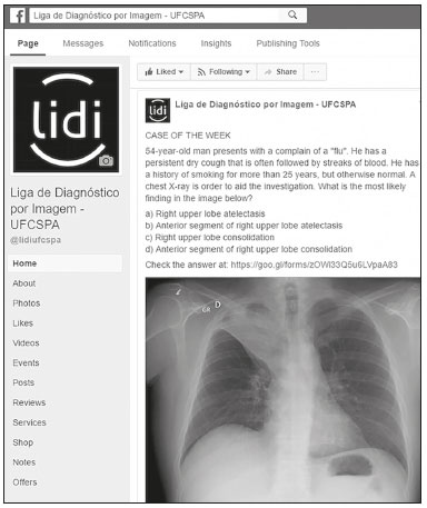 Figure 1. Example of one of the clinical cases posted on our page. The original data was posted in Portuguese. For this case, there were 1637 clicks on the post; the post reached 4764 people; and 103 users "liked", "commented on", or "shared" the content. Most topics and imaging modalities were chosen on the basis of the curriculum framework for undergraduate education in radiology proposed by the European Society of Radiology (ESR)(11). We set an easy to moderate level of difficulty, as rated by the senior radiologist. In addition to the clinical cases, one extra weekly post per week focused on radiology teaching topics geared to medical students and residents was also planned. To reach a larger audience, the medical student responsible for preparing the case of the week also shared the images to a public Latin-American Facebook group with 28,182 participants, composed mainly of physicians and medical students, that was created with the goal of sharing scientific papers, reviews, and case reports. We did not solicit any publicity via Facebook to gain visibility, likes, or number of subscribers. Data analysis From August 2015 to October 2016, we prospectively analyzed user feedback on the content posted on our page. Public response was assessed using Facebook Audience Insights, a tracking tool that provides data about user demographics, engagement, likes, top posts, and number of visits. A "like" establishes a connection between a user and a Facebook page, the user thereafter receiving notifications about all the information posted, including photos, videos, links, status updates, and polls(3). Other measures of user feedback about a page are "reach" and "engagement". The former indicates the number of people who have visualized any content, not necessarily interacting with the post, whereas the latter is a more precise measure of user reactions to the content, analyzing the number of likes, views, and shares(3). Because of Facebook''s privacy policies, it is not possible to collect any user data other than sex, age, language, and location. Therefore, using Google Forms, we created a questionnaire that we shared on our Facebook page and encouraged users to complete. We thus collected data regarding the occupation of the users, as well as their opinions on the quality of the cases, topics, and imaging modalities they would like to see addressed. The questionnaire was live (i.e., responses were accepted) for a month. Statistical analysis Statistical analysis was performed with the IBM SPSS Statistics software package, version 21.0 (IBM Corporation; Armonk, NY, USA). Data analysis included descriptive statistics, and variables are reported as mean ± standard deviation, range, or relative frequency, as appropriate. In addition, we calculated the number of views proportional to that of engaged users over time. That was done to evaluate whether any growth in the number of users was accompanied by a proportional increase in the interactions with the content. Group comparisons were made using one-way analysis of variance. For all statistical analyses, the level of significance was set at p ≤ 0.05. Due to the nature of this study, institutional review board approval was not required. RESULTS Thirty-five cases were posted in the period. As shown in Table 1, X-ray (n = 15) and CT (n = 13) were the most common imaging methods, chest X-ray being the topic most often addressed (n = 9). Before we began posting the clinical cases, our page had 286 likes. By October 2016, that number had risen to 4244, representing an increase of approximately 1484% and eight times the size of the medical student community at our institution (n = 530). The demographic characteristics of the subscribers are also shown in Table 1. Most were 18–24 years of age (n = 2037; 48%) or 25–34 years of age (n = 1867; 44%), and 58% (n = 2461) were women. Most of the users (n = 3919; 90%) accessed the page via a Desktop computer. Initially, the users were concentrated in Porto Alegre, where our university is located. During the period analyzed, the number of users increased and their distribution expanded to all of Brazil, as well as to other countries, such as Bolivia. Figure 2 provides a quantitative depiction of the geographic distribution of subscribers to the LiDi Facebook page over time. Users in Brazil accounted for 93% of the subscribers (n = 3943), whereas those in other South American countries (Argentina, Bolivia, Ecuador, Peru, Uruguay, and Venezuela) collectively accounted for 3% (n = 121). The other 4% were mostly in the European Union or United States. 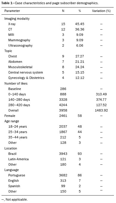 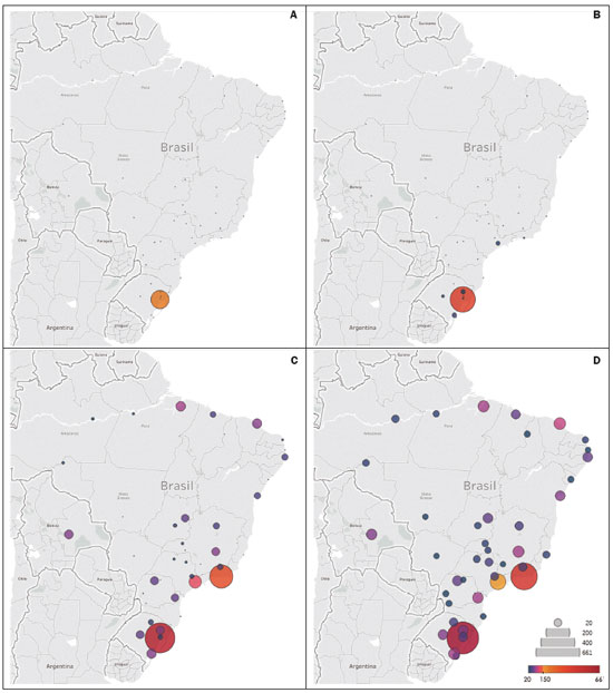 Figure 2. Quantitative maps showing the geographic distribution of subscribers to the LiDi Facebook page over time. Only cities with more than 20 users are represented. Initially, subscribers were concentrated in Porto Alegre, where our university is located. During the period analyzed, the number of users increased and the reach extended to the rest of Brazil, as well as to other countries, such as Bolivia. A: baseline; B: 0–140 days; C: 140–280 days; D: 280–420 days. The Google Forms questionnaire comprised 121 questions designed to assess user opinions and characteristics (Table 2). Undergraduate medical students made up the majority of the respondents (76%), followed by radiology residents (5.8%). Figure 3 presents user opinions about the quality and level of difficulty of the cases. When the respondents were asked what they would like to see featured in future cases, the most commonly requested topics were neuroradiology (n = 73; 60.3%), chest X-ray (n = 68; 56.2%), and abdominal X-ray (n = 63; 52.1%), whereas the most commonly requested imaging modalities were CT (n = 97; 80.2%), MRI (n = 90; 74.4%), X-ray (n = 70; 57.9%), and ultrasound (n = 61; 50.4%). Most (n = 76; 62.8%) of the users reported that they had read the explanation provided along with the cases through Google Docs. However, 34 (28.1%) did not even know that option existed. An excellent or moderate contribution to personal image interpretation skills was reported by 79 (65.3%) and 40 (33.1%) of the users, respectively. Only two users (1.7%) reported that the contribution was slight, and none of the users reported that there was no contribution. 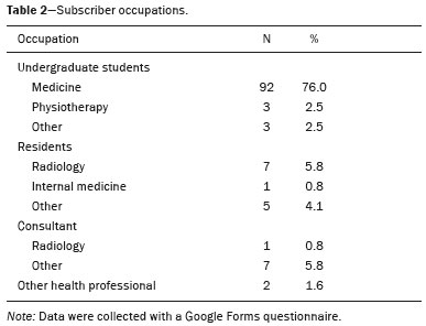 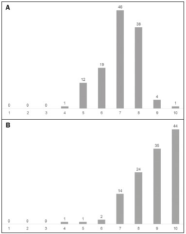 Figure 3. User evaluations (n = 121) of the level of difficulty and quality of the clinical cases (A and B, respectively), on a scale of one to ten. Analysis of the engaged users per total reach ratio during the period assessed demonstrated a significant increase in subscriber interaction over time (p < 0.001). It is noteworthy that the peaks in interaction occurred on the dates when clinical cases were posted on the page (Figure 4). 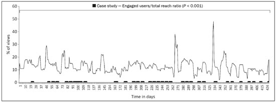 Figure 4. Distribution of engaged users per total reach ratio over time. There was a statistically significant increase in user engagement over the period analyzed (p < 0.001), and the peaks corresponded to the dates when clinical cases were posted. DISCUSSION We found that Facebook was a valuable tool to promote our radiology education project and to compensate for the potential lack of undergraduate training in the subject, because most users reported a significant positive impact on their personal image interpretation skills. In addition, the weekly posting of a clinical case with a multiple-choice question was found to improve the LiDi page''s growth rate and subscriber interaction with the posts. Other authors have recognized the utility of Facebook as a tool to promote research projects, deliver health information, and facilitate medical education(3–5). Social media other than Facebook (e.g., Twitter, Instagram, and YouTube) have also contributed to the dissemination of medical education(6,7). Because they are free resources, social media facilitate interactivity and information sharing. For instance, Hoang et al.(8) demonstrated that a specific educational point is viewed six times more frequently in blogs, such as Radiopedia.org (a collaborative blog on Radiology), than in scientific papers from the American Journal of Roentgenology or the American Journal of Neuroradiology. In our experience, X-rays were the most common type of examination addressed in the clinical cases. As a common first-line imaging modality, we believed that this would be the method of greatest interest for undergraduate medical students. According to a publication by the ESR on undergraduate education in radiology, students should display a systematic approach to the comprehensive interpretation of X-rays, being able to detect abnormalities on X-rays of the chest, abdomen, and skeleton, as well as being able to relate the findings to clinical management(11). Although there was a slightly superior prevalence of cases focusing on chest conditions (n = 9), we tried to maintain an equal distribution of topics related to conditions affecting the abdomen (n = 7) and skeleton (n = 8), the same areas recommended by the ESR guidelines. Other imaging modalities, such as MRI and mammography, and other areas of study, such as neuroradiology and gynecology/obstetrics, were also discussed, although they were given less emphasis. Our LiDi Facebook page also provided other content, such as educational videos/lessons, curiosities about glossary terms in radiology, and information about the latest advances in imaging technology. However, those posts garnered less engagement of the public than did the clinical cases, in accordance with the findings of a previous study(3). One limitation of our study is that student performance after exposure to the content was not assessed. Initiatives disposed to create similar Facebook pages should try to use free online platforms, such as Google Forms, in order to monitor user performance prospectively. Use of Facebook compared with other learning and teaching environments or in comparison with different social media tools should also be investigated. We demonstrated that radiology education could also benefit from the use of social media. Posting cases weekly helped attract more users and engage subscribers with the content. We conclude that creating a Facebook page based on weekly clinical cases can be a valuable method to promote radiology training. REFERENCES 1. Poot JD, Hartman MS, Daffner RH. Understanding the US medical school requirements and medical students’ attitudes about radiology rotations. Acad Radiol. 2012;19:369–73. 2. Heptonstall NB, Ali T, Mankad K. Integrating radiology and anatomy teaching in medical education in the UK—the evidence, current trends, and future scope. Acad Radiol. 2016;23:521–6. 3. Lugo-Fagundo C, Johnson MB, Thomas RB, et al. New frontiers in education: Facebook as a vehicle for medical information delivery. J Am Coll Radiol. 2016;13:316–9. 4. Pander T, Pinilla S, Dimitriadis K, et al. The use of Facebook in medical education—a literature review. GMS Z Med Ausbild. 2014; 31:Doc33. 5. Junhasavasdikul D, Srisangkaew S, Sukhato K, et al. Cartoons on Facebook: a novel medical education tool. Med Educ. 2017;51:539–40. 6. Ranschaert ER, van Ooijen PMA, Lee S, et al. Social media for radiologists: an introduction. Insights Imaging. 2015;6:741–52. 7. Kelly BS, Redmond CE, Nason GJ, et al. The use of Twitter by radiology journals: an analysis of Twitter activity and impact factor. J Am Coll Radiol. 2016;13:1391–6. 8. Hoang JK, McCall J, Dixon AF, et al. Using social media to share your radiology research: how effective is a blog post? J Am Coll Radiol. 2015;12:760–5. 9. Chojniak R, Carneiro DP, Moterani GSP, et al. Mapping the different methods adopted for diagnostic imaging instruction at medical schools in Brazil. Radiol Bras. 2017;50:32–7. 10. Nobre LF. Teleradiology, the Internet, and the development of multidisciplinary professional networks: new times for the specialty? Radiol Bras. 2017;50(3):v. 11. European Society of Radiology (ESR). Undergraduate education in radiology. A white paper by the European Society of Radiology. Insights Imaging. 2011;2:363–74. 1. Department of Diagnostic Methods, Universidade Federal de Ciências da Saúde de Porto Alegre (UFCSPA), Laboratório de Pesquisa em Imagens Médicas (Labimed), Porto Alegre, RS, Brazil 2. Department of Radiology, Universidade Federal do Rio de Janeiro (UFRJ), Rio de Janeiro, RJ, Brazil Study conducted in the Department of Diagnostic Methods, Universidade Federal de Ciências da Saúde de Porto Alegre (UFCSPA), Porto Alegre, RS, Brazil. Mailing address: Gabriel Sartori Pacini Laboratório de Pesquisa em Imagens Médicas – Santa Casa de Misericórdia de Porto Alegre Avenida Independência, 75, Independência Porto Alegre, RS, Brazil, 90035-072 E-mail: gabrielsartorip@gmail.com Received July 4, 2017 Accepted after revision July 28, 2017 |
|
Av. Paulista, 37 - 7° andar - Conj. 71 - CEP 01311-902 - São Paulo - SP - Brazil - Phone: (11) 3372-4544 - Fax: (11) 3372-4554