Radiologia Brasileira - Publicação Científica Oficial do Colégio Brasileiro de Radiologia
AMB - Associação Médica Brasileira CNA - Comissão Nacional de Acreditação
 Vol. 49 nº 1 - Jan. /Feb. of 2016
|
ICONOGRAPHIC ESSAY
|
|
Hyperechoic breast lesions: anatomopathological correlation and differential sonographic diagnosis |
|
|
Autho(rs): Marcelo Menezes Medeiros1; Luciana Graziano2; Juliana Alves de Souza2; Camila Souza Guatelli2; Miriam Rosalina B. Poli2; Rafael Yoshitake2 |
|
|
Keywords: Hyperechoic breast lesions; Ultrasonography; Breast neoplasms; Differential diagnosis. |
|
|
Abstract: INTRODUCTION
Hyperechoic breast lesions are uncommon findings(1), corresponding to 5.6% of alterations identified at ultrasonography (US), with high predictive value for benignity. Such lesions correspond to 0.6% of all biopsied lesions and only 0.4% of all malignant lesions(2). Breast nodules with fatty or fibrous contents, either of vascular origin or with high cellularity may present increased echogenicity at US (Table 1). The knowledge of the characteristics of the main hyperechoic lesions, as well as the recognition of characteristics suggestive of malignancy to avoid late diagnosis might avoid many unnecessary invasive procedures(3). In most cases, hyperechoic lesions are detected for being palpable or for presenting suspicious findings at mammography or magnetic resonance imaging (MRI)(2,4). 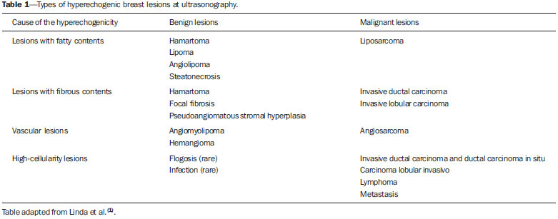 The analysis of sonographic characteristics has shown that non parallel orientation and non circumscribed margins are more frequently found in malignant hyperechoic nodules than in benign ones. Such results suggest that the same sonographic characteristics utilized to evaluate hypo- or isoechoic nodules (such as spiculated margins, association with microcalcifications) should be applied in cases of hyperechoic nodules to differentiate between malignant and benign lesions(4,5). Additionally, the presence of focal hypoechoic areas within hyperechoic findings increases the risk of malignancy(2). In the present study, the authors describe cases of hyperechoic breast lesions observed at US, with emphasis on the relevance of possible differential diagnoses for the correct clinical approach. BENIGN LESIONS Adenosis Adenosis represents a wide spectrum of benign alterations of the breast tissue. In simple adenosis, there is a major preservation of the breast architecture, despite the presence of histological alterations. At US, hyperechoic areas with little or no architectural distortion are observed, since there is no stromal fibrosis (Figure 1). The sclerosing form may present architectural distortion and be associated with proliferative lesions such as intraductal papilloma, fibroadenomas, and coexist with invasive carcinomas in situ(6,7). 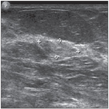 Figure 1. Simple adenosis. Ultrasonography showing hyperechoic, ovoid nodule with circumscribed margins and largest axis parallel to the skin. Hamartomas Hamartomas are constituted of glandular, fatty and fibrous tissues, with estimated incidence of 0.1-0.7%. In most cases, they present as a mobile, barely painful nodule in middle-aged women. At US, they are nodules with circumscribed margins, peripheral halo and compressible by the transducer (Figure 2). They may be hyperechoic in 12-43% of cases, or even heterogeneous, hypoechoic and isoechoic(8). 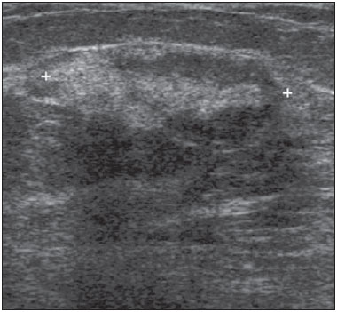 Figure 2. Hamartoma. Ultrasonography showing heterogeneous, ovoid, predominantly hyperechoic nodule with circumscribed margins, largest axis parallel to the skin, and subtle posterior acoustic shadowing. Steatonecrosis It is a common entity that may result from trauma, but in most cases it occurs after surgery or radiotherapy. Its appearance depends upon the presence of histiocytic infiltrate, hemorrhage, fibrosis or calcification(4). At US it presents with varied aspects, as a focal hyperechoic subcutaneous area, anechoic mass with posterior acoustic shadowing, solid or cystic mass with internal echoes, or a cystic mass with mural nodules (Figure 3)(4). 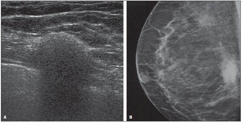 Figure 3. Steatonecrosis. On A, ultrasonography showing hyperechoic, ovoid nodule with indistinct margins and posterior acoustic shadowing. On B, correlation with mammographic study - focal asymmetry is observed in the posterior third of the breast. Fibroadenoma Fibroadenoma is the third most common cause of biopsy in cases of benign breast conditions. The maximum incidence occurs at the third decade of life, with a second peak at the fifth decade. At US, it presents with an elliptical or slightly lobulated shape, and the axis with orientation parallel to the skin, isoechoic or slightly hypoechoic echogenicity, a fine, mobile and slightly compressible echogenic capsule. In 3.1% of cases, fibroadenomas are remarkably hypoechoic, and in 0.9%, either completely or partially hyperechoic (Figure 4). This is due to the presence of either smaller or greater proportions of epithelial and stromal elements(9). As it degenerates, internal, gross (popcorn) or peripheral (halo sign) calcifications are observed(8,9). 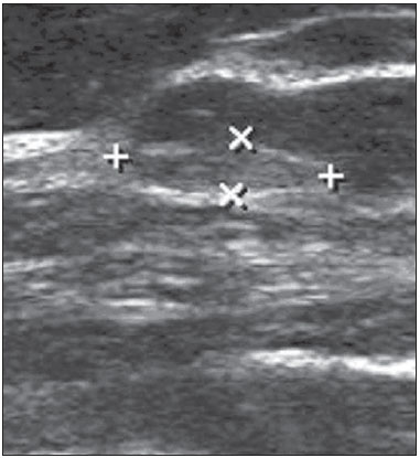 Figure 4. Fibroadenoma. Follow-up ultrasonography showing hyperechoic, ovoid nodule with circumscribed margins and largest axis parallel to the skin. Phyllodes tumor It is responsible for 0.3-1.0% of breast tumors, affecting women aged between 35 and 55 years, as a fast-growing, palpable mass. At US it presents as a hypoechoic and less frequently hyperechoic, solid, well delimited lesion with lobulated margins, occasionally with cystic components, and related to the degree of necrosis and fibrosis (Figure 5)(8,10). 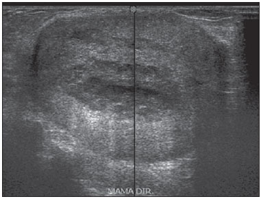 Figure 5. Malignant phyllodes tumor. Ultrasonography showing ovoid, echogenic mass with circumscribed and heterogeneous margins, with a central cystic component and posterior acoustic enhancement. Hemangioma A superficial vascular lesion located in the dermis or in the subcutaneous layer, rarely affecting the breast, with higher incidence in middle-aged women. At US, hemangiomas present with a lobulated or ovoid shape, with well circumscribed margins. Most hemangiomas are either hypoechoic or isoechoic, and may be complex. However, in 33% of cases, they appear as hypoechoic lesions with distal attenuation (Figure 6)(11). 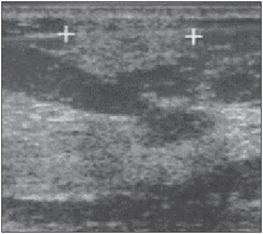 Figure 6. Hemangioma. Ultrasonography showing subcutaneous, palpable, hyperechoic nodule with microlobulated contours. Intraductal papilloma Intraductal papillomas are polypoid lesions within the breast duct. Generally, they affect perimenopausal women, and the most frequent symptoms are sanguinolent, serous or serosanguinolent discharge. At US, intraductal papillomas are seen as a hypoechoic, solid, round or lobulated nodule, but its echogenicity may be variable. In cases of ductal obstruction, the papilloma may be surrounded by fluid, mimicking a mural nodule within a cyst (Figure 7)(8,12). 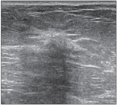 Figure 7. Intraductal papiloma. Ultrasonography showing irregular nodule with orientation perpendicular to the skin, and posterior acoustic shadowing. Myofibroblastoma It is a rare benign, mesenchymal, spindle cell tumor with varied histological aspect and cellularity, representing differential diagnosis of sarcomatous tumor. This tumor is predominantly reported in men as a circumscribed nodule, generally smaller than 3 cm(11). The radiological characteristics are variable, and, at US. It presents as a solid, well delimited mass that may be hypoechoic, isoechogenic or hyperechoic, depending on the fatty component (Figure 8)(13). 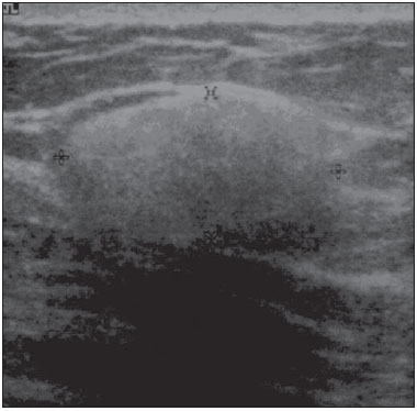 Figure 8. Myofibroblastoma. Ultrasonography showing ovoid, hyperechoic nodule with indistinct margins and posterior acoustic shadowing. Pseudoangiomatous stromal hyperplasia It is a mesenchymal tumor commonly found in perimenopausal women or those undergoing hormone replacement therapy, representing 0.4% of breast lesions. Clinically, it may present as either a palpable nodule or as a diffuse involvement of the breast. At US, they appear as ovoid, heterogeneous and occasionally hyperechogenic lesions (Figure 9)(5,11). 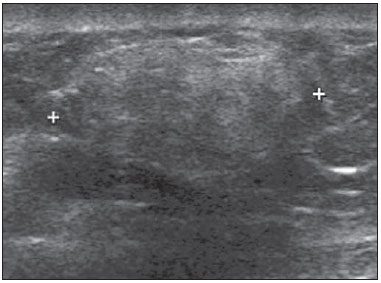 Figure 9. Pseudoangiomatous stromal hyperplasia. Ultrasonography demonstrating heterogeneous, predominantly hyperechogenic, ovoid nodule with indistinct posterior margin and subtle posterior acoustic shadowing. MALIGNANT LESIONS Ductal carcinoma in situ The hyperechogenic presentation at US is uncommon, and is reported in less than 0.8% of cases. Certain histological patterns, such as cribriform carcinoma and solid subtypes, together with tumor heterogeneity, are associated with the lesion hyperechogenicity(13). Thus, despite the high negative predictive value for malignancy of hyperechogenic nodules, the lesions should be carefully evaluated as regards shape, margin and hypervascularization, indicating histological evaluation in the presence of suspicious findings (Figure 10)(13). 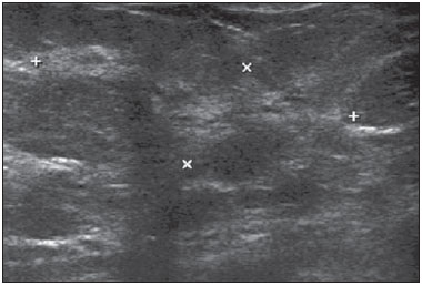 Figure 10. Ductal carcinoma in situ in a patient with Paget disease. Ultrasonography showing heterogeneous, irregular area with subtle parenchymal disorganization associated with intermingled microcysts. Lymphoma It corresponds to 0.1-0.5% of malignant breast lesions. Clinically, it may manifest as a palpable mass. At US, it presents as a hypoechogenic mass with well defined or irregular margins, but the pattern may be heterogeneous with a hyperechogenic halo (Figure 11)(12). 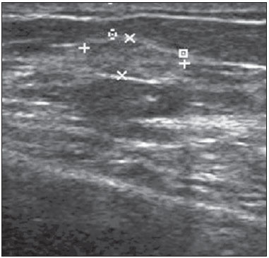 Figure 11. Lymphoma. Ultrasonography showing a slightly heterogeneous, regular nodule parallel to the skin. Lesion identified at follow-up of a patient undergoing treatment for non-Hodgkin lymphoma. Invasive ductal carcinoma It represents 75% of invasive breast tumors. At US it presents as a hypoechogenic image with non-circumscribed margins, and in 2% of cases it may be hyperechogenic. Probably, the hyperechogenicity is due to reflective interfaces caused by growth and infiltration of the cellular component, and fatty inclusions involving a poorly perceptible hypoechogenic, hyalinized center (Figure 12)(4,14). 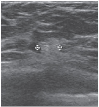 Figure 12. Invasive ductal carcinoma. Ultrasonography showing hyperechoic, rounded nodule with indistinct posterior margin. Metastases Metastases represent 0.5-2% of malignant breast nodules. The most common primary tumors are lymphoma, melanoma and rhabdomyosarcoma. At US, they present as bilateral, fast-growing, palpable, painless, hypoechogenic nodules with irregular margins (Figure 13)(11,15). 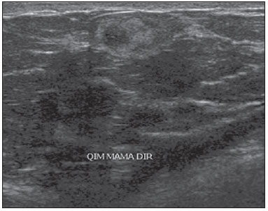 Figure 13. Metastasis from leiomyosarcoma. Ultrasonography identifying heterogeneous, palpable nodule with microlobulated margins. CONCLUSION Hyperechogenic breast nodules are uncommon and, despite the high predictive value for benignity, all the sonographic characteristics should be taken into consideration(16). A full appreciation of the most suspicious imaging findings such as non-circumscribed margins and posterior acoustic shadowing, together with appropriate mammographic correlation and clinical context, contribute to determine the most appropriate clinical/surgical approach(17). REFERENCES 1. Linda A, Zuiani C, Lorenzon M, et al. The wide spectrum of hyperechoic lesions of the breast. Clin Radiol. 2011;66:559-65. 2. Linda A, Zuiani C, Lorenzon M, et al. Hyperechoic lesions of the breast: not always benign. AJR Am J Roentgenol. 2011;196:1219-24. 3. Barra FR, Barra RR, Barra Sobrinho A. Novel functional methods in the evaluation of breast lesions. Radiol Bras. 2012;45:340-4. 4. Gao Y, Slanetz PJ, Eisenberg RL. Echogenic breast masses at US: to biopsy or not to biopsy? Radiographics. 2013;33:419-34. 5. Badan GM, Roveda Júnior D, Ferreira CAP, et al. Positive predictive values of Breast Imaging Reporting and Data System (BI-RADS®) categories 3, 4 and 5 in breast lesions submitted to percutaneous biopsy. Radiol Bras. 2013;46:209-13. 6. Gill HK, Ioffe OB, Berg WA. When is a diagnosis of sclerosing adenosis acceptable at core biopsy? Radiology. 2003;228:50-7. 7. Melhado VC, Alvares BR, Almeida OJ. Radiological and histological correlation of non-palpable breast lesions in patients submitted to preoperative marking according to BI-RADS classification. Radiol Bras. 2007;40:9-11. 8. Stavros AT. Ultrassonografia da mama. 1ª ed. Rio de Janeiro, RJ: Guanabara Koogan; 2005. 9. Goel NB, Knight TE, Pandey S, et al. Fibrous lesions of the breast: imaging-pathologic correlation. Radiographics. 2005;25:1547-59. 10. Yabuuchi H, Soeda H, Matsuo Y, et al. Phyllodes tumor of the breast: correlation between MR findings and histologic grade. Radiology. 2006;241:702-9. 11. Chung EM, Cube R, Hall GJ, et al. From the archives of the AFIP: breast masses in children and adolescents: radiologic-pathologic correlation. Radiographics. 2009;29:907-31. 12. Da Costa D, Taddese A, Cure ML, et al. Common and unusual disease of the nipple-areolar complex. Radiographics. 2007;27 Suppl 1:S65-77. 13. Adrada B, Wu Y, Yang W. Hyperechoic lesions of the breast: radiologic-histopathologic correlation. AJR Am J Roentgenol. 2013;200:W518-30. 14. Coeli GNM, Reis HF, Bertinetti DR, et al. Mucinous carcinoma of the breast: iconographic essay with histopathological correlation. Radiol Bras. 2013;46:242-6. 15. Moreira BL, Lima ENP, Bitencourt AGV, et al. Breast metastasis from ovarian carcinoma: a case report and literature review. Radiol Bras. 2012;45:123-5. 16. Roveda Junior D, Piato S, Oliveira VM, et al. Predictive values of BI-RADS categories 3, 4 and 5 in non-palpable breast masses evaluated by mammography, ultrasound and magnetic resonance imaging. Radiol Bras. 2007;40:93-8. 17. Pardal RC, Abrantes AFL, Ribeiro LPV, et al. Screening of breast lesions: a comparative study between mammography, B-mode ultrasonography, sonoelastography and histological results. Radiol Bras. 2013;46:214-20. 1. MD, Resident in Radiology, Department of Radiology and Imaging Diagnosis - A.C.Camargo Cancer Center, São Paulo, SP, Brazil 2. Physicians, Department of Radiology and Imaging Diagnosis - A.C.Camargo Cancer Center, São Paulo, SP, Brazil Mailing Address: Dra. Luciana Graziano Rua Taquari, 956, ap. 152, Moóca São Paulo, SP, Brasil, 03166-001 E-mail: lugraziano79@gmail.com Received April 19, 2014. Accepted after revision December 11, 2014. Study developed at Department of Radiology and Imaging Diagnosis - A.C.Camargo Cancer Center, São Paulo, SP, Brazil. |
|
Av. Paulista, 37 - 7° andar - Conj. 71 - CEP 01311-902 - São Paulo - SP - Brazil - Phone: (11) 3372-4544 - Fax: (11) 3372-4554