Radiologia Brasileira - Publicação Científica Oficial do Colégio Brasileiro de Radiologia
AMB - Associação Médica Brasileira CNA - Comissão Nacional de Acreditação
 Vol. 47 nº 6 - Nov. / Dec. of 2014
|
CASE REPORT
|
|
Uncommon primary tumors of the orbit diagnosed by computed tomography-guided core needle biopsy: report of two cases |
|
|
Autho(rs): Chiang Jeng Tyng1; João Paulo Kawaoka Matushita Jr.2; Almir Galvão Vieira Bitencourt3; Flávia Branco Cerqueira Serra Neves4; Maurício Kauark Amoedo5; Paula Nicole Vieira Barbosa6; Rubens Chojniak7 |
|
|
Keywords: Orbital neoplasms; Needle biopsy; Computed tomography; Diagnosis. |
|
|
Abstract: INTRODUCTION
Imaging-guided biopsy, either utilizing ultrasonography (US) or computed tomography (CT), has been widely accepted as an effective and safe procedure, with high accuracy in the diagnosis of tumors in different organs(1,2). However, few reports are found in the literature about the utilization of such procedures as a minimally invasive diagnostic method in cases of orbital tumors. The present report describes two cases of primary orbital tumor diagnosed by CT-guided percutaneous biopsy. CASES REPORT Case 1 - A male, 56-year-old patient with history of papilliferous thyroid carcinoma whose follow-up facial CT detected the presence of a right intraorbital expansile process (Figure 1A). At physical examination, the patient presented with no visual complaint and with normal external ocular movements. Orbital magnetic resonance imaging (MRI) was requested and demonstrated the presence of a retrobulbar solid cystic, extraconal lesion in the posterior and superolateral region of the right orbit, leading to inferomedial displacement of the superior rectus muscle and of the optic nerve (Figures 1B and 1C). The lesion was ovoid, with regular contours, presenting with delayed contrast-enhancement of the solid portions. The lesion measured about de 28 × 16 × 12 mm. Thinning of the adjacent orbital roof bone was observed. 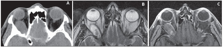 Figure 1. Case 1 Imaging findings in a 56-year-old patient with an orbital lesion at right. A: Non-contrast-enhanced computed tomography demonstrating isointense retrobulbar, ovoid lesion with regular contours at right, in close contact with the apex of the orbit. B,C: Magnetic resonance imaging demonstrating the same lesion medially displacing the extraocular muscles, with heterogeneous high signal intensity on T2-weighted sequence (B), and enhancement after paramagnetic contrast injection in the areas with lower signal intensity on T2-weighted sequences (C). Case 2 - A male, 32-year-old patient with no previous history of cancer, complaining of right ocular proptosis for six months. During the investigation with facial CT, an intraorbital expansile process was diagnosed (Figure 2). At physical examination, the patient presented with no visual complaint and with normal external ocular movements. Orbital MRI (Figure 2) demonstrated the presence of an extraconal, expansile lesion at right in the lower and lateral portions of the orbit, displacing the inferior and lateral rectus muscles, with intraconal extension and with no clear involvement of the optic nerve whose morphology and signal intensity were normal. The lesion was well delimited, presenting with a low-signal halo at all the sequences, with high signal intensity on T1-weighted sequences, intermediate signal intensity on T2-weighted sequences and intense paramagnetic contrast uptake. 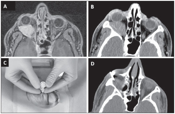 Figure 2. Case 2 A 32-year-old patient with an orbital lesion at right. A: Magnetic resonance imaging demonstrating extraconal lesion at right, with regular contours. Anterior displacement of the ocular globe and medial displacement of the extraocular muscles and optic nerve, with intense enhancement after paramagnetic contrast injection. B: Noncontrast enhanced computed tomography demonstrating the same lesion. C,D: Positioning of the coaxial needle by inferior transpalpebral approach (C) and tomographic section confirming the location of the needle end adjacent to the lesion (D). Both patients were submitted to CT-guided percutaneous biopsy of their orbital lesions, under local anesthesia and conscious sedation. The procedures were performed by means of the coaxial method, with transpalpebral lateral insertion of a 17-gauge needle between the ocular globe and the major sphenoid wing in a pathway paralleling the optic nerve (case 1), and by lower transpalpebral approach, between the ocular globe and the zygoma, inferiorly to the lateral rectus muscle (case 2). After the coaxial needles positioning, 18-gauge cutting needles were inserted, and three to five specimens were collected after 1.5 cm triggering, respectively in cases 1 and 2. Both procedures were uneventful (Figures 2 and 3). The anatomopathological results demonstrated schwannoma with no sign of malignancy and intermediate melanocytic neoplasm (melanocytoma), respectively, for cases 1 and 2. 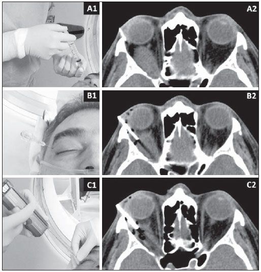 Figure 3. Case 1 Computed tomography-guided core biopsy of orbital lesion at right. A: Local anesthesia with 2% lidocaine in the subcutaneous tissue (A1) and tomographic section demonstrating the location and angulation of the needle (A2) for procedure planning. B: Positioning of coaxial needle by lateral transpalpebral approach (B1) and tomographic section confirming the location of the needle end adjacent to the lesion (B2). C: Insertion of the biopsy needle through the coaxial needle for samples collection (C1) and tomographic section demonstrating the presence of foci of gas within the lesion (C2), confirming that the samples were collected from this area. In both cases, the option was for surgical resection. In the case 1, right frontal craniotomy with orbitotomy was performed for radical resection of the tumor which was apparently related to the intraorbital trigeminal neural complex. In the case 2, lateral orbitotomy was performed with removal of the bone wall and resection with careful detachment of the lesion. Both patients did not present any postoperative complication, and the analyses of the surgical specimens demonstrated results coinciding with those from biopsies, confirming schwannoma in case 1 and melanocytoma in case 2. DISCUSSION Schwannoma is an uncommon, usually noninvasive tumor of the orbit, and is most frequently found in adult individuals, with an insidious onset. Its clinical presentation varies and may include optic neuropathy, proptosis and diplopia. The treatment is surgical(3). At CT, schwannomas are shown as retrobulbar ovoid lesions with regular contours, either intra- or extraconally located. At MRI, such tumors are generally isointense to the extraocular muscles on the T1-weighted sequences, with heterogeneous hypersignal on T2-weighted sequences and variable contrast enhancement. Areas of high signal intensity on T2-weighted sequences are associated with areas of cystic or mucinous degeneration(3,4). The definitive diagnosis is hardly based just on imaging findings considering the superimposition of findings with other orbital tumors. Thus, in most cases the lesion is diagnosed after surgical resection. However, in some cases, the preoperative diagnosis is essential for an appropriate therapeutic planning. Melanocytomas are rare pigmented tumors of the central nervous system, most frequently found in metastatic diseases. Most common locations of such tumors include the posterior fossa and the spinal cord. Orbital involvement is rarely found. At MRI, the imaging finding of melanocytomas is hypersignal on T1- and T2-weighted sequences, with marked enhancement after paramagnetic contrast injection. It is important to differentiate primary melanocytic lesions from those of metastatic origin, considering that they require different therapeutic approaches. The absence of a known malignant melanoma is useful in the differentiation of such tumors, and a complete investigation of these patients is essential in the search for a diagnosis(5,6). In this context, imaging-guided biopsy may be useful for collecting appropriate material for histological analysis, with less morbidity than surgical biopsy. Both fine needle aspiration biopsy (FNAB) and core biopsy have already been described for the diagnosis of orbital lesions(7,8). The advantages of the US-guided as compared with the CT-guided procedure include absence of ionizing radiation and real-time monitoring of the needle positioning. However, studies involving CT-guided procedures have not demonstrated a higher number of complications, and the Access to the lesion may be easier, depending upon the lesion location(8-11). Chojniak et al. have demonstrated that core biopsies present better results than FNABs in the collection of appropriate material and determination of specific diagnoses, without increasing the number of complications(9). In a study developed by Gupta et al.(7), evaluating 37 US-guided FNABs of orbital tumors, the samples were insufficient for the diagnosis in 22% of cases. However, few reports in the literature report the use of CT-guided core biopsy for the diagnosis of orbital lesions(8,10). In the literature, the first reports about core biopsy of orbital lesions occurred in the nineties(11,12). In spite of this procedure being uncommonly requested, considering that most of times the diagnosis is suggested by imaging findings, our small experience demonstrates that the procedure is feasible and well tolerated by the patients. Retrobulbar hemorrhage is the most common complication, but most of times it is small and reabsorbed without sequelae(8). Reports about more severe complications from imaging (either US or CT) guided procedures such as ocular globe and optic nerve injuries are not found in the literature. Such procedures, provided they are performed by radiologists with experience in percutaneous procedures and core biopsy, do not offer major risks since the needle pathway can be appropriately controlled by CT. Some techniques may be utilized to reduce the risk for complications, such as the utilization of lower caliber needles (for example, 20 gauge) and short-reach triggering (1.5 cm), which also produce satisfactory diagnoses in most cases. In a study developed by Yarovoy et al.(8), evaluating 50 core biopsies of orbital tumors utilizing 18- and 20-gauge needles (11 US-guided biopsies and the other procedures without imaging guidance), only two patients presented mild retrobulbar hematoma, with no visual acuity impairment. Additionally, no case of ocular globe or optic nerve injuries, impairment of ocular movements or inflammation was reported. In such study, the histopathological diagnosis was successfully established in 94% of the procedures(8). In both cases described in the present report, the decision to perform CT-guided core biopsy was based on multidisciplinary discussion with the assisting physicians, including oncologists and surgeons. The option for core biopsy and not FNAB was made due to the non-availability of a cytopathologist in the room during the procedure to confirm the presence of sufficient material for analysis, besides the great professional experience of the interventional radiology team in the service. In the specific case of suspicion of lymphoma in an orbital lesion, the histological evaluation is preferred for an appropriate diagnosis and tumor classification, considering the high number of divergent results in FNAB(13). Finally, in this small sample, CT-guided core biopsy was a safe and effective alternative for collection of material for histological analysis of selected orbital lesions in cases where a preoperative diagnosis was relevant for an appropriate therapeutic planning. However, one should ever take the cost/benefit ratio of the procedure into consideration, exhausting all the possibilities of diagnosis by means of noninvasive methods before indicating biopsy. REFERENCES 1. Chojniak R, Pinto PNV, Tyng CJ, et al. Computed tomography-guided transthoracic needle biopsy of pulmonary nodules. Radiol Bras. 2011;44:315-20. 2. Chojniak R, Grigio HR, Bitencourt AGV, et al. Percutaneous computed tomography-guided core needle biopsy of soft tissue tumors: results and correlation with surgical specimen analysis. Radiol Bras. 2012;45:259-62. 3. Kapur R, Mafee MF, Lamba R, et al. Orbital schwannoma and neurofibroma: role of imaging. Neuroimaging Clin N Am. 2005;15:159-74. 4. Rawlings NG, Brownstein S, Robinson JW, et al. Orbital schwannoma: histopathologic correlation with magnetic resonance imaging. Can J Ophthalmol. 2007;42:326-8. 5. Smith AB, Rushing EJ, Smirniotopoulos JG. Pigmented lesions of the central nervous system: radiologic-pathologic correlation. Radiographics. 2009;29:1503-24. 6. Mathai AM, Naik R, Pai MR, et al. Orbital melanocytoma. Orbit. 2008;27:383-7. 7. Gupta S, Sood B, Gulati M, et al. Orbital mass lesions: US-guided fine-needle aspiration biopsy - experience in 37 patients. Radiology. 1999;213:568-72. 8. Yarovoy AA, Bulgakova ES, Shatskikh AV, et al. CORE needle biopsy of orbital tumors. Graefes Arch Clin Exp Ophthalmol. 2013;251:2057-61. 9. Chojniak R, Isberner RK, Viana LM, et al. Computed tomography guided needle biopsy: experience from 1,300 procedures. Sao Paulo Med J. 2006;124:10-4. 10. Ortiz O, Bastug D, Ellis B. CT-guided percutaneous lateral suprazygomatic approach for posterior orbital wall biopsy. Skull Base Surg. 1996;6:249-51. 11. Shields JA, Shields CL. Biopsy techniques for orbital tumors. Int Ophthalmol Clin. 1993;33:175-80. 12. Alter C, Heywang-Köbrunner SH, Beck R. Diagnosis of intraorbital meningioma. Aktuelle Radiol. 1996;6:232-4. 13. Landgren O, Porwit MacDonald A, Tani E, et al. A prospective comparison of fine-needle aspiration cytology and histopathology in the diagnosis and classification of lymphomas. Hematol J. 2004;5:69-76. 1. Master, Titular Physician and Responsible for the Unit of Percutaneous Intervention, Imaging Department at A.C.Camargo Cancer Center, São Paulo, SP, Brazil 2. Master, Fellow, Imaging Department, A.C.Camargo Cancer Center, São Paulo, SP, Brazil 3. PhD, Titular Physician at Imaging Department, A.C.Camargo Cancer Center, São Paulo, SP, Brazil 4. MD, Fellow, Division of Ophthalmology, Hospital do Servidor Público Estadual, São Paulo, SP, Brazil 5. MD, Fellow, Imaging Department, A.C.Camargo Cancer Center, São Paulo, SP, Brazil 6. Master, Titular Physician and Responsible for the Unit of Computed Tomography, Imaging Department at A.C.Camargo Cancer Center, São Paulo, SP, Brazil 7. PhD, Titular Physician and Director, Department of Imaging, A.C.Camargo Cancer Center, São Paulo, SP, Brazil Mailing Address: Dr. Almir Galvão Vieira Bitencourt Rua Antônio Prudente, 211, Liberdade São Paulo, SP, Brazil, 01509-010 E-mail: almirgvb@yahoo.com.br Received August 20, 2013. Accepted after revision January 17, 2014. Study developed at A.C.Camargo Cancer Center, São Paulo, SP, Brazil. |
|
Av. Paulista, 37 - 7° andar - Conj. 71 - CEP 01311-902 - São Paulo - SP - Brazil - Phone: (11) 3372-4544 - Fax: (11) 3372-4554