Radiologia Brasileira - Publicação Científica Oficial do Colégio Brasileiro de Radiologia
AMB - Associação Médica Brasileira CNA - Comissão Nacional de Acreditação
 Vol. 47 nº 2 - Mar. / Apr. of 2014
|
REVIEW ARTICLE
|
|
The carpal boss: a review of different sonographic findings |
|
|
Autho(rs): Carlos Frederico Arend |
|
|
Keywords: Ultrasonography; Carpal boss; Cyst; Ganglion; Wrist. |
|
|
Abstract: INTRODUCTION
Palpable masses on the dorsum of the wrist are very frequently found in the clinical practice and generally result from cysts which present as a primarily cosmetic disorder, with no functional repercussion, although occasionally they may cause some discomfort. Their clinical presentation, however, is nonspecific and the presumptive clinical diagnosis of dorsal cyst may impair and delay the identification of other less prevalent lesions such as, for example, carpal boss. Such a differentiation is important, considering that a specific management results in higher chances of clinical complaint resolution, mostly because failure in attempts to blindly perform needle aspiration puncture of the presumed cyst may result in injury by the needle and iatrogenic worsening of the carpal boss. In such a context, ultrasonography is a useful adjuvant tool since it allows for a specific diagnosis and helps to appropriately guide procedure(1). The present study is aimed at reviewing the different sonographic presentations of carpal boss. PHYSIOPATHOGENESIS Carpal boss is a bony prominence located on the dorsum of the hand, originally described as carpe bossu by the French surgeon Fiolle(2). The actual incidence of such a condition is unknown, certainly underestimated and frequently confused with other causes of tumor-like masses in the dorsal carpal bone, but still admittedly most common in the right hand, between the third and fourth decades of life, and undefined predilection for gender. Physiopathologically, carpal boss may represent degenerative osteophytes in the carpometacarpal joint of the second or third digit and/or the presence of os styloideum, an accessory ossicle that is formed at the embryonic stage of life(3,4). Such ossicle, which was firstly described by Saltzmann in 1725, is dorsally located between the trapezoid, the capitate, second and third metacarpal bones(5). In only 2% of cases, the os styloideum is completely isolated from the circumjacent bones, and is more commonly fused with the second or third metacarpal bones, which occurs in 94% of the individuals(6). CLINICAL PRESENTATION Although carpal boss is classified into acquired (caused by osteophytes), congenital (due to the presence of os styloideum), or mixed (caused by a combination of osteophytes and os styloideum), the clinical manifestation seems not to diverge from one group to another(7). In most cases, carpal boss presents itself as a merely cosmetic disorder, with no functional repercussion, although it may occasionally cause discomfort. In symptomatic patients with the acquired or mixed presentation of the condition, local pain is usually caused by the degenerative alterations characteristic of the lesion. In the other individuals, the complaint is frequently resulting from the development of dorsal cysts, neobursae or frictional tendinopathy(3,7,8). The degenerative presentation is the most common(9) and the congenital presentation is the most rare, because the presence of the os styloideum interfere affects the usual biomechanics of the adjacent joints, potencializing the development of secondary degenerative changes, generating the mixed presentation of the condition(10). In the authors' experience, a relevant group of their patients is constituted of boxers due to the repeated mechanical overload they apply over the metacarpal joints of the second and third digits during the punch movement, predisposing such joints to osteoarthrosis(11). From the clinical point of view, the main obstacle to the recognition of the carpal boss is the nonspecificity of the symptoms frequently attributed to dorsal cysts, considering that both share a very similar location on the carpal bone(1). In fact, the differentiation between carpal boss and dorsal cyst by means of a physical examination is frequently difficult, if not impossible. The stony consistency of the mass is not a reliable indicator for the diagnosis of bone lesion, considering that cysts frequently present as hard nodules and tense content. Also, both types of lesion tend to be clinically exacerbated by wrist flexion and, in symptomatic cases, there is local discomfort which intensifies with manual activity and decreases with rest. Transillumination may be a useful adjuvant tool in the process of differentiation, but cystic lesions should have a minimum size to be appropriately evaluated by this technique and, sometimes, such a minimum size is much larger than the size of the lesions observed in the clinical practice. The anamnesis may also contribute in such cases, as cysts typically present cyclic variations, increasing, decreasing or even disappearing, while carpal boss remains stable, with no report of spontaneous remission(12). In dubious cases, supplementary imaging evaluation allows for specific diagnosis which is useful to appropriately guide the approach to be adopted. However, routine radiographic images usually utilized in the assessment of the wrist are not appropriate to demonstrate the boss. A specific lateral view acquired with a supine hand position at 30º and minimal ulnar deviation is preferable for this purpose(13). SONOGRAPHIC DIAGNOSIS In the authors' routine, the assessment is performed with the patient's hand comfortably resting over the examination table, with a single focal zone adjusted for the carpal bone depth. Usually it is necessary to apply a generous amount of conducting gel over the skin surface in order to bring the region of interest into the focal zone. Then, the transducer is longitudinally positioned over the palpable bulging (Figure 1), so the bone prominence is readily detected, whether it is degenerative (Figure 2), congenital (Figure 3) or mixed (Figure 4). Once the diagnosis is established, the management tends to conservative, with anti-inflammatory drugs and eventual immobilization. Surgical excision is reserved for those refractory cases. 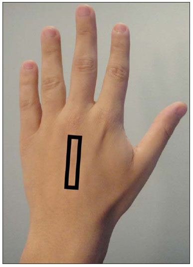 Figure 1. Transducer positioning for longitudinal carpal boss evaluation. 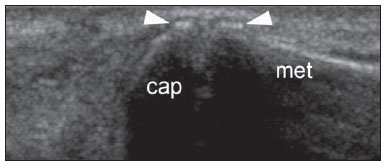 Figure 2. Carpal boss (acquired presentation). Longitudinal image demonstrating the capitate bone (cap), metacarpal bone (met) and osteophytes on the margins of the metacarpal joint (arrowheads), the latter, secondary to osteoarthrosis. The bone prominence determined by the osteophytes characterizes the acquired carpal boss. 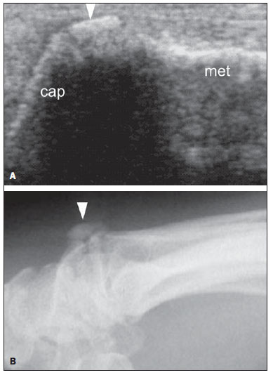 Figure 3. Carpal boss (congenital presentation). A: Longitudinal image demonstrating os styloideum (arrow head), corresponding to the palpable clinical finding. Also, observe the capitate (cap) and metacarpal (met) bones forming the carpometacarpal joint. B: Lateral radiography confirming the presence of an accessory ossicle. 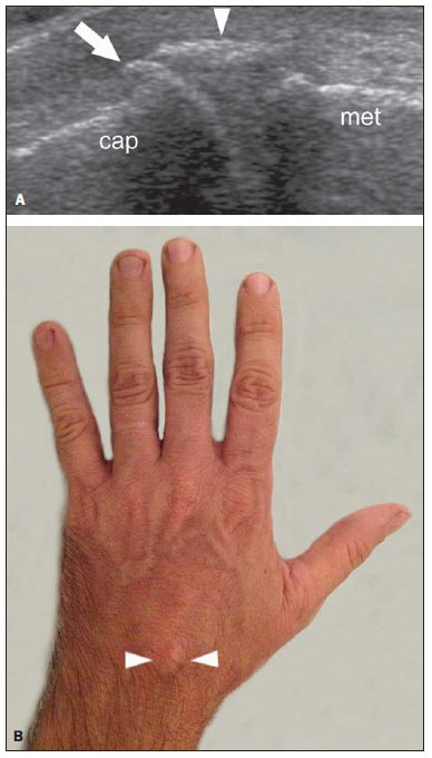 Figure 4. Carpal boss (mixed presentation). A: Longitudinal image demonstrating the capitate bone (cap), the metacarpal bone (met) and the osteophyte on the carpal margin of the carpometacarpal joint (arrow). Also, note the presence of os styloideum (arrowhead). The combination of acquired and congenital findings characterizes the mixed presentation of carpal boss. B: Clinical presentation demonstrating a prominence of stony consistency on the dorsal aspect of the metacarpophalangeal joint of the third digit (arrowheads). CONCLUSION Carpal boss is an uncommon entity, but its incidence is underestimated at clinical evaluation and is frequently confused with dorsal cysts or other causes of development of tumor-like masses on the dorsal aspect of the carpal bone. Supplementary evaluation with ultrasonography allows for fast diagnosis, appropriately guiding the approach to be adopted. REFERENCES 1. Arend CF. MASTER Ultrassonografia musculoesquelética. 2ª ed. São Paulo: Revinter; 2012. 2. Fiolle J. Le "carpe bossu". Bull Mem Soc Natl Chir. 1931;57:1687-90. 3. Cuono CB, Watson HK. The carpal boss: surgical treatment and etiological considerations. Plast Reconstr Surg. 1979;63:88-93. 4. Zimmer EA. Eine krankhafte Veränderung am Os styloideum. Fortschr Geb Rontgenstr. 1940;61:187-92. 5. Grumbach A. Das Handskelett im Lichte den Röntgenstrahlen. Vienna: Braumüller, 1921. 6. Bizzaro AH. On sesamoid and supernumerary bones of the limb. J Anat. 1921;55(Pt 4):256-68. 7. Conway WF, Destouet JM, Gilula LA, et al. The carpal boss: an overview of radiographic evaluation. Radiology. 1985;156:29-31. 8. Koostra G, Huffstadt JC, Kauer JM. The styloid bone. A clinical and embryological study. Hand. 1974;6:185-9. 9. Zanetti M, Saupe N, Nagy L. Role of MR imaging in chronic wrist pain. Eur Radiol. 2007;17:927-38. 10. Park MJ, Namdari S, Weiss AP. The carpal boss: review of diagnosis and treatment. J Hand Surg Am. 2008;33:446-9. 11. Melone CP Jr, Polatsch DB, Beldner S. Disabling hand injuries in boxing: boxer's knuckle and traumatic carpal boss. Clin Sports Med. 2009;28:609-21. 12. Kissel P. Conservative management of symptomatic carpal bossing in an elite hockey player: a case report. J Can Chiropr Assoc. 2009;53:282-9. 13. Bhat AK, Kumar B, Acharya A. Radiographic imaging of the wrist. Indian J Plast Surg. 2011;44:186-96. MD, Radiologist, Radimagem Diagnóstico por Imagem, Porto Alegre, RS, Brazil Mailing Address: Dr. Carlos Frederico Arend Avenida Cristóvão Colombo, 1691, Floresta Porto Alegre, RS, Brazil, 90560-004 E-mail: carlos_arend@hotmail.com Received May 7, 2013. Accepted after revision July 22, 2013. * Study developed at Radimagem Diagnóstico por Imagem, Porto Alegre, RS, Brazil. |
|
Av. Paulista, 37 - 7° andar - Conj. 71 - CEP 01311-902 - São Paulo - SP - Brazil - Phone: (11) 3372-4544 - Fax: (11) 3372-4554