Radiologia Brasileira - Publicação Científica Oficial do Colégio Brasileiro de Radiologia
AMB - Associação Médica Brasileira CNA - Comissão Nacional de Acreditação
 Vol. 46 nº 2 - Mar. / Apr. of 2013
|
ORIGINAL ARTICLE
|
|
Estimation of MSAD values in computed tomography scans using radiochromic films |
|
|
Autho(rs): Bruno Beraldo Oliveira1; Arnaldo Prata Mourão2; Teógenes Augusto da Silva3 |
|
|
Keywords: Kerma profiles; Computed tomography; Radiochromic films; MSAD. |
|
|
Abstract: INTRODUCTION
The main objective of patient dosimetry in computed tomography (CT) is determining the dosimetric quantities for the establishment and utilization of the diagnostic reference levels (DRL) in addition to risk assessment. In many situations it is desirable to make measurements directly; however, it is preferable to utilize a patient phantom(1). In the clinical practice, the doses to patients are specified on the basis of measurements performed on standard phantoms for CT dosimetry. The kerma index (Ca,100) and the weighted kerma index (CW) are the basic dosimetric quantities utilized in CT. The Ca,100, measured in the air for a single rotation of the CT X-ray tube, is the quotient of the integral of air kerma along a line parallel to the rotation axis for a length of 100 mm and the nominal slice thickness. The notation CPMMA,100 is utilized for measurements performed within polymethylmethacrylate (PMMA) phantoms. The CW combines the values of CPMMA,100 measured in the center with the mean value of the four peripheral positions of the phantom. The CW is utilized in combination with the scanning parameters to calculate volumetric kerma index (CVOL), which provides a better mean value of the volume. The air kerma length product (PKL,CT) is determined for a complete CT scan, and is analogous to the dose length product introduced by the European Commission(2). Multiple scan average dose (MSAD) has also been utilized for dosimetry in Brazilian CT apparatuses, and is intimately related to CVOL(1). In compliance with international recommendations(4), the Brazilian regulations have included DRL in terms of MSAD. The following values are adopted: 50 mGy for the head, 35 mGy for the lumbar spine and 25 mGy for the abdomen in a typical adult(3). The DRLs must be utilized for quality control of CT apparatuses in order to optimize the procedures as the doses exceed the specified values(3). The relevance of improving the methods of dosimetry and radiological protection has been object of a number of recent studies published by Brazilian authors in different areas of knowledge(5-11). In CT, measurements of dosimetric quantities are performed by means of a calibrated pencil-type ionization chamber. Previous studies have also suggested the utilization of thermoluminescent dosimeters (TL)(12,13) and radiochromic films(12,14-16) in order to obtain kerma profiles and calculate the MSAD values from such alternative materials for dosimetry in CT. Several other studies(17-20) have established the response to the dose and to the energy dependence of the radiochromic films, XR-CT model, designed for measurements in CT. The present study proposes estimating the MSAD of three standard imaging protocols for CT abdomen studies, with different pitch values, in order to verify the viability of radiochromic films as an alternative to ionization chamber and TL dosimeters. Results for MSAD calculated from kerma profiles were compared with the values established by the Brazilian regulations, with the purpose of undertaking a critical analysis of such DRL. MATERIALS AND METHODS The measurements were performed with Gafchromic XR-CT radiochromic films in 100 mm x 9 mm strips, in a four-channel GE Bright Speed apparatus located at a hospital in the city of Belo Horizonte, MG, Brazil. The metrological reliability of radiochromic films was demonstrated by means of tests of homogeneity and repeatability and by their calibration at a specific CT reference radiation (RQT9), reproduced in the Calibration Laboratory of Centro de Desenvolvimento da Tecnologia Nuclear (CDTN/CNEN). The film calibrations and measurements were obtained in an abdomen PMMA phantom with 320 mm in diameter and 150 mm in length, with five parallel bores (one in the center and the others peripherally located, at 3h00, 6h00, 9h00 and 12h00 positions. For the calibration, radiochromic film strips were inserted into the chamber cap(16) and positioned inside the phantom bores, reproducing exactly the position of the 10X5-3CT 100 mm pencil-type ionization chamber (Radcal Corporation) calibrated according to the established procedures(21). The obtained results were compared with the purpose of obtaining the respective calibration coefficients. The phantom with the inserted radiochromic films was positioned at the isocenter of the CT apparatus (Figure 1). 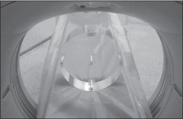 Figure 1. Positioning of the phantom at the isocenter of the CT scanner. A topogram (scout view) was acquired with the purpose of verifying the materials positioning and limiting the irradiation area to the length of the phantom. A single scanner rotation was selected and the irradiation was performed according to the standard protocol for adult abdomen in the center of the chamber cap, i.e., where the sensitive region of the ionization chamber would be. The following parameters were utilized: 120 kV, 4 x 2.5 mm slices and 242.4 mA.s. For the measurements, the chamber cap was replaced by new radiochromic film strips positioned along the PMMA cylinders (Figure 2) and distributed in the center and four peripheral bores of the phantom, according to the following parameters: I) 120 kV, 4 x 2.5 mm slices, 242.4 mA.s and 0.75 pitch; II) 120 kV, 4 x 2.5 mm slices, 240.0 mA.s and 1.5 pitch; III) 120 kV, 2 x 0.625 mm slices, 80.0 mA.s and 1.00 pitch. Such parameters were based on a study developed in the hospital itself aimed at changing imaging protocols in order to reduce the radiation dose, and to utilize a pitch equal to 1.00. 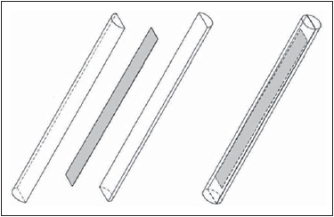 Figure 2. Positioning of the radiochromic film in the PMMA cylinder. The exposed films were digitized with a Microtek ScanMaker 9800XL scanner in the reflection mode. Digitization parameters were the following: RGB color mode (48 bits per color) and 300 dpi. The red channel was selected for calibration because such radiochromic films have a main absorption peak in the red region of the visible spectrum (636 nm). The MSADs were calculated from the values of the defined integrals obtained in the center of the phantom for the three protocols with different pitch values. The utilized integration limits corresponded to the length of the radiochromic films (100 mm). RESULTS All measurements were converted into kerma values in the PMMA, utilizing the ratio between the attenuation coefficients of air and of the PMMA, corresponding to approximately 1.07(22). The radiochromic films presented homogeneity of 6.9% and repeatability of 4.3%. Their calibration coefficients in terms of air kerma for RQT9 (120 kV) are shown in Table 1. 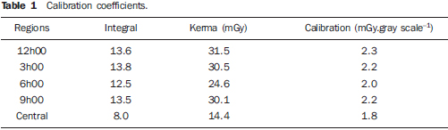 The kerma profiles along the longitudinal axis, in the five regions where the cylinders were inserted in the phantom's bores, utilizing three different scanning parameters, are shown in Figures 3 to 7. The relative expanded uncertainty (k = 2.03) for the measurements utilizing radiochromic films is approximately 5.2%. The calculated MSAD values were 13.6 ± 0.7, 13.5 ± 0.7 and 18.7 ± 1.0 mGy for the three imaging protocols with pitch values of 0.75, 1.00 and 1.50, respectively. 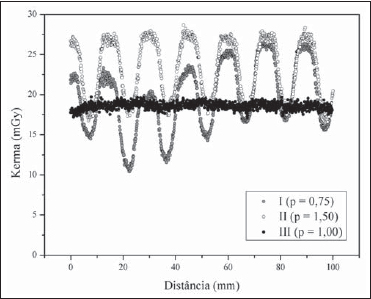 Figure 3. Kerma profiles of the 12h00 region for three different parameters. 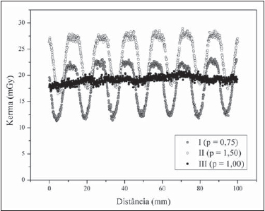 Figure 4. Kerma profiles of the 3h00 region for three different parameters. 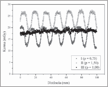 Figure 5. Kerma profiles of the 6h00 region for three different parameters. 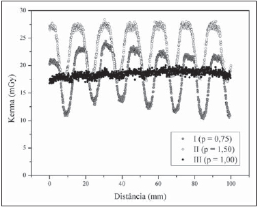 Figure 6. Kerma profiles of the 9h00 region for three different parameters. 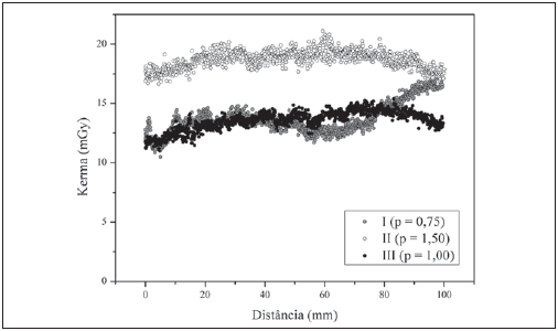 Figure 7. Kerma profiles of the central region for three different parameters. DISCUSSION The Gafchromic XR-CT radiochromic films constitute a good alternative in dose evaluations. Such films are not sensitive to visible light and darken as exposed to ionizing radiation, the change in the color tone being associated with the radiation dose(15,20). Such characteristic facilitates the visualization of irradiation regions, on account of different pitch values utilized in the standard imaging protocols. Oscillatory curves with pulse heights of up to 10 mGy were observed in the peripheral measurements at the phantom, depending upon the pitch value utilized in the protocols. The peripheral regions of the phantom presented higher kerma values than the central region, because of the high attenuation of the x rays through the phantom. The region at 6h00 presented the lowest dose, as compared with other peripheral regions, due to the beam attenuation caused by the table of the CT scanner. The MSAD values are associated with the characteristics of the apparatuses, besides their specific performances in the quality control tests and protocols adopted in each service(23). The obtained MSADs demonstrate that a dose reduction occurs as the protocol I is adopted instead of protocol II, mainly due to the utilized pitch value (pitch = 0.75). As the protocol III was adopted, the dose value decreases a little further on account of the new pitch value (pitch = 1.00) and mainly on account of the tube loading value, extending the X-ray tube useful life, thus reducing costs for the institution(24). It is clear that CT scan protocols should utilize reduced values for voltage and tube loading, provided the image quality is maintained to allow a safe medical diagnosis utilizing dose values as low as reasonably achievable(3,25). In spite of the fact that the calculated MSAD values resulted < 25 mGy for DRL established by the Brazilian regulations for abdomen studies(3), it is suggested that there is room for procedure optimization, also considering the need for an appropriate evaluation of image quality for each protocol. CONCLUSIONS The advantages of the radiochromic films for measurements of kerma profile are the following: continuous reading, which facilitates the observation of the oscillatory curves on account of different pitch values; ease of manipulation, which decreases the need for long interruptions in analyses of the clinical practice; and the acceptable uncertainty (~ 5.2%). Therefore such materials seem to be appropriate for CT quality control, in spite of their high cost and non-reusability. The obtained MSAD values suggest the need for reviewing the DRL for abdomen studies, as its value is very high and it is possibly not representing the reality of the practice in Brazil. The MSAD values also suggest that it would be better to adopt a standard imaging protocol with a pitch value equal or close to 1 (one) and reduced tube loading values in order to reduce the doses, avoid differently irradiated regions and increase the X-ray tubes useful life. It would be more appropriate to adopt, together with the established MSAD values, the DRLs in terms of the other CT dosimetric quantities. The knowledge of such values would be important for the optimization of clinical procedures, besides facilitating the comparison with internationally recommended values. Considering the relevance of optimization, in addition to the calculation of dosimetric quantities, an evaluation of the image quality for each protocol should be made. As radiochromic films have not yet been utilized for dosimetry in radiology in Brazil, the present study contributes to the dissemination of the procedure for calculation of the MSADs from kerma profiles measured with radiochromic films among hospitals and radiologists. Acknowledgements The authors wish to thank Coordenação de Aperfeiçoamento de Pessoal de Nível Superior (Capes), for the Master's scholarship granted to Bruno Beraldo Oliveira. The present study was supported by Instituto Nacional de Ciência e Tecnologia (INCT) em Metrologia das Radiações em Medicina. REFERENCES 1. International Atomic Energy Agency. Dosimetry in diagnostic radiology: an international code of practice. Technical Reports Series no. 457. Vienna: IAEA; 2007. 2. European Commission. European guidelines for quality criteria for computed tomography. EUR 16262 EN. Luxembourg: European Commission; 2000. 3. Brasil. Ministério da Saúde. Secretaria de Vigilância Sanitária. Portaria nº 453, de 01 de junho de 1998. Diretrizes de proteção radiológica em radiodiagnóstico médico e odontológico. Brasília, DF: Diário Oficial da União; 02 de junho de 1998. 4. International Atomic Energy Agency. International basic safety standards on protection against ionizing radiation and for safety of radiation sources. Safety Series No. 115. Vienna: International Atomic Energy Agency; 1996. 5. Silva MSR, Khoury HJ, Borrás C, et al. Dosimetria de pacientes e médicos em intervenções coronárias percutâneas em Recife, Pernambuco, Brasil. Radiol Bras. 2011;44:90-6. 6. Soares FAP, Pereira AG, Flôr RC. Utilização de vestimentas de proteção radiológica para redução de dose absorvida: uma revisão integrativa da literatura. Radiol Bras. 2011;44:97-103. 7. Sabino T, Rodrigues LN, Furnari L, et al. Caracterização dosimétrica de feixes de fótons com detector de diamante. Radiol Bras. 2012;45:35-8. 8. Hoff G, Andrade JRM, Fischer ACFS, et al. Ferramenta computacional para avaliação de kerma no ar em aplicações de radioproteção em áreas de internação de pacientes: proposição de um método simples para avaliação experimental. Radiol Bras. 2012;45:71-81. 9. Figueiredo FM, Gama ZAS. Melhoria da proteção radiológica mediante um ciclo de avaliação interna da qualidade. Radiol Bras. 2012;45:87-92. 10. Navarro VCC, Navarro MVT, Maia AF, et al. Avaliação de exposições médicas em procedimentos pediátricos de radiologia intervencionista. Radiol Bras. 2012;45:210-4. 11. Rodrigues S, Abrantes AF, Ribeiro LP, et al. Estudo da dose nos exames de tomografia computadorizada abdominal em um equipamento de 6 cortes. Radiol Bras. 2012;45:326-33. 12. Oliveira BB, Mourão AP, Da Silva TA. Kerma profile measurements in CT chest scans - a comparison of methodologies. World Academy of Science, Engineering and Technology. 2011;80:88. 13. Tsai HY, Tung CJ, Huang MH, et al. Analyses and applications of single scan dose profiles in computed tomography. Med Phys. 2003;30:1982-9. 14. Gorny KR, Leitzen SL, Bruesewitz MR, et al. The calibration of experimental self-developing Gafchromic HXR film for the measurement of radiation dose in computed tomography. Med Phys. 2005;32:1010-6. 15. Martin CJ, Gentle DJ, Sookpeng S, et al. Application of Gafchromic film in the study of dosimetry methods in CT phantoms. J Radiol Prot. 2011;31:389-409. 16. Rampado O, Garelli E, Ropolo R. Computed tomography dose measurements with radiochromic films and a flatbed scanner. Med Phys. 2010;37:189-96. 17. Butson MJ, Cheung T, Yu PKN. Measurement of energy dependence for XRCT radiochromic film. Med Phys. 2006;33:2923-5. 18. Butson MJ, Cheung T, Yu PKN. Absorption spectra of irradiated XRCT radiochromic film. Phys Med Biol. 2006;51:3099-103. 19. Butson MJ, Cheung T, Yu PKN, et al. Dose response of irradiated XRCT radiochromic film. Iran J Sci Technol Trans A. 2008;32:161-5. 20. Liao YL, Kao HC, Chuang KS, et al. CT dose estimation using Gafchromic XR-CT film: models comparison. Radiat Meas. 2011;46:2052-5. 21. Maia AF, Caldas LVE. Calibração das câmaras de ionização para feixes de tomografia computadorizada no Brasil: a realidade atual. Radiol Bras. 2006;39:209-13. 22. National Institute of Standards and Technology. NIST Physical Measurement Laboratory. [acessado em 15 de setembro de 2010]. Disponível em: http://www.nist.gov/pml/data/xraycoef/index.cfm. 23. Pina DR, Duarte SB, Ghilardi Netto T, et al. Controle de qualidade e dosimetria em equipamentos de tomografia computadorizada. Radiol Bras. 2009;42:171-7. 24. Marconato JA, Mazzola AA, Sanvitto PC, et al. Redução de dose e aumento na vida útil do tubo de raios X em tomografia computadorizada. Radiol Bras. 2004;37:351-6. 25. Dalmazo J, Elias Júnior J, Brocchi MAC, et al. Otimização da dose em exames de rotina em tomografia computadorizada: estudo de viabilidade em um hospital universitário. Radiol Bras. 2010;43:241-8. 1. Master, Fellow PhD degree, Course of Sciences and Technology of Radiations, Minerals and Materials - Centro de Desenvolvimento da Tecnologia Nuclear/Comissão Nacional de Energia Nuclear (CDTN/CNEN), Belo Horizonte, MG, Brazil. 2. PhD, Professor, Centro Federal de Educação Tecnológica de Minas Gerais (CEFET-MG), Belo Horizonte, MG, Brazil. 3. PhD, Full Researcher Professor, Comissão Nacional de Energia Nuclear (CNEN), Belo Horizonte, MG, Brazil. Mailing Address: Bruno Beraldo Oliveira Centro de Desenvolvimento da Tecnologia Nuclear (CDTN/CNEN) Avenida Presidente Antônio Carlos, 6627, Campus UFMG, Pampulha Belo Horizonte, MG, Brazil, 31270-90 E-mail: bbo@cdtn.br Received October 2, 2012. Accepted after revision January 10, 2013. * Study developed in the Program of Post-graduation in Sciences and Technology of Radiations, Minerals and Materials - Centro de Desenvolvimento da Tecnologia Nuclear/Comissão Nacional de Energia Nuclear (CDTN/CNEN), Belo Horizonte, MG, Brazil. |
|
Av. Paulista, 37 - 7° andar - Conj. 71 - CEP 01311-902 - São Paulo - SP - Brazil - Phone: (11) 3372-4544 - Fax: (11) 3372-4554