Radiologia Brasileira - Publicação Científica Oficial do Colégio Brasileiro de Radiologia
AMB - Associação Médica Brasileira CNA - Comissão Nacional de Acreditação
 Vol. 45 nº 4 - July / Aug. of 2012
|
CASE REPORT
|
|
Complete renal fusion in a child with recurrent urinary tract infection |
|
|
Autho(rs): Saul Gun1; Guilherme Lippi Ciantelli2; Marília Akemi Uzuelle Takahashi2; Alexandre Mineto Brabo2; Lívea Athayde de Morais2; Caio Barros Figueiredo2 |
|
|
Keywords: Kidney; Malformation; Cake kidney. |
|
|
Abstract: INTRODUCTION
Cake kidney is a rare congenital abnormality of the genitourinary tract, with barely more than twenty cases described in the literature(1). The term "cake kidney" or fused pelvic kidney was defined by Glenn in 1958 as "an abnormality in which all the renal system tissue are fused into a single mass lying at the bottom of the pelvis and which two ureters drain separately into the vesical trigone"(2). The early diagnosis and the recognition of potential complications associated with such abnormality constitute relevant factors to prevent permanent renal injury(3). In the present paper, the authors report a case of cake kidney drained by two ureters diagnosed at a tertiary health care unit specialized in urology. CASE REPORT A 12-year-old male patient with a history of recurrent urinary tract infections since childhood was referred to the urology unit of a tertiary school hospital for investigation of a mass on right kidney topography discovered on a ultrasonography examination. In his city of origin, the patient had already been hospitalized three times, because of acute pyelonephritis. A previous pelvic ultrasound report brought by the patient demonstrated a mass in the right kidney and absence of the left kidney. Contrast-enhanced computed tomography of the pelvis was requested and demonstrated right-sided renal ectopia in the lower pelvis and presence of a fused cake kidney (Figure 1) drained by two distinct ureters (Figure 2), without any further alteration. 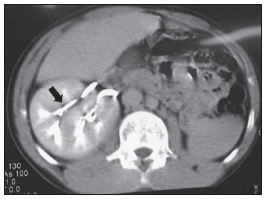 Figure 1. Computed tomography showing the presence of right-sided renal ectopia located in the lower pelvis, in association with absence of the left kidney and complete renal fusion with drainage by two distinct ureters. 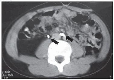 Figure 2. Computed tomography demonstrating right-sided renal fusion drained by two distinct ureters.. Considering the possibility of coexisting abnormalities, supplementary imaging studies were requested but no other alteration was found. Additionally, creatinine testing (0.6 mg/dl) and DMSA renal scan were performed and did not demonstrate renal function compromise. The patient is currently undergoing prophylactic treatment with nitrofurantoin and has not presented further episode of urinary tract infection and is asymptomatic since. DISCUSSION Cake kidney is a congenital abnormality defined as a complete fusion of both kidneys, representing only 2% of all renal fusion cases. Such abnormality may be diagnosed at any age range and likewise other renal fusion abnormalities, is most frequently found in men at a 2-3:1 ratio(4). Such abnormality occurs at the early phases of the embryonic development. Under normal conditions, two masses of metanephrogenic tissue existing in the lower pelvis develop until taking their definitive positioning in the lumbar region after complex movements involving lateral and ascending migration, axial deflection and internal rotation. It is believed that during the formation of a cake kidney, nephrogenic blastemas are compressed by the umbilical arteries at the beginning of the cranial migration of ureteral buds, which could be the cause of the fusion. The fused kidneys do not ascend as they should in a normal development, remaining in an ectopic pelvic position(5). Partial renal fusion is more frequently found, being principally represented by horseshoe kidney and crossed fused renal ectopia. Horseshoe kidneys correspond to 90% of all the renal abnormalities, with an incidence of up to 0.25%(1,4). Anatomically, the cake kidney presents a lobulated anterior aspect while the posterior facet is smooth and homogeneous(2). The renal pelvis is located anteriorly to the kidney and except for some few cases, there are two ureters which drain into the bladder in the normal anatomical regions of the vesical trigone(6). Such congenital abnormalities may present some histological alterations, namely: immature glomeruli; cystic changes; widening and dilatation of tubules; or even evidences of chronic renal disease(3). In other cases, there may be signs of infarct or ischemia secondary to a blood supply abnormality(4). This renal fusion abnormality can remain asymptomatic, or even be detected only in an autopsy exam(1,5). The presence of a cake kidney does not indicate a poor prognosis with kidney malfunction or possible progressive deterioration of its function. The follow up is important for the patient for early diagnosis of complications such as: obstruction, calculus, infection, hematuria and uremia. These conditions also can be present in other urinary tract fusions. Besides, it´s important to exclude other congenital abnormalities and perform constant evaluation of the renal function, which reduces the chances of associated morbidity and mortality(1,3,4). The vascular supply to the kidneys is compatible with their migration, and the arterial irrigation can be provided directly from the aorta in a region proximal to its bifurcation or from the common iliac arteries and the venous drainage usually occurs towards the distal segment of the inferior vena cava or to the common iliac veins(1,3). Such anomalous blood irrigation is considered a risk factor for renal vascular compromise due to pelvic trauma, vascular disease, pregnancy and atherosclerosis(7). Typically, the cake kidney coexists with other anomalies, such as: abnormal testicular descent, Tetralogy of Fallot, vaginal atresia, sacral agenesis, caudal regression syndrome, bifid spine and anal abnormalities(6). Such renal fusion anomaly may remain asymptomatic or even be detected at autopsy(1,5). But, in some cases, there may be infections secondary to the obstruction and calculosis or localized pain resulting from the renal vessels' traction caused by the weight of the organ; in other cases, a misdiagnosis of renal tumor may lead to unnecessary nephrectomy(5). The finding of a cake kidney is not indicative of a worse prognosis, renal dysfunction, or possible progressive renal function deterioration(1,3,4), but it is important that the patient is followed-up to allow the early diagnosis of possible complications such as obstruction, calculosis, infection, hematuria and uremia, which may also be present in other urinary tract fusions. Besides, ruling out other concomitant congenital abnormalities and perform constant evaluation of the renal function is important to reduce the associated morbidity. REFERENCES 1. Calado AA, Macedo Jr A, Srougi M. Cake kidney drained by single ureter. Int Braz J Urol. 2004; 30:321-2. 2. Glenn JF. Fused pelvic kidney. J Urol. 1958;80:7-9. 3. Türqkvatan A, Demir D, Olçer T, et al. Cake kidney: MDCT urography for diagnosis. Clin Imaging. 2006;30:420-2. 4. Kaufman MH, Findlater GS. An unusual case of complete renal fusion giving rise to a 'cake' or 'lump' kidney. J Anat. 2001;198(Pt 4):501-4. 5. Srivastava RN, Singh M, Ghai OP, et al. Complete renal fusion ("cake"/"lump" kidney). Br J Urol. 1971;43:391-4. 6. Goren E, Eidelman A. Pelvic cake kidney drained by single ureter. Urology. 1987;30:492-3. 7. Brock JW 3rd, Braren V, Phillips K, et al. Caudal regression with cake kidney and a single ureter: a case report. J Urol. 1983;130:535-6. 1. PhD, Associate Professor, Department of Surgery, Faculdade de Medicina de Sorocaba, Urologist at Conjunto Hospitalar de Sorocaba, Sorocaba, SP, Brazil. 2. Graduate Students (6th year) of Medicine, Faculdade de Ciências Médicas e da Saúde da Pontifícia Universidade Católica de São Paulo (FCMS/PUC-SP), Sorocaba, SP, Brazil. Mailing Address: Guilherme Lippi Ciantelli Avenida Moreira César, 39, ap. 112, Centro Sorocaba, SP, Brazil, 18010-010 E-mail: gui_lippi@hotmail.com Received January 27, 2012. Accepted after revision March 23, 2012. Study developed at Faculdade de Ciências Médicas e da Saúde da Pontifícia Universidade Católica de São Paulo (FCMS/ PUC-SP), Sorocaba, SP, Brazil. |
|
Av. Paulista, 37 - 7° andar - Conj. 71 - CEP 01311-902 - São Paulo - SP - Brazil - Phone: (11) 3372-4544 - Fax: (11) 3372-4554