Radiologia Brasileira - Publicação Científica Oficial do Colégio Brasileiro de Radiologia
AMB - Associação Médica Brasileira CNA - Comissão Nacional de Acreditação
 Vol. 45 nº 1 - Jan. /Feb. of 2012
|
ORIGINAL ARTICLE
|
|
Dosimetric characterization of photon beams using a diamond detector |
|
|
Autho(rs): Talita Sabino1; Laura Natal Rodrigues2; Laura Furnari3; Érika Yumi Watanabe4; Gisela Menegussi5 |
|
|
Keywords: Diamond; Dosimetry; Radiotherapy. |
|
|
Abstract: INTRODUCTION
In photon beam dosimetry, ideally a detector with high sensitivity, good spatial resolution, low energy dependence, good stability and tissue equivalence should be utilized. Frequently, ionization chambers are utilized; but other types of detectors, such as diamond detectors, may also present desirable characteristics for this type of dosimetry. The diamond detector is practically tissue-equivalent, since the atomic number of carbon and that of tissue are very similar. So, the signal from the detector can be considered as being directly proportional absorbed dose in tissue without the need to perform any correction(1,2). Additionally, such a detector is quite appropriate for relative absorbed dose measurements(3), because of its intrinsic characteristics which favor such type of measurement, namely: high sensitivity (approximately 0.5 mC/Gy), good spatial and temporal resolution, low energy dependence in photon and electron beams, good stability with temperature (better than approximately 1%.ºK1), and good irradiation stability, that is, it is practically insensitive to damage caused by radiation. It is important to highlight that some authors have introduced the utilization of synthetic diamonds, comparing their performances with that of one of the early natural diamond prototypes: they have described the synthetically developed crystal growth process(4,5). Most recently, some authors(6,7) investigated the utilization of natural diamonds in small field dosimetry, more specifically for the measurement of total scatter factor for the Cyberknife system and for dose measurements of radiation beams in intensity-modulated radiation therapy (IMRT), respectively. Other authors(8) have also analyzed the utilization of the diamond detector for determining the total scatter factor comparatively with semiconductor diodes. The detector is constituted by a 0.2 mm thick natural diamond plate inserted in a polymethyl-methacrylate capsule, at a depth of 1.0 mm, with a special contact system. The external dimensions of the detector are almost the same as those of the silicon diode, with a diameter of 7.3 mm and a length of 20 mm. The polarization voltage is +100 V. As soon as the voltage is applied to the detector, one recommends that an approximately 10 Gy pre-irradiation dose be applied to the detector so that its sensitivity reaches certain stability. MATERIALS AND METHODS The detector was vertically irradiated and a reference chamber was positioned at the border of the irradiation field in order to detect eventual linear accelerator output fluctuations. Subsequently, depth dose distributions were obtained by utilizing the automatic PTW MP3 scanning system. The measurements were performed at Hospital Alemão Oswaldo Cruz with a Varian 21EX linear accelerator on a 6 MV photon beam. In the determination of the depth dose distributions, the effective point of the detectors were positioned on the measurement points as follows: for the diamond, the effective point is located at 1.0 mm from the external surface, in the diamond center; for the 31010 PTW chamber (volume of 0.125 cm3, equivalent to the 31002 PTW chamber which is listed on the TRS 398 table), it is 1.7 mm above the central axis and at 4.5 mm from its extremity, according to the International Atomic Energy Agency (IAEA) protocol(9). The measurements were performed with the detectors being moved from greater to smaller depths, in order to eliminate the meniscus effect. The analysis of dose linearity parameters was based upon measurements performed at a rate of 300 cGy/min, field of 10 × 10 cm2 at a depth of 5.0 cm, and monitoring units ranging from 20 UM to 1000 UM. The dose-rate dependence was investigated by means of measurements performed on 5 × 5 cm2, 10 × 10 cm2 and 20 × 20 cm2 fields, at a depth of 5.0 cm with 50 UM and dose rates of 100 cGy/min, 200 cGy/min, 300 cGy/min and 400 cGy/min. Deep dose rates and profiles were determined on the transverse axis with 400 UM/min at a depth of 1.5 cm for the 1 × 20 cm2, 3 × 20 cm2, 10 × 20 cm2 and 20 × 20 cm2 fields, source-surface distance of 100 cm. RESULTS The diamond detector presented a linear behavior for the dose, with a correlation coefficient R2 = 99.99%. The dose-rate dependence was obtained by comparing the measurements with those performed with the model 31010 PTW ionization chamber, and by normalizing the results for a rate of 300 cGy/min, which is the most common dose rate utilized in radiotherapy (Figure 1). 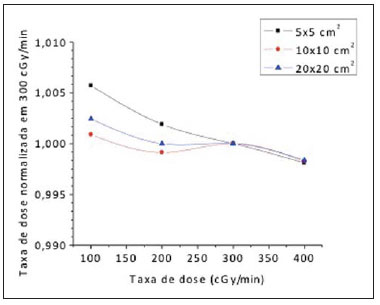 Figure 1. Values for dose rate dependence normalized at 300 cGy/min for all measured fields. The maximum observed variation for the dose-rate dependence was 0.57%, as shown on Table 1. Such dependence was established by comparison with the ionization chamber. 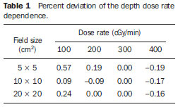 The diamond detector and the ionization chamber readings ratio for the 20 × 20 cm2 field normalized for a dose rate of 300 cGy/min are shown on Figure 2. 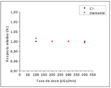 Figure 2. Comparison between diamond detector and ionization chamber responses. Obtained response with a CC125 chamber and with a diamond detector in relation to dose rate in the 20 × 20 cm2 field. In order to analyze the deep dose rates, in all field sizes, the charts were separately presented for the two detectors (Figure 3 for the ionization chamber and Figure 4 for the diamond detector). 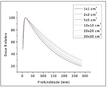 Figure 3. Depth dose rate measured with the ionization chamber. 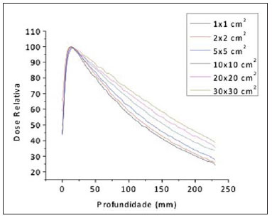 Figure 4. Depth dose rate measured with the diamond detector. As the depth-dose curves for the two detectors at Figure 5 are observed, one verifies that for a 10 × 10 cm2 field, both detectors presented a same behavior, with good agreement between the obtained values. The selection of the 10 × 10 cm2 field in this analysis is based on the IAEA(9) protocol which recommends such a field as a reference in dosimetry of high energy photon beams. This same behavior was observed for other field sizes. 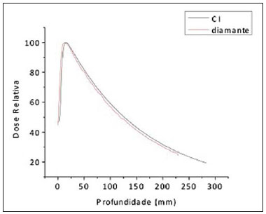 Figure 5. Depth dose rate for the ionization chamber and the diamond detector in a 10 × 10 cm2 field. Profiles were also obtained for the two detectors, with different field sizes for comparison, as shown on Figure 6. 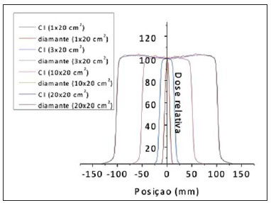 Figure 6. Dose profiles obtained with the ionization chamber and the diamond detector. DISCUSSION As regards the linear dose dependence, one observed good linearity of the diamond detector, with a correlation coefficient R2 = 99.99%, demonstrating the appropriateness of its utilization in all the clinically relevant ranges in radiotherapy. As the dose rate dependence results of the diamond detector are compared with those reported in the literature(10), a similar behavior was observed, but with a smaller variation, 0.5% instead of 2%. With the purpose of allowing a quantitative evaluation in the comparison between the diamond detector and the ionization chamber responses, an analysis was performed considering some specific depths typical for high energy photon beams, whose percentage differences are presented on Table 2. On such Table, one verifies that the differences between the diamond detector and the ionization chamber are acceptable for small fields, except for greater depths. It is important to highlight that the measurements of depth dose distribution for small fields (0.6 × 0.6 cm2) are extremely sensitive for the alignment of the incident beam in relation to the detector(11). Those authors have found an error of approximately 5% in the depth dose rate values at greater depths (> 15 cm) in cases where there is a small misalignment, about 0.1 cm, between the central axis of the beam and an ionization chamber with a sensitive volume of 0.01 cm3. On the other hand, for larger fields, the differences observed in the present study between the two detectors are within the expected values. Such data show that the diamond detector, because of its high resolution, is quite appropriate for the characterization of small fields. 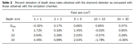 Based on the analysis of the profiles measured by the diamond detector as compared with those measured by the ionization chamber, one verifies that such detector presents a difference of approximately 2% in terms of field flatness (1 × 20 cm2 and 3 × 20 cm2, respectively), while for the square fields such difference is reduced to less than 1%. On the other hand, as regards symmetry, such behavior is reversed, with the greatest differences (of approximately 1%) occurring for the conventional fields. Such differences may be attributed to the different orientations of each detector in these measurements, particularly in what concerns the determination of the penumbra threshold: the diamond detector was positioned with its axis perpendicularly to the water surface, while the ionization chamber had its axis perpendicular to the radiation beam. Some authors(12,13) have also observed similar differences in the dose profile measurement because of the detector positioning in relation to the radiation beam: such a difference may be minimized by positioning the diamond detector with its longitudinal axis parallel to the beam, in order to increase its spatial resolution. Additionally, it is necessary to take the finite size of the ionization chamber into consideration, since the measurement along the central ray is performed along the length of the ionization chamber's cavity(2). Based on the authors' observations in the present study and on the conclusions drawn by other authors, one could say that measurements with both the ionization chamber and the diamond detector present an inherent uncertainty caused by several factors such as chamber orientation, alignment accuracy, and detector volume. Thus small differences between the results should be expected. However, it should be highlighted that the utilization of a diamond detector is only recommended for relative dosimetry purposes, since calibration services for such type of detector are not available yet, and its performance is still highly dependent on the appropriate selection of the crystal in the course of its construction. CONCLUSIONS The dosimetric characterization undertaken in the present study demonstrates that the diamond detector is an appropriate measurement system for small field measurements. The present results were validated by studies published in the literature. Acknowledgements To the Department of Radiology of Hospital das Clínicas da Faculdade de Medicina da Universidade de São Paulo (InRad/HC-FMUSP) and to the Department of Radiotherapy of Hospital Alemão Oswaldo Cruz. REFERENCES 1. Rustigi SN. Evaluation of the dosimetric characteristics of a diamond detector for photon beam measurements. Med Phys. 1995;22:56770. 2. Bucciolini M, Buonamici FB, Mazzocchi S, et al. Diamond detector versus silicon diode and ion chamber in photon beams of different energy and field size. Med Phys. 2003;30:214954. 3. Vatnisky S, Järvinen H. Application of natural diamond detector for the measurement of relative dose distributions in radiotherapy. Phys Med Biol. 1993;38:17384. 4. Bucciolini M, Borchi E, Bruzzi M, et al. Diamond dosimetry: outcomes of the CANDIDO and CONRAD INFN projects. Nucl Instrum Meth Res A. 2005;552:18996. 5. Descamps C, Tromson D, Guerrero MJ, et al. Nitrogen-doped diamond: thermoluminescence and dosimetric applications. Diamond Relat Mater. 2006;15:8337. 6. Barnett E, MacKenzie M, Fallone BG. IMRT point dose measurements with diamond detector. Radiol Oncol. 2005;39:718. 7. Francescon P, Cora S, Cavedon C. Total scatter factors of small beams: a multidetector and Monte Carlo study. Med Phys. 2008;35:50413. 8. McKerracher C, Thwaites DI. Phantom scatter factors for small MV photon fields. Radiol Oncol. 2008;86:2725. 9. IAEA. Absorbed dose determination in external beam radiotherapy: an international code of practice for dosimetry based on standards of absorbed dose to water. Technical Reports Series No. 398. Vienna, Austria: IAEA; 2001. 10. Hoban PW, Heydarian M, Beckham WA, et al. Dose rate dependence of a PTW diamond detector in the dosimetry of a 6 MV photon beam. Phys Med Biol. 1994;39:121929. 11. Ding GX, Duggan DM, Coffey CM. Commissioning stereotactic radiosurgery beams using both experimental and theoretical methods. Phys Med Biol. 2006;51:254966. 12. Rustgi SN, Frye DMD. Dosimetric characterization of radiosurgical beams with a diamond detector. Med Phys. 1995;22:211721. 13. Laub WU, Wong T. The volume effect of detectors in the dosimetry of small fields used in IMRT. Med Phys. 2003;30:3417. 1. Master of Applied Nuclear Technology, Instituto de Pesquisas Energéticas e Nucleares (Ipen/CNEN), São Paulo, SP, Brazil. 2. Doctor of Science, D.Sc., Researcher at Instituto de Pesquisas Energéticas e Nucleares (Ipen/CNEN), São Paulo, SP, Brazil. 3. Master of Nuclear Physics, Medical Physicist, Instituto de Física da Universidade de São Paulo (IFUSP), São Paulo, SP, Brazil. 4. Master of Applied Nuclear Physics, Medical Physicist, Hospital Alemão Oswaldo Cruz, São Paulo, SP, Brazil. 5. Specialist in Medical Physics, Medical Physicist, Unit of Radiotherapy, Instituto de Radiologia Hospital das Clínicas da Faculdade de Medicina da Universidade de São Paulo (InRad/HC-FMUSP), São Paulo, SP, Brazil. Mailing Address: Dra. Laura Natal Rodrigues Instituto de Radiologia Setor de Radioterapia Avenida Doutor Enéas de Carvalho Aguiar, 255, 3º andar, Cerqueira César São Paulo, SP, Brazil, 05403-001 E-mail: lnatal@usp.br Received November 9, 2010. Accepted after revision December 1st, 2011. Study developed at Instituto de Pesquisas Energéticas e Nucleares (Ipen/CNEN), Unit of Radiotherapy Hospital Alemão Oswaldo Cruz, and at Instituto de Radiologia Hospital das Clínicas da Faculdade de Medicina da Universidade de São Paulo (InRad/HC-FMUSP), São Paulo, SP, Brazil. Financial support: INCT Metrologia das Radiações em Medicina, Conselho Nacional de Desenvolvimento Científico e Tecnológico (CNPq), Fundação de Amparo à Pesquisa do Estado de São Paulo (Fapesp), Coordenação de Aperfeiçoamento de Pessoal de Nível Superior (Capes) and Agência Internacional de Energia Atômica (AIEA). |
|
Av. Paulista, 37 - 7° andar - Conj. 71 - CEP 01311-902 - São Paulo - SP - Brazil - Phone: (11) 3372-4544 - Fax: (11) 3372-4554