Radiologia Brasileira - Publicação Científica Oficial do Colégio Brasileiro de Radiologia
AMB - Associação Médica Brasileira CNA - Comissão Nacional de Acreditação
 Vol. 44 nº 6 - Nov. / Dec. of 2011
|
CASE REPORT
|
|
Breast hemangioma mimicking metastasis at PET-CT |
|
|
Autho(rs): Sabas Carlos Vieira1; Jucélia Saraiva e Silva2; Eveline Brandão Madeira3; Júlio César Queiroz de França3; Sebastião Nunes Martins Filho3 |
|
|
Keywords: PET/CT; FDG-18F; Hemangioma; Breast. |
|
|
Abstract: INTRODUCTION
Hemangiomas are benign vascular tumors, rarely found in the breast, which present low 18F-fluoro-2-deoxy-D-glucose (FDG) uptake at positron emission tomography (PET), being differentiable from malignant tumors(1). Positron emission tomography with FDG has been utilized to differentiate malign from benign lesions, since this imaging modality can detect the glucose metabolism that is generally greater in malignant than in benign tumors(2). However, such a method does not present the same anatomical accuracy of computed tomography (CT), so a technique combining both scanners (PET/CT) is utilized. Even so, false-negative and false-positive results have been reported(3). The present study reports a case of breast hemangioma in a woman whose PET/CT scan has demonstrated increased FDG uptake (simulating a malignant tumor). A brief review of factors leading to false positive and false negative PET results is also undertaken. CASE REPORT At clinical examination, a female, 63-year-old patient presented a nodule measuring about 1.5 cm in the junction of the lower quadrants of her right breast. The nodule had partially defined contours and fibroelastic consistency. The axilla was clinically negative. The patient presented a previous history of surgery for a colon tumor, with adjuvant chemotherapy and that later progressed with development of pulmonary metastasis. Mammography demonstrated a partially delimited, lobulated nodular lesion in the right breast, and ultrasonography revealed a solid lesion measuring 1.8 × 0.7 cm, that was isoechoic in relation to the breast tissue, classified as BI-RADS 4. The patient was submitted to FDG-PET/CT that demonstrated increased FDG uptake by the breast nodule (Figure 1). Such finding determined the nodule resection. The final histopathological study revealed the presence of a benign vascular neoplasm measuring 1.6 × 1.5 × 0.5 cm, consisting of thin-walled, ectatic, congested blood vessels inserted in the conjunctival stroma, and absence of atypias, compatible with capillary hemangioma. 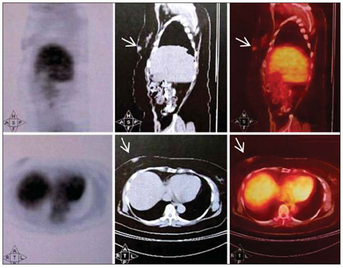 Figure 1. Positron emission tomography/computed tomography (FDG-PET/TC) demonstrating increases FDG uptake in a breast (arrows). DISCUSSION Capillary hemangiomas are benign vascular tumors characterized by proliferation of capillary vessels(4). Breast hemangiomas primarily affect post-menopausal women and may increase in size in the setting of hormone replacement therapy(5). Several imaging methods are available for detection, diagnosis and decision making on the approach to be adopted in the setting of breast diseases. However, mammography still remains as the most relevant imaging technique for breasts. In order to standardize mammographic findings reporting, the BI-RADS classification was developed, subdividing imaging findings into five classes as follows: negative, benign, probably benign, suspicious and highly suspicious of malignancy (6). Some studies have reported the finding of breast hemangiomas as well circumscribed, macrolobular lesions that may contain calcification(7,8). However, such findings are nonspecific, which may explain the significant number of hemangiomas classified either as BI-RADS 3 or 4 and the non differentiation from fibroadenomas or cysts(8). PET/CT detects glycolytic hyperactivity of malignant cells through FDG (a glucose analogue) uptake. Such uptake is caused by an increase in the number of glucose transporter proteins and in hexokinase and phosphofructokinase levels which promote glycolysis(3). Once phosphorylated at FDG-6-phosphate by hexokinase, structural changes prevent FDG to be catabolized or transported back into the extracellular space, being selectively accumulated within tumor cells(2). False-negative results may be observed at PET scan in tumors with low glycolytic activity such as adenomas, low-grade lymphomas and small-sized lesions(9). Additionally, in cases of disease adjacent to sites of physiological uptake (heart, kidneys, bladder and liver), FDG‑PET should be supplemented with other imaging modalities to confirm the results(3). FDG-PET scan performed within up to one month following chemotherapy may present decreased sensitivity because of the reduced number of metabolically active tumor cells, which is not always predictive of a good response(2). On the other hand, false-positive results may be observed in infectious and inflammatory diseases where activated macrophages and neutrophils show increased FDG accumulation, since they utilize glucose as a source of energy for chemotaxis and phagocytosis(10). It has been suggested that inflammatory cells utilize more glucose under hyperglycemic than under euglycemic conditions and, therefore, lesions containing such cells are most frequently interpreted as malignant lesions under hyperglycemic conditions(3). Accumulation of FDG in hemangiomas may be related to blood retention in the lesion, resulting in focal ischemia. Then, the secondary hypoxia may accelerate the anaerobic glycolysis, leading to a high FDG uptake(11). CONCLUSION Despite its significant contribution to the evaluation, diagnosis and treatment of cancer patients, FDG-PET/CT may present false-positive and false-negative results. Therefore, the understanding of the causes of false results is critical to avoid equivocal diagnoses. REFERENCES 1. Sakurai K, Haram M, Ozawa Y, et al. Thoracic hemangiomas: imaging via CT, MR, and PET along with pathologic correlation. J Thorac Imaging. 2008;23:11420. 2. Selzner M, Hany TF, Wildbrett P, et al. Does the novel PET/CT imaging modality impact on the treatment of patients with metastatic colorectal cancer of the liver? Ann Surg. 2004;240:102734; discussion 10356. 3. Chang JM, Lee HJ, Goo JM, et al. False positive and false negative FDG-PET scans in various thoracic diseases. Korean J Radiol. 2006;7:5769. 4. Courcoutsakis NA, Hill SC, Chow CK, et al. Breast hemangiomas in a patient with Kasabach-Merritt syndrome: imaging findings. AJR Am J Roentgenol. 1997;169:13979. 5. Mariscal A, Casas JD, Balliu E, et al. Breast hemangioma mimicking carcinoma. Breast. 2002;11:3578. 6. Vieira AV, Toigo FT. Classificação BI-RADS: categorização de 4.968 mamografias. Radiol Bras. 2002;35:2058. 7. Siewert B, Jacobs T, Baum JK. Sonographic evaluation of subcutaneous hemangioma of the breast. AJR Am J Roentgenol. 2002;178:10257. 8. Mesurolle B, Sygal V, Lalonde L, et al. Sonographic and mammographic appearances of breast hemangioma. AJR Am J Roentgenol. 2008;191:W1722. 9. Carter KR, Kotlyarov E. Common causes of false positive F18 FDG PET/CT scans in oncology. Braz Arch Biol Technol. 2007;50(special number):2935. 10. Shim SS, Lee KS, Kim BT, et al. Focal parenchymal lung lesions showing a potential of false-positive and false-negative interpretations on integrated PET/CT. AJR Am J Roentgenol. 2006;186:63948. 11. Hatayama K, Watanabe H, Ahmed AR, et al. Evaluation of hemangioma by positron emission tomography: role in a multimodality approach. J Comput Assist Tomogr. 2003;27:707. 1. MD, Oncologist and Breast Specialist, President of Sociedade Brasileira de Mastologia, Full Professor, Discipline of Oncology at Faculdade de Medicina da Universidade Federal do Piauí (UFPI), Teresina, PI, Brazil. 2. MD, Pathologist, Physician at Department of Pathology and Medical Practice, MedImagem, Teresina, PI, Brazil. 3. Graduate Students of Medicine, Universidade Federal do Piauí (UFPI), Teresina, PI, Brazil. Mailing Address: Eveline Brandão Madeira Rua Fidalma Martins de Carvalho, 4355, Bl-06, ap. 204, Ininga Teresina, PI, Brazil, 64048-480 E-mail: evelinemadeira@hotmail.com Received January 14, 2011. Accepted after revision June 21, 2011. * Study developed at Universidade Federal do Piauí (UFPI), Teresina, PI, Brazil. |
|
Av. Paulista, 37 - 7° andar - Conj. 71 - CEP 01311-902 - São Paulo - SP - Brazil - Phone: (11) 3372-4544 - Fax: (11) 3372-4554