Radiologia Brasileira - Publicação Científica Oficial do Colégio Brasileiro de Radiologia
AMB - Associação Médica Brasileira CNA - Comissão Nacional de Acreditação
 Vol. 44 nº 5 - Sep. / Oct. of 2011
|
CASE REPORT
|
|
Gorhams disease of scapula and clavicle: case report and two-year follow-up of a rare disorder |
|
|
Autho(rs): Paulo Cesar Rocha Oliveira1; Fabio Peixoto Alcantara1; Paola Lima Pasini Judice1; Giovani Rodrigues Batista1; Ilailson de Goes Teles1; Julio Salgado Antunes2 |
|
|
Keywords: Gorhams disease; Massive osteolysis; Computed tomography; Magnetic resonance imaging. |
|
|
Abstract: INTRODUCTION
Gorhams disease is a rare disorder characterized by extensive osteolysis and anomalous vascular proliferation, whose diagnosis is difficult and the prognosis, unpredictable. Such condition was firstly described by Jackson in 1838, and its clinical and pathological characteristics were established by Gorham and colleagues (1954) and Gorham & Stout (1955)(13). It is also known by several terms such as Gorhams syndrome, Gorham-Stout syndrome, Morbus Gorham-Stout disease, massive osteolysis, and vanishing bone disease(2,4). The authors report a case of Gorhams disease affecting the right scapula and, two years after, the ipsilateral clavicle of a female, 20-year-old patient. A brief literature review is carried out. CASE REPORT In July 2007, a female 18-year old patient was admitted with complaints of intermittent pain in her left shoulder for one year, with progressive incapacity of the ipsilateral upper limb. No history of fever, focal inflammatory signs, previous trauma, surgery or radiotherapy was reported. The clinical examination demonstrated pain at palpation and functional incapacity, similar to a massive rotator cuff tear, without a palpable mass. Neurological tests were normal. Laboratory tests only demonstrated a subtle increase in C-reactive protein levels (0.9 mg/dl). Shoulder radiographs revealed cortical rupture and bone fragmentation affecting the upper segment of the scapula, with no sign of bone expansion (Figure 1). Computed tomography (CT) clearly demonstrated the involvement of the whole scapula (Figure 2). Magnetic resonance imaging (MRI) revealed heterogeneous bone signal intensity, with areas of spontaneous hyperintense signal intensity on T1-weighted images and predominance of hyperintensity on STIR images, without bone expansion or extraosseous component, with irregular post-contrast enhancement (Figure 3AC). An expansile lesion was observed in the soft tissues of the left supraclavicular region, with a signal similar to the one of the scapular abnormality, but without continuity, measuring 4.6 cm in the longest axis (Figure 3A), that was not biopsied because it was considered as a probable formation of lymphatic nature. Chest CT did not demonstrate any other alteration. Gorhams disease was the main radiological hypothesis in this case. Bone biopsy was performed and histopathology demonstrated trabecular bone destruction with prominent bone formation and resorption by osteoblastic and osteoclastic activity, respectively. The bone marrow was replaced by loose fibrous tissue, with extensive vascular proliferation (Figure 3D). 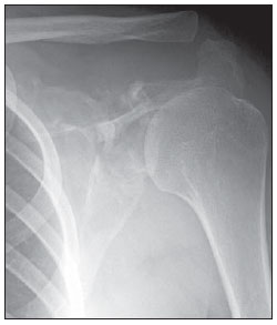 Figure 1. Frontal radiograph of left shoulder demonstrating cortical bone rupture and fragmentation of the upper half of the scapula, with a relative preservation of the distal segment. Note the normal appearance of the distal extremity of the clavicle. 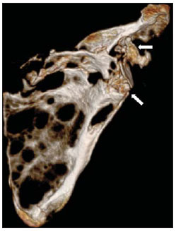 Figure 2. Computed tomography with 3D reconstruction demonstrating extensive areas of osteolysis in practically the whole scapula, with deformity of the choracoid process and glenoid neck (arrows). 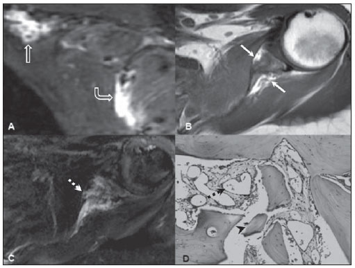 Figure 3. Magnetic resonance imaging and histopathology. A: Sagittal STIR image demonstrating marked hypersignal on the scapular body (open circle arrow) and an also hyperintense expansile extraosseous suprascapular component (straight open-headed arrow), considered as a probable lymphatic lesion. B: Axial, T1-weighted image demonstrating heterogeneous signal intensity on the glenoid neck, with spontaneously hyperintense areas (white arrows) suggestive of fatty replacement of bone. C: Axial contrastenhanced T1-weighted image with fat suppression submitted to subtraction technique, slightly caudal as related to B, demonstrating heterogeneous contrast enhancement of the glenoid neck (dashed white arrow). D: Photomicrograph (bone biopsy) revealing areas of bone destruction (arrow head) and vascular proliferation (black dashed arrow) hematoxylin-eosin stain, 400 x magnification. Two years later, follow-up radiographs and CT demonstrated distal clavicular osteolysis and progression of scapular resorption (Figure 4). The patient has been conservatively treated with oral analgesics and muscle strength exercises, and pain management has been achieved. 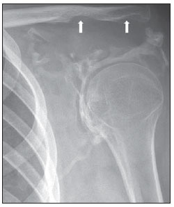 Figure 4. Frontal radiograph of left shoulder acquired two years after the patient admission, demonstrating unequivocal progression of scapular resorption and appearance of osteolysis on the inferior aspect of the distal segment of the clavicle (arrows). DISCUSSION Gorhams disease may affect any bone, particularly the scapular and pelvic girdles. Unifocal involvement is usual, while multifocal involvement is rarely observed(2,5). Osteolysis may involve joints(4,6) in the present case, osteolysis extended from the scapula towards the clavicle. In most of cases this disease occurs under the age of 40, with neither sex predilection nor hereditary predisposition(2,5). Clinical presentation depends on location, including sudden or insidious pain onset, pathological fractures, progressive functional limb disability like in the present case and soft tissue atrophy(2,7). Laboratory tests are non-specific and generally present normal results. The clinical disease course may be progressive, relatively stable or rapidly leading to death. Spontaneous resolution is extremely rare(2,5). Severe complications such as chylothorax, pleural or pericardial effusion, osteomyelitis, septicemia and spinal cord compression are not frequently observed(2,7). The Gorhams disease etiopathogeny still remains obscure. The histopathological substrate for the disease is the replacement of healthy bone by an aggressively expansile, non-neoplastic vascular tissue similar to hemangioma and lymphangioma, which is progressively replaced by fibrous connective tissue. Also, fatty replacement of bone marrow may occur(13,8). Specific and effective treatment is not available yet. Radiotherapy, surgery, anti-osteoclastic medication (bisphosphonates) and interferon alpha-2b have been utilized with diverging results(2,5,8). Imaging methods, particularly radiography and CT, play a relevant role in the initial evaluation of patients with this disease. The initial suspicion, the clinical progression and the post-therapy follow-up are based on findings of such imaging methods. The radiographic findings demonstrate the sequential disease progression with a clear absence of new bone formation(1,6,7). Initially multiple radiolucent foci are seen in a relatively normal bone, followed by progression of deformity and bone mass loss with later osteolysis dissemination toward adjacent tissues. Subsequently, cortical bone erosion and focal invasion of soft tissues by the angiomatous mass are observed, culminating in the disappearance of the remaining bone(1,6,7). CT can confirm the radiological findings and, additionally evaluates soft tissues and guides the biopsy(7), better demonstrating the disease extent in complex structures such as the scapular girdle. MRI is an adjuvant method with great variability in signal intensity and post-contrast enhancement, reflecting the spectrum of neovascular and fibrous progression(1,7). Differential diagnoses for Gorhams disease include osteomyelitis, metastasis, osteolysis secondary to rheumatoid arthritis and hyperparathyroidism, besides several osteolytic syndromes. The diagnosis is based on clinical, radiological and mainly histopathological findings(1,2,5,7,8). It is important to highlight the relevance of imaging methods in the setting of an initial diagnostic suspicion, as well as in the evaluation of the disease progression and in the post-therapy follow-up in patients with this disease. Acknowledgements The authors thank to the librarians of the institution for their usual efficiency, promptness and cordiality. REFERENCES 1. Chung C, Yu JS, Resnick D, et al. Gorham syndrome of the thorax and cervical spine: CT and MRI findings. Skeletal Radiol. 1997;26:559. 2. Patel DV. Gorhams disease or massive osteolysis. Clin Med Res. 2005;3:6574. 3. Gorham LW, Stout AP. Massive osteolysis (acute spontaneous absorption of bone, phantom bone, disappearing bone); its relation to hemangiomatosis. J Bone Joint Surg Am. 1955;37:9851004. 4. Glass-Royal M, Stull MA. Musculoskeletal case of the day. Gorham syndrome of the right clavicle and scapula. AJR Am J Roentgenol. 1990;154:13356. 5. Rubel IF, Carrer A, Gilles JJ, et al. Progressive Gorham disease of the forearm. Orthopedics. 2008;31:284. 6. Torg JS, Steel HH. Sequential roentgenographic changes occurring in massive osteolysis. J Bone Joint Surg Am. 1969;51:164955. 7. Cano B, Insa S, Cifrián C, et al. Radiologic findings in Gorham-Stout syndrome. Radiologia. 2006;48:336. 8. Ruggieri P, Montalti M, Angelini A, et al. Gorham-Stout disease: the experience of the Rizzoli Institute and review of the literature. Skeletal Radiol. 2010 Oct 25 [Epub ahead of print]. [cited 2011 Mar 1st]. Available from: http://www.springerlink.com/content/1x34043902023828/fulltext.pdf 1. MD, Diagnostic Imaging Department, SARAH Network of Rehabilitation Hospitals, Brasília, DF, Brazil. 2. MD, Surgical Pathology Laboratory, SARAH Network of Rehabilitation Hospitals, Brasília, DF, Brazil. Mailing Address: Paulo Cesar Rocha Oliveira, MD. Diagnostic Imaging Department, Hospital SARAH-Brasilia, SARAH Network of Rehabilitation Hospitals. SMHS Qd 501, Bl A. Brasilia, DF, Brazil, 70335-901. E-mail: bsbpaulocesar@gmail.com. Received December 4, 2010. Accepted after revision April 18, 2011. * Study developed at SARAH Hospital Brasília Center of SARAH Network of Rehabilitation Hospitals, Brasília, DF, Brazil. |
|
Av. Paulista, 37 - 7° andar - Conj. 71 - CEP 01311-902 - São Paulo - SP - Brazil - Phone: (11) 3372-4544 - Fax: (11) 3372-4554