Radiologia Brasileira - Publicação Científica Oficial do Colégio Brasileiro de Radiologia
AMB - Associação Médica Brasileira CNA - Comissão Nacional de Acreditação
 Vol. 44 nº 4 - July / Aug. of 2011
|
CASE REPORT
|
|
Verrucous carcinoma of the hand: a rare presentation evaluated by magnetic resonance imaging |
|
|
Autho(rs): Jailson Rodrigues Lopes1; Marcelo Bordalo Rodrigues2; Laercio Alberto Rosemberg3; Rafael Burgomeister Lourenço4; Giovanni Guido Cerri5 |
|
|
Keywords: Verrucous; Carcinoma; Spiculated; Sea-urchin; Squamous; Hand. |
|
|
Abstract: INTRODUCTION
Verrucous carcinoma is a well differentiated variant of squamous cell carcinoma seen in the skin and mucosae and rarely found in the hand. We report a case of two lesions on the dorsum of the hand, with no contact to each other, which underwent en-block resection and were confirmed as verrucous carcinoma. The largest lesion presented a sea-urchin-like exophytic growth pattern and an enhancement pattern described at magnetic resonance imaging (MRI) as spiculated or teased cotton wool. CASE REPORT A 24-year-old, male patient, agriculturist, presenting two exophytic lesion on the dorsum of his hand for one year, with a more expressive growth over the last two months. The patient also presented pain and a persistent difficulty in extending the interphalangeal joints of his fourth and fifth fingers for two weeks. The lesions had been empirically treated as probably fungal skin lesions, because of their insidious growth and based on the report, by the patient, of contact with soil during his working activities. As therapeutic success had not been achieved, biopsy was requested to guide a decision-making regarding a new therapeutic approach. The biopsy result was compatible with verrucous squamous cell carcinoma. Hand MRI was requested for locoregional staging of the tumor. At MRI, two skin lesions were visualized, with subcutaneous invasion, localized on the dorsum of the hand (Figure 1). The smallest lesion measured 2.0 cm, was superficial and restricted to the cutaneous and subcutaneous planes in the fourth metacarpal region. The largest one measured 3.0 cm and was located on the surface of the fifth metacarpal region, without infiltrating the extensor tendons of the fourth and fifth fingers (Figure 2). Both lesions presented intermediate signal intensity on T1-weighted sequences, a spiculated pattern with high signal intensity on T2-weighted sequences (Figure 3A) and also a spiculated enhancement pattern on post-gadolinium T1-weighted sequences (Figure 3B). The external contours of the lesions resembled the shape of a sea-urchin. 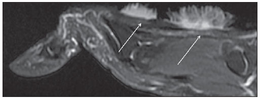 Figure 1. Sagittal MRI post-gadolinium T1-weighted image with fat saturation demonstrates two contrast-enhanced superficial lesions (arrows) in the extensor tendons, with no contact to each other. 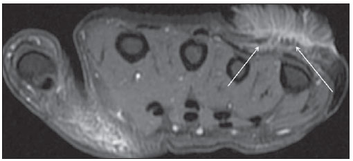 Figure 2. Axial MRI T2-weighted image with fat saturation demonstrates the largest, noninvasive lesion (arrows) in contact with the extensor tendons of the fourth and fifth fingers of the hand. 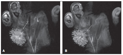 Figure 3. Coronal MRI T2-weighted (A) and T1-weighted, post-gadolinium fat saturated (B) images demonstrate a spiculated pattern (arrows) similar to a sea-urchin. Bone invasion and distant lesions were not observed. Both lesions underwent en-block resection in the next day following the MRI study, with free surgical margins. DISCUSSION Verrucous carcinoma is an indolent variant of squamous cell carcinoma that is the second most common type of skin cancer(1). Verrucous carcinomas are not frequently seen on hands. As far as the authors are concerned, less than twenty cases have been reported up to the end of 2010. Much more frequently, verrucous carcinomas are seen on the feet (more than 90% of cases in the skin)(2). The verrucous denomination is adopted because of the lesions similarity with viral warts(3). The etiology of such condition is still to be completely understood. Immunosuppression and greater Sun exposure increase the risk for disease development. Also, there are indications of association with human papilloma virus infection(4,5). Generally, verrucous carcinomas are single lesions; and cases of multiple lesions on the hand had not been reported yet. Rarely, such lesions produce metastases, but they may present deep tissue invasion, which is related to relapse(3). Although extensor tendons invasion has not been observed in the present case, the observed perilesional edema might be implicated in the difficulty to extend the fingers and in the presence of pain. The spiculated enhancement pattern on post-gadolinium T1-weighted sequences had already been described(6), which is also confirmed on T2-weighted images. Wasserman et al.(7) have reported that such image pattern resembled teased cotton wool and would be a result from inflammatory infiltrate as a response to the stromal tissue of the tumor. What the present report emphasizes is a particular tumor presentation, with typical MRI findings and location which are useful in the development of the diagnostic rationale. However, the main role of the radiologist is still in the locoregional staging of the disease. REFERENCES 1. Amaral ACN, Azulay RD, Azulay DR. Neoplasias epiteliais. In: Azulay RD, Azulay DR, editores. Dermatologia. 4ª ed. Rio de Janeiro, RJ: Guanabara Koogan; 2006. p. 510-26. 2. Kao GF, Graham JH, Helwig EB. Carcinoma cuniculatum (verrucous carcinoma of the skin): a clinicopathologic study of 46 cases with ultrastructural observations. Cancer. 1982;49:2395-403. 3. Gertler R, Werber KD. Management of verrucous carcinoma of the hand: a case report. Int J Dermatol. 2009;48:1233-5. 4. Noel JC, Peny MO, Detremmerie O, et al. Demonstration of human papillomavirus type 2 in a verrucous carcinoma of the foot. Dermatology. 1993;187:58-61. 5. Zemtsov A, Koss W, Dixon L, et al. Anal verrucous carcinoma associated with human papilloma virus type 11: magnetic resonance imaging and flow cytometry evaluation. Arch Dermatol. 1992;128:564-5. 6. Theodorou SJ, Theodorou DJ, Bona SJ, et al. Primary squamous cell carcinoma: an incidental toe mass. AJR Am J Roentgenol. 2005;184(3Suppl):110-1. 7. Wasserman PL, Taylor RC, Pinillia J, et al. Verrucous carcinoma of the foot and enhancement assessment by MRI. Skeletal Radiol. 2009;38:393-5. 1. MD, Radiologist, Collaborator at Unit of Musculoskeletal Imaging, Instituto de Radiologia do Hospital das Clínicas da Faculdade de Medicina da Universidade de São Paulo (InRad/HC-FMUSP), São Paulo, SP, Brazil. 2. MD, Radiologist, Director for Instituto de Ortopedia e Traumatologia do Hospital das Clínicas da Faculdade de Medicina da Universidade de São Paulo (IOT/HC-FMUSP), São Paulo, SP, Brazil. 3. PhD, MD, Radiologist, Assistant, Unit of Radiology and Instituto de Radiologia do Hospital das Clínicas da Faculdade de Medicina da Universidade de São Paulo (InRad/HC-FMUSP), São Paulo, SP, Brazil. 4. MD, Radiologist, Assistant, Instituto de Radiologia do Hospital das Clínicas da Faculdade de Medicina da Universidade de São Paulo (InRad/HC-FMUSP), São Paulo, SP, Brazil. 5. Titular Professor, Division of Radiology, Head of the Unit of Radiology, Instituto de Radiologia do Hospital das Clínicas da Faculdade de Medicina da Universidade de São Paulo (InRad/HC-FMUSP), São Paulo, SP, Brazil. Mailing Address: Dr. Jailson Rodrigues Lopes Rua Cristiano Viana, 116, ap. 133, Jardim América São Paulo, SP, Brazil, 05411-000 E-mail: jailsonlopes@hotmail.com Received January 20, 2011. Accepted after revision March 31, 2011. Study developed at Instituto de Radiologia do Hospital das Clínicas da Faculdade de Medicina da Universidade de São Paulo (InRad/HC-FMUSP), São Paulo, SP, Brazil. |
|
Av. Paulista, 37 - 7° andar - Conj. 71 - CEP 01311-902 - São Paulo - SP - Brazil - Phone: (11) 3372-4544 - Fax: (11) 3372-4554