Radiologia Brasileira - Publicação Científica Oficial do Colégio Brasileiro de Radiologia
AMB - Associação Médica Brasileira CNA - Comissão Nacional de Acreditação
 Vol. 44 nº 3 - May / June of 2011
|
ORIGINAL ARTICLE
|
|
Collaborative environment for nuclear medicine training |
|
|
Autho(rs): Cláudia Régio Brambilla1; Gabriel Goulart Dalpiaz2; Ana Maria Marques da Silva3; Neivo da Silva Júnior4; Lucia Maria Martins Giraffa5; Tiago Coelho Ferreto6; Cesar Augusto Fonticielha De Rose7; Vinicius Duval da Silva8 |
|
|
Keywords: Collaborative environment; Nuclear medicine; Medical education; Distance education. |
|
|
Abstract: INTRODUCTION
Traditional ways for medical learning in nuclear medicine generally comprise discussions over clinical image banks, monitoring of specialists in the images analysis, classroom or distance learning courses, and studies on the available literature(1). Within this reality, the following problem arises with respect to personnel training in nuclear medicine: How to promote the primary and continued education of nuclear medicine professionals, considering the small number of training centers in the country? Currently, the availability of digital and virtual technologies allows the access to virtual environments where instructional contents and learning support platforms are available(1). Some of those are targeted at medical teaching. However, normally such contents are offered in the form of open or restricted access online courses, such as the case of the initiative from the American Association of Physicists in Medicine (AAPM), together with the Radiological Society of North America (RSNA), in which modules were developed for the education of residents in radiology or nuclear medicine(2,3). The self-explanatory modules were developed by interdisciplinary specialist groups, comprising at least a physicist and a radiologist. The virtual medical learning environments usually utilize static and sequential contents, without incorporating interaction capabilities. In such environments, interactivity is usually restricted to individual users browsing, without allowing interaction between users or the insertion of questionings or new contents into the environment. The available information and communication technologies can be utilized to enhance the understanding of the processes involved in medical images acquisition and processing. The present study presents a proposal of development and validation of a virtual collaborative environment that allows interactivity, coupled with computational power, for the education of nuclear medicine professionals. MATERIALS AND METHODS Initially, exploratory meetings were held with multi-disciplinary teams comprising medical physicists, nuclear medicine physicians and computer scientists, in order to identify the organizational, environmental and external assumptions and restrictions to the collaborative environment. Aspects related to infrastructure of partner institutions and hospitals as well as matters related to data accessibility and security were also addressed by the information technology team. From that preliminary study, the following requirements for the development of the collaborative environment were defined: provide the users with a friendly interface, allowing the interaction among users and access to dynamic contents; allow users to interact in discussion forums; provide contents of interest to nuclear medicine, such as image banks and literature-based support contents; allow user-friendly submission of nuclear medicine computer simulations, by means of the utilization of high-performance computing resources. Based on the social constructivist learning theory(4) that permeates the proposal of collaborative environments, materials were made available in the environment, relying on the assumption that the construction and expansion of the environment´s resources would be accomplished by the collaboration among users. For the construction of a collaborative environment prototype and subsequent validation, the myocardial perfusion study was selected as the initial application because of its high demand at nuclear medicine centers. Discussion topics, such as tomographic images acquisition and reconstruction parameters were selected as discussion starters in the environment, as well as clinical case studies. Free digital image visualization and processing tools were also made available. Concomitantly, a prototype was developed for the submission of computer simulations by utilizing the resources of a high-performance laboratory. The computer simulation of images by means of the Monte Carlo method is widely utilized to simulate the effects produced by parameter changes in the acquisition of nuclear medicine images(57). In order to create a simulated images bank, a nuclear medicine equipment was modeled by utilizing the resources available at the GATE package (geant4 application for tomographic emission), which simulates PET and SPECT systems(8,9). As the contents of the collaborative environment is dynamic and dependent on the users interest, a limited number of images and documents was initially made available. The management of the contents and security of the collaborative environment was carried out by an anonymous administrator that authorized user registrations under pre-established rules. Experienced professionals in the field of nuclear medicine acted as moderators, coordinating the discussions and selecting themes to be discussed in the forums. The users sample for validation of the proposed collaborative environment was intentionally recruited, and the users were nuclear medicine specialists in order to interact in the collaborative environment. A pilot study was developed with 10 medical physicists and 5 nuclear physicians. At the end of the interaction, the users filled out a semi-structured questionnaire (Likert scale) evaluating the relevance and ease on different aspects of functionalities and interaction with the environment, indicating suggestions for improvements in the proposal. The interaction test was carried out over a one-month period. The analysis was initially performed on the closed part of the questionnaire, allowing the evaluation of available functionalities in the environment by means of the opinion from users after the interaction test (Likert scale). A contents analysis based on Moraes & Galiazzi was also performed in the open sections of the questionnaire. Such an analysis approach has an operations cycle that initiates by the unitarization of materials in the textual corpus, moving to the categorization of analysis units. From such a process new comprehensions emerge, constituted by the self-organization of the results interpretation text(10). RESULTS Development of the environment The collaborative environment in nuclear medicine (Figure 1) developed at Moodle, is hosted by a virtual computer at a cluster in the High Performance Laboratory of Pontifícia Universidade Católica do Rio Grande do Sul (PUCRS) and can be accessed at http://marfim.lad.pucrs.br:58080/moodle/. 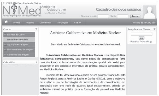 Figure 1. Homepage of the Collaborative Environment in Nuclear Medicine. The environment is divided into categories at the main menu (Figure 1) as follows: Project, Images, Documents, Simulation and Contact. The approached topics (Courses) are located on the bar at left, as follows: Case Studies, Images Acquisition, Images Processing, Images Corrections. Such categories are divided into sub-categories with the themes and discussion topics. At all the sub-categories, discussion forums are enabled to allow themes discussions among users. The moderators and users can utilize the image banks and support documentation in the discussions, besides adding other materials and new questions. A prototype of computational simulations submission in the collaborative environment is currently under development, which will allow the choice of parameters for the simulation of nuclear medicine studies by means of the GATE application. Figure 2 depicts a diagram demonstrating the process of simulation submission in the environment. 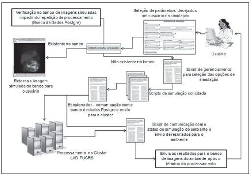 Figure 2. Diagram of user interaction with the environment of simulations submission and communication of the environment with the cluster for processing and uploading of the simulated images (returned to the environment/user). Clinical image banks, both experimental and simulated, as well as free softwares for images processing and visualization in the environment are accessed at Menu> Images. Validation of the collaborative environment proposal in the pilot study The interaction sample in the pilot test of the nuclear medicine collaborative environment prototype comprised 15 subjects, with 10 of them being medical physicists (67%) and 5, nuclear medicine physicians (33%). Among the subjects, 67% were male individuals (6 medical physicists and 4 nuclear medicine physicians) and 33% were female individuals (4 medical physicists and one nuclear medicine physician). The mean graduation time was 9.4 years (median of 6 years). In the sample, 67% of the participants in the interaction had completed post-graduation, with a mean post-graduation time of 3.5 years. Mean time in the current position or function in the sample was 6.27 years (median of 5 years). Mean age in the sample was 33.87 years (median of 32 years). Data corresponding to the responses to the questionnaire are presented below, and evaluate the relevance of the following topics: Menu Items; Image Banks; Possibility of Contributions by the Users (cases, questions and materials); Document Banks (articles, support materials, dissertations and theses, published papers and reports); Availability of Free Softwares (images visualization and processing); Interaction with the Collaborative Environment performed in the discussion forums (quality of the discussions and moderators performance); and Possibility of Submitting Nuclear Medicine Simulations by means of the Collaborative Environment (prototype). Figure 3 shows the result of users opinions with respect to relevance of the categories on the evaluation questionnaire. 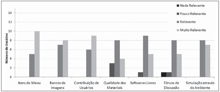 Figure 3. Users opinions on the relevance of items in the menu, image banks, users contributions, quality of the materials, free softwares, discussion forums and simulations submission. As regards Evaluation of Materials Available in the Environment, the quantitative data represented by the first five categories on Figure 3, show that all the users considered as being relevant the items available in the menu, the image banks and the possibility provided to users to contribute for the environment with cases, questions and materials. As regards the quality of the available materials, some users considered them of little relevance. One of the users suggested: the inclusion of other topics, not only those related with the area of image quality. (FM-01). The free softwares were considered of little relevance by one of the users, and some comments on the complexity in their utilization were reported. As far as the Discussion Forums are concerned, whose quantitative data are represented by the sixth category on Figure 3, which analyzes the quality of discussions and moderators performances, two participants rated them as little or no relevant at all. Even among those who considered the discussion forums as relevant, the participants in the interaction test presented several suggestions, as the following user opinion transcripts demonstrate: ...the moderator did not provide an attractive and interesting environment, which caused a small number of participations... the moderator should promote discussions from the beginning and also encourage access to the website (FM-04). As regards the possibility of Submission of Simulations by means of the collaborative environment, the quantitative data represented by the seventh category on Figure 3, all users considered such functionality as relevant or very relevant. The qualitative data on the Interface Resources are represented by the three categories on Figure 4, evaluating the organization of the elements in the collaborative environment, according to the following requirements: Localization of the Data (icons and files); Contact with the Research Group and prototype of the Submission of Simulations (icons for modeling, submission of simulations and simulation status progress bar). 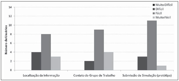 Figure 4. Userss opinion on the interface resources; information, group contact and simulation submission prototype. As regards Localization of Data, some of the users considered it to be difficult. From an excerpt from a users opinion, one notices the anxiety towards the initial interaction with the environment: For several moments I was disoriented with respect to the website routine, and had some operational difficulties, that I was finally able to overcome. Posting as well as task execution could be made a little easier...the difficulties were not lasting, but they were a hindrance (MN-02). As regards the Contact with the Research Team, in spite of some reports of difficulties in contacting the team, no e-mails were received by the group contact available in the environment. No postings on the environments doubts forum were made on this theme. No justification was found in the open questions, thus making the comprehension of such difficulty, quite troublesome. As regards the interface of the Submission of Simulations interface prototype, the users did not offer criticism or suggestions, a fact that also impairs the proper understanding of the reasons leading to the option by difficulties with such an interface. The analysis of the contents of the open questions on the last section of the questionnaire on the Utilization of this Environment by a Supervisor and his Team for Continued Education is presented next. Such analysis results from the opinions on the utilization of the environment in the field of nuclear medicine, both for initial and continued education purposes. The users suggest that the collaborative environment in nuclear medicine be utilized for case discussions and for the demonstration of artifacts on images, providing the possibility of clarifying doubts related to relevant themes. In the opinions, the tool is considered as potentially useful in the evaluation/discussion of difficult cases and in the discussion of conflicting opinions in medical diagnosis. Additionally, this tool allows the solution of daily problems that can be discussed/solved by the group, by means of interaction and exchange of experiences among users in the virtual community. The availability of forums and chats allows the exchange of knowledge and experiences, particularly in relation to different levels of competence in the field of nuclear medicine. The users point out that by means of the collaboration among users, it will be possible to generate data and image banks for reference regarding pathologies, false-positive and false-negative results. Thus, as the number of active participants in the collaborative environment grows, the data banks can be updated and increased by means of the insertion of rare cases, reducing the loss of data/cases due to geographical limitations. Multicenter studies may also benefit from this environment by means of the ease of access to images and exchange of opinions on cases. The virtual models for the simulation of medical images could be utilized for research in the field of nuclear medicine. In the users opinion, with the high demand and data flow in the current labor market, the professionals updating and capacitation are of utmost importance, as are the rapid and efficient solving of cases. The professionals face time limitations for studying, besides increase in workloads and information flow. The environment could be utilized for continued individual learning at ones own rhythm. One observed that most of the subjects that effectively interacted in the environment became familiar with the social constructivist approach inherent to collaborative environments, providing significant contributions. DISCUSSION This study presents the development and validation of a collaborative environment in nuclear medicine for personnel education, by means of a pilot interaction study with experienced users acting in the field. The functionalities required for the implementation of a collaborative environment that allowed primary and continued learning by nuclear medicine professional groups were analyzed. By analyzing the profile of users that considered the quality of available materials as being of little relevance, one observes that such users were those with a longer experience in the field, a fact that may have generated little motivation/interest towards the subjects discussed in the environment, as well as those users that were interested in other specific areas of nuclear medicine. As regards the option for initially making available a limited number of materials in the environment meets the recommendations from specialists in collaborative environments found in the literature(4), in order to encourage the users to contribute with the environment construction. Aretio et al.(4) highlight the need to gradually increase the complexity of the collaborative environments, starting them only with the baseline materials. Considering the limitations of the free softwares that were available in the environment, it would be interesting to develop software for images visualization and processing similar to those utilized in the workstations used in the clinical nuclear medicine routine as the present study proposal is expanded. Another possibility would be entering in an agreement with the software developers in order to provide the environment with a suitable version of their own image processing software to be used in the collaborative environment. Although all the moderators that acted in the environment were experienced professionals in the field of nuclear medicine, they were not initially trained to participate in virtual collaborative environments. After the evaluation, such training proved to be a necessity. Another important aspect was the short time span of the interaction test (one month). A longer period of interaction with the environment would allow a greater participation of users, greater use of the available materials and a more effective contribution from users. In spite of some criticism, the functionalities available in the collaborative environment were generally accepted. The presented suggestions, such as the online chat, inclusion of new topics and materials can be easily implemented on account of functionalities available at Moodle. It should be highlighted that on account of a research strategy option, no preferential path was indicated to users at the beginning of the interaction, thus letting users feel free to define their own pathways in the environment, which allows the evaluation of the need for the implementation of guidance in the website and initial instructions in the environment for new users. The aim was assessing whether the environments interface would be susceptible to cognitive understanding without any initial help. The users opinions suggest the need for the planning of a map of the environment, explaining on how the user can interact by means of a summary of the features with multimedia explanations. After the assessment of users perceptions on the environments interface resources, one realized that users accepted the available resources and functionalities. The criticism and suggestions present good indications for improvements on the proposal, particularly with respect to the need of providing more information on the possibilities in the initial interaction to promote a greater familiarization with the collaborative environment. CONCLUSION The developed environment was evaluated as relevant by a community of nuclear medicine professionals, with respect to its usefulness in assisting the education of personnel. The available functionalities, the materials and discussion topics were considered as being relevant by most of the users. It is possible to conclude that the method utilized in the virtual environment may allow the exchange of experiences and case discussions among professionals located in institutions from different regions of the country, which would enable greater professional proximity and collaboration among such professionals. REFERENCES 1. Wallis JW, Miller MM, Miller TR, et al. An internet-based nuclear medicine teaching file. J Nucl Med. 1995;36:15207. 2. RSNA/AAPM Online Physics Modules. [cited 2009 Dec 10]. Available from: http://www.aapm.org/education/webbasedmodules.asp 3. Diagnostic radiology residents physics curriculum AAPM Subcommittee of the Medical Physics Education of Physicians Committee - May 2009. [cited 2009 Dec 10].Available from: http://www.aapm.org/education/documents/Curriculum.pdf 4. García Aretio L, Ruíz Corbella M, Domínguez Figaredo D. El profesor y el formador en los sistemas digitales de enseñanza y aprendizaje. In: García Aretio L, Ruíz Corbella M, Domínguez Figaredo D, editores. De la educación a distancia a la educación virtual. Barcelona: Ariel; 2007. p. 16386. 5. Brambilla CR. Impacto da determinação da profundidade renal na quantificação renal absoluta em estudos de cintilografia plana com 99mTc-DMSA [trabalho de conclusão de curso]. Porto Alegre, RS: Pontifícia Universidade Católica do Rio Grande do Sul; 2007. 6. Silva AM. Reconstrução quantitativa de SPECT: avaliação de correções [tese]. São Paulo, SP: Universidade de São Paulo; 1998. 7. Zaidi H. Relevance of accurate Monte Carlo modeling in nuclear medical imaging. Med Phys. 1999;26:574608. 8. Strulab D, Santin G, Lazaro D, et al. GATE (geant4 application for tomographic emission): a PET/SPECT general-purpose simulation platform. Nucl Phys B (Proc Suppl). 2003;125:759. 9. De Beenhouwer J, Staelens S, Kruecker D, et al. Cluster computing software for GATE simulations. Med Phys. 2007;34:192633. 10. Moraes R, Galiazzi MC. Análise textual discursiva. Ijuí, RS: Unijuí; 2007. 1. Medical Physicist, Master in Health Sciences, Pontifícia Universidade Católica do Rio Grande do Sul (PUCRS), Porto Alegre, RS, Brazil. 2. Graduate Student of Engineering, Developer of Research Projects for Pontifícia Universidade Católica do Rio Grande do Sul (PUCRS), Porto Alegre, RS, Brazil. 3. PhD of Physics, Director of the Department of Physics Pontifícia Universidade Católica do Rio Grande do Sul (PUCRS), Porto Alegre, RS, Brazil. 4. PhD, Nuclear Physician at Hospital São Lucas Pontifícia Universidade Católica do Rio Grande do Sul (HSL-PUCRS), Porto Alegre, RS, Brazil. 5. PhD, Titular Professor at School of Information Technology, Researcher for the Program of Mastership in Education, Sciences and Mathematics and Program of Post-graduation at School of Education of Pontifícia Universidade Católica do Rio Grande do Sul (PUCRS), Porto Alegre, RS, Brazil. 6. PhD, Assistant Professor, Instituto de Informática da Pontifícia Universidade Católica do Rio Grande do Sul (PUCRS), Porto Alegre, RS, Brazil. 7. PhD, Professor at Instituto de Informática da Pontifícia Universidade Católica do Rio Grande do Sul (PUCRS), Porto Alegre, RS, Brazil. 8. PhD, Professor, Department of Pathology and Radiations, School of Medicine, Pontifícia Universidade Católica do Rio Grande do Sul (Famed-PUCRS), Porto Alegre, RS, Brazil. Mailing Address: MSc. Cláudia Régio Brambilla Rua Major Ismael Alves, 74, Centro Gravataí, RS, Brazil, 94010-350 E-mail: claudinharb@gmail.com Received December 14, 2010. Accepted after revision May 6, 2011. Financial support: Fundo Regional para a Inovação Digital na América Latina e Caribe FRIDA, Project B80. Study developed at Pontifícia Universidade Católica do Rio Grande do Sul (PUCRS), Porto Alegre, RS, Brazil. |
|
Av. Paulista, 37 - 7° andar - Conj. 71 - CEP 01311-902 - São Paulo - SP - Brazil - Phone: (11) 3372-4544 - Fax: (11) 3372-4554