Radiologia Brasileira - Publicação Científica Oficial do Colégio Brasileiro de Radiologia
AMB - Associação Médica Brasileira CNA - Comissão Nacional de Acreditação
 Vol. 44 nº 2 - Mar. / Apr. of 2011
|
ORIGINAL ARTICLE
|
|
Acceptability of a future phantoms bank for quality control in nuclear medicine |
|
|
Autho(rs): Fernanda Carla Lima Ferreira1; Divanizia do Nascimento Souza2 |
|
|
Keywords: Nuclear medicine; Quality control; Phantom. |
|
|
Abstract: INTRODUCTION
Simulators, also known as phantoms, are utilized in nuclear medicine for evaluating the performance of collimation systems in scintillation chambers, in the calibration of such devices, in the analysis of parameters for images reconstruction and capacity of lesions detection, and in the definition of the sensitive volume and other properties or characteristics of such systems. In this field, the following types of phantoms are most frequently utilized: quadrant bar phantoms; homogeneity phantoms, such as the Jaszczak type, with artifacts of different dimensions for the simulation of radiopharmaceuticals uptake, allowing a separate evaluation of several tomographic conditions of nuclear medicine imaging systems( 1,2); the Rollo phantom, utilized for evaluating contrast and spatial resolution; brain phantoms, such as the Hoffman 3-D Brain Phantom; cardiac phantoms and thyroid phantoms(3). Certain phantoms may also function as dosimeters, whenever they contain materials such as thermoluminescent detectors or special polymer gels(49). Some of these objects, such as the Jaszczak type, are efficient for use in quality control of single photon emission computed tomography (SPECT) and positron emission tomography (PET) systems(2,6). Although several types of phantoms are already commercially available, new models are being developed to meet specific requirements or to improve nuclear medicine devices quality control processes(1013). For example, in 2008, based on the perception that the protocols for the study of the thyroid function would change as a result of the availability of iodine-123, an isotope that allows improved image quality and greater agility in results of thyroid scintigraphy, Andrade et al.(10) developed a thyroid phantom for validating the use of iodine-123 in the determination of the capacity of iodine uptake by the thyroid gland. In such a study, the authors focused on eliminating possible errors caused by the adaptation of the iodine-131 usage protocols for the use of the iodine-123 radioisotope. Debrun et al.(14) and Lee et al.(15) have also developed studies with phantoms for quality control in nuclear medicine. Debrun et al.(14) have utilized a dynamic cardiac phantom as a reference in the comparison and validation of volume measurements in gated-SPECT and 4D echocardiography. Lee et al.(15), utilizing a Korean adult voxel phantom, developed a study to calculate the specific absorbed fraction for estimating internal organ dose, and compared their results with data obtained with an adult phantom (Oak Ridge National Laboratory (ORNL), Oak Ridge, USA). Most of the phantoms utilized in Brazil are imported, with a relatively high financial cost. Although these devices can be considered as efficient for use in quality control, their high cost and related bureaucratic import procedures probably represent limiting factors for the acquisition of such devices by nuclear medicine centers. Difficulties in the acquisition of such devices are also observed in Cuba. For example, Varela et al.(16) have observed that the main difficulty at the Cuban nuclear medicine centers was the lack of phantoms for quality control tests. Because of the high cost of such devices in that country, nuclear medicine centers are not required to have their own phantoms. Thus, the Cuban State Center for Medical Equipment (CCEEM) has decided to create a national phantoms bank. The creation of such bank has allowed the quantification of the phantoms utilized at nuclear medicine services in Cuba. Any interested specialist can access the bank´s electronic portal and find out the types and quantities of available phantoms and also information on the users and institutions that own such phantoms( 16). It is important to highlight that, according to the data reported on the study developed by Varela et al.(16), in 2004 Cuba had 18 nuclear medicine centers. Just for comparison purposes, in 2009 Brazil had more than 300 operational nuclear medicine centers. In Brazil, nuclear medicine services must comply with the Standard CNEN-NE-3.05(17) and with the Resolution MS-Anvisa-RDC No. 38(18). According to such regulations, every nuclear medicine center must have at least one quadrant bar phantom set, also known as orthogonal bar phantom. This type of phantom must to be readily available at the nuclear medicine services for mandatory quality control tests, according to such regulations. Thus, phantoms banks would allow the sharing of less usual, more complex and expensive phantoms. The possibility of sharing other phantoms could play an important role in the improvement of the services provided by such institutions, increasing the availability of technical resources for quality control and for professional education. Based on the need to study the possibility of making phantoms that are less frequently found at nuclear medicine centers more accessible, and based on the observation of the increase in technological incentives in Brazil, a questionnaire was prepared in order to assess the acceptability of the deployment of a national phantoms bank or regional phantoms banks for shared use of such devices in quality control activities by nuclear medicine centers. The questionnaire served the purpose of evaluating the opinions of radioprotection supervisors and medical physicists on the possibility of the creation of such banks. The results are presented below. MATERIALS AND METHODS A questionnaire to evaluate the acceptability of the creation of a phantoms bank for nuclear medicine in Brazil was answered by medical physicists and/or radioprotection supervisors working at medical clinics and hospitals providing services of nuclear medicine diagnosis and therapy. The present study was developed in the period from January to April of 2009, based on the assumption that the organization of such phantoms bank would allow, besides the improvement in the quality control of nuclear medicine images, a greater exchange of data amongst professionals involved in the radioprotection of such centers. Additionally, the deployment of a phantoms bank would help to increment the cooperation among such centers, making a set of data on clinical cases and quality control tests available for a wide number of professionals. The questionnaire was initially applied in the city of Aracaju, SE, in the Northeast region of Brazil. After the definition of the questionnaire´s contents, format, evaluation scale and the planning of the qualitative e quantitative analyses of the questions, two radioprotection supervisors in nuclear medicine centers were interviewed for evaluation and validation of the questionnaire. Subsequently, the questionnaire was applied to other radioprotection supervisors in nuclear medicine centers and hospitals in the five Brazilian geographic regions. In the North region, the questionnaires were answered by professionals in the states of Pará, Amapá and Tocantins; in the Northeast region, by professionals in the states of Ceará, Rio Grande do Norte, Paraíba, Pernambuco, Alagoas, Sergipe, Piauí and Maranhão; in the South region, by professionals from the states of Paraná, Santa Catarina and Rio Grande de Sul; in the southeastern region, by professionals in the states of Minas Gerais, Espírito Santo, São Paulo and Rio de Janeiro; and in the Midwest region, by professionals in the state of Goiás and Brasília. It is important to highlight that the states with the highest number of nuclear medicine centers are Rio de Janeiro and São Paulo. The questionnaire comprised nine questions on quantitative and qualitative matters, allowing the respondents to express the advantages and disadvantages of a future phantoms bank and also their interest in acquiring such devices. The questionnaires were sent to 188 nuclear medicine institutions, although, at the time, the Comissão Nacional de Energia Nuclear (CNEN) (National Committee for Nuclear Energy) had approximately 320 institutions on its records(19). The application of the questionnaires was done by means of interviews with radioprotection supervisors and medical physicists at the centers, through email or phone. Table 1 presents the Brazilian regions and number of professionals participating in the sample. 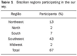 RESULTS As expected, some of the radioprotection supervisors responded that high cost and complex bureaucratic import procedures discouraged the adoption of more complex quality control procedures in nuclear medicine centers. Additionally, on some of the answers, the respondents mentioned the fact that some center administrators do not understand the actual need for the acquisition of phantoms. Figure 1 presents the degree of interest by the professionals in acquiring phantoms for quality control and training. In the Northeast region, of all 13 participants, 10 (77%) declared their interest in the acquisition of phantoms for quality control in nuclear medicine; in the Midwest region, such interest was declared by one of the two respondents. In the South region, five (72%) of seven respondents intended to acquire additional phantoms; in the Southeast region, interest was demonstrated by 36 (84%) of 43 respondents; and in the North region, a result similar to the Midwest region was observed, with one of two respondents declaring interest in the acquisition of phantoms. 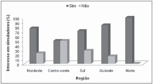 Figure 1. Degree of interest in acquiring some phantoms. Those professionals who reported no interest in acquiring new phantoms said that the devices available at their nuclear medicine centers were appropriate for their needs with respect to the quality control test requirements set by the CNEN Standards( 17). Such professionals declared that their services owned quadrant bar type phantoms. Based on the results of the questionnaire, it was also possible to evaluate the acceptance of the proposal to set up a future phantoms bank in Brazil, as demonstrated by Figure 2. 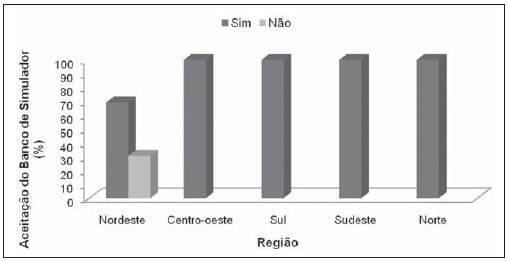 Figure 2. Index of professional acceptability of the creation of a phantoms bank. Only in the Northeast region the respondents were not unanimous in relation to the creation of such bank. Only nine (70%) of 13 respondents in the region supported the creation of such bank. In the other regions, the acceptability achieved 100%. Figure 3 presents the degree of interest of the supervisors in sharing the phantoms bank. In such a sharing process, each institution would make their phantoms available to other institutions. 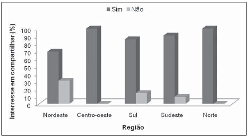 Figure 3. Level of interest of nuclear medicine professionals in sharing the phantoms by means of a phantoms bank in Brazil. In general, the majority of the respondents demonstrated interest in sharing the phantoms bank. In the Northeast region, such interest was demonstrated by nine (70%) of the respondents, in the Midwest region, by two (100%), in the South region, by six (86%), in the Southeast region, by 39 (91%), and in the North region, by two (100%) of the respondents. DISCUSSION According to the respondents, the most relevant phantoms for quality control that should be included in the bank would be those utilized for evaluating image homogeneity, which would be utilized for heart, kidneys, lungs and liver simulations, provided that such phantoms allowed the evaluation of scintigraphic images of organs with different degrees of abnormalities. Examples of phantoms that could be utilized for such purposes would be the Jaszczak-type, Rollo and Hoffman 3-D Brain phantoms. Some professionals also mentioned that it would be important to have the phantoms locally produced. In their answers, such professionals demonstrated preoccupation with the phantoms that have been utilized for sensitivity tests, because of the way such tests are normally performed, subjecting the professionals and equipment to contamination. Such preoccupation is justifiable, as one of the ways to perform this test is by means of the deposition of the radioactive sample over a Petri dish(6). However, as the sample does not always remain concealed, in the manipulation of the sample or at the moment of the homogenization of the radioactive material, it may contaminate the hands of the professional that is performing the test, or even parts of the scintillation chamber. Although other quality control tests may lead to contamination, the sensitivity test presents greater possibility of such an event. The Northeast region professionals who did not accept the proposed phantom bank pointed out that administrative problems might be a hindrance to the proper functioning of the bank. Even believing in such hindrances, such professionals would accept being users of such bank. In general, the respondents mentioned that the deployment of the bank would be a good alternative for the improvement of quality control in nuclear medicine, as it would lead to costs reduction, allowing improvements in the required procedures, besides encouraging the exchange of technical and organizational data among the professionals of Brazilian nuclear medicine services. After all, the availability of access to different types of phantoms would simplify the testing procedures and would allow the sharing of methods and problems solving solutions amongst medical physicists and supervisors, besides the early detection of avoidable problems such as those that are normally observed in linearity, field uniformity and spatial resolution tests in image acquisition systems. Considering the fact that all Brazilian nuclear medicine services have a quadrant bar type phantom, the institutions would only borrow other types of phantoms useful for quality control improvements. In addition, the bank would provide data on the types of tests and scintillation chambers in which the registered phantoms could be utilized. CONCLUSION The present study demonstrates that there is significant interest in the possibility of the deployment of a phantoms bank for nuclear medicine. The responses to the phone interviews and by e-mail also included suggestions given by the radioprotection supervisors for improvement in quality control and on the method for implementing the phantoms bank. Among those suggestions, the most frequent one was the initial deployment of the bank on a regional basis, with the justification that this would optimize the access to the bank, considering the reduced transportation costs and smaller number of users as compared with a national centralized phantoms bank. In order to set the bank up, it is necessary to survey the types, models and quantities of the phantoms that may be available at the institutions. With the results of such survey, a system for identifying the centers, the possible users as well as the availability and borrowing or exchange procedures, with the definition of usage rules, utilization scheduling and transportation of the phantoms. Additionally, even before such survey is carried out, it would be necessary to define which institution would be responsible for the management of the phantoms bank. Possibly, the Agência Nacional de Vigilância Sanitária (National Health Surveillance Agency) might assist in the indication of the managing agents for the national or regional phantoms banks. It is likely that the demand for the phantoms and transportation difficulties may cause initial hindrances to the efficiency in availability of phantoms; however, these shall not be taken as an obstacle, but as a stage to be overtaken towards the success and advances in quality control at nuclear medicine centers in Brazil. After all, the bank would allow a greater exchange of information amongst nuclear medicine professionals at regional or even at national levels. The present study demonstrates that a motivation to share existing phantoms in Brazil does exist. If such sharing comes to fruition, it will allow an effective improvement in quality control of nuclear medicine equipment, in the images acquisition in this medical specialty and in the continued education of professionals in this field. Acknowledgements The authors wish to express their gratitude to Coordenação de Aperfeiçoamento de Pessoal de Nível Superior (Capes) and to Conselho Nacional de Desenvolvimento Científico e Tecnológico (CNPq) (National Council for the Scientific and Technological Development) for the financial support, and to Clínica de Medicina Nuclear, Endocrinologia e Diabetes Ltda. (Climedi) for the assistance in the validation of data. REFERENCES 1. EANM Physics Committee, Buseman Sokole E, P»achcínska A, et al. Routine quality control recommendations for nuclear medicine instrumentation. Eur J Nucl Med Mol Imaging. 2010;37: 66271. 2. Sandborg M, Althén JN, Gustafsson A. Efficient quality assurance programs in radiology and nuclear medicine in Ostergotland, Sweden. Radiat Prot Dosimetry. 2010;139:4107. 3. Jacob J, Luo J, Mistretta M, et al. Comparison of gamma camera spatial resolution measurements with use of a point source, a line source and a Jaszczak phantom. J Nucl Med. 2006;47(Suppl 1): 554P. 4. Gear JI, Charles-Edwards E, Partridge M, et al. A quality-control method for SPECT-based dosimetry in targeted radionuclide therapy. Cancer Biother Radiopharm. 2007;22:16674. 5. De Deene Y. Essential characteristics of polymer gel dosimeters. J Phys: Conf Ser 3. 2004;3:3457. 6. Magalhães ACC, Decker HH, Castro Júnior A, et al. Influência da espessura do cristal da câmara de cintilação na qualidade das imagens. Rev Imagem. 2004;26:436. 7. Normatização dos equipamentos e técnicas de exames para realização de procedimentos em cardiologia nuclear. Controle de qualidade e desempenho da instrumentação. Arq Bras Cardiol. 2006;86(Supl 1):102. 8. Castro AA, Moraes ER, Araújo DB, et al. Reconstrução e volumetria de imagens de SPECT para aplicação clínica diagnóstica do estômago. [acessado em 8 de setembro de 2009]. Disponível em: http://www.abfm.org.br/c2006/trabalhos/ B_adelso_51.pdf 9. SabbirAhmed AS, Demir M, Kabasakal L, et al. A dynamic renal phantom for nuclear medicine studies. Med Phys. 2005;32:5308. 10. Andrade JRM, Solla CA, Moraes IV, et al. 4.02 Utilização de um simulador de tireóide para validação do uso de iodo-123 para determinação da captação pela tireóide. Alasbimn J. 2002;4(14). 11. Ng AH, Ng KH, Dharmendra H, et al. A low-cost phantom for simple routine testing of single photon emission computed tomography (SPECT) cameras. Appl Radiat Isot. 2009;67:18648. 12. Graham MM. The Clinical Trials Network of the Society of Nuclear Medicine. Semin Nucl Med. 2010;40:32731. 13. Ferreira FCL, Peixoto JA, Maciel MF, et al. Development of a simulator with thyroid nodules for nuclear medicine. Scientia Plena. 2009;5(10). 14. Debrun D, Thérain F, Nguyen L, et al. Volume measurements in nuclear medicine gated SPECT and 4D echocardiography: validation using a dynamic cardiac phantom. Int J Cardiovasc Imaging. 2005;21:23947. 15. Lee C, Park S, Lee JK. Specific absorbed fraction for Korean adult voxel phantom from internal photon source. Radiat Prot Dosimetry. 2007;123: 3608. 16. Varela C, Díaz M, Torres LA, et al. Development of a national phantom bank for quality control tests on Cuban nuclear medicine services. [acessado em 5 de outubro de 2009]. Disponível em: http://irpa11.irpa.net/pdfs/412.pdf 17. Brasil. Ministério da Ciência e Tecnologia. Comissão Nacional de Energia Nuclear. CNEN-NN- 3.05. Requisitos de radioproteção e segurança para serviços de medicina nuclear. [acessado em 5 de outubro de 2009]. Disponível em: http:// www.cnen.gov.br/seguranca/normas/mostranorma.asp?op=305 18. Brasil. Ministério da Saúde. Agência Nacional de Vigilância Sanitária. Resolução da Diretoria Colegiada RDC Nº 38, de 4 de junho de 2008. [acessado em 4 de outubro de 2009]. Disponível em: http://portal.anvisa.gov.br/wps/wcm/connect/ b218cb804319dcf4a951ab9c579bb600/ RDC+N%C2%BA+38,+DE+4+DE+JUNHO+ DE+2008.pdf?MOD=AJPERES 19. Brasil. Ministério da Ciência e Tecnologia. Comissão Nacional de Energia Nuclear. Entidades autorizadas e registradas em medicina nuclear. [acessado em 7 de janeiro de 2010]. Disponível em: http://www.cnen.gov.br/seguranca/cons-entprof/ lst-entidades-aut-cert.asp?p_ent=mnu 1. Master of Sciences in the field of Condensed Matter Physics, Fellow PhD degree, Program of Post-Graduation in Physics at Universidade Federal de Sergipe (UFS), São Cristóvão, SE, Brazil. 2. PhD of Sciences in the field of Nuclear Technology, Professor of the Program of Post-Graduation in Physics at Universidade Federal de Sergipe (UFS), São Cristóvão, SE, Brazil. Mailing Address: Fernanda Carla Lima Ferreira / Divanizia do Nascimento Souza Departamento de Física, Universidade Federal de Sergipe Avenida Marechal Rondon, s/nº, Jardim Rosa Elze São Cristóvão, 49100-000, SE, Brazil E-mail: fernacarlaluan@gmail.com Received January 16, 2010. Accepted after revision January 14, 2011. Study developed at Department of Physics, Universidade Federal de Sergipe (UFS), São Cristóvão, SE, Brazil. |
|
Av. Paulista, 37 - 7° andar - Conj. 71 - CEP 01311-902 - São Paulo - SP - Brazil - Phone: (11) 3372-4544 - Fax: (11) 3372-4554