Radiologia Brasileira - Publicação Científica Oficial do Colégio Brasileiro de Radiologia
AMB - Associação Médica Brasileira CNA - Comissão Nacional de Acreditação
 Vol. 44 nº 2 - Mar. / Apr. of 2011
|
ORIGINAL ARTICLE
|
|
Level of evidence and grade of recommendation of articles on the diagnostic accuracy of ultrasonography in carpal tunnel syndrome |
|
|
Autho(rs): Kátia Maria Diniz de Carvalho1; Evelyne Pessoa Soriano2; Marcus Vitor Diniz de Carvalho3; Clóvis César de Mendoza4; Humberto Gomes Vidal5; Ana Beatriz Vasconcelos Lima Araújo6 |
|
|
Keywords: Ultrasonography; Carpal tunnel syndrome; Sensitivity and specificity; Evidence-based medicine. |
|
|
Abstract: INTRODUCTION
Carpal tunnel syndrome stands out as the most common compressive neuropathy of the upper limbs, with an estimated incidence ranging from 0.125% to 1% and prevalence ranging from 5% to 15%, depending on the criteria adopted for the diagnosis( 1,2). More than 80% of the patients are above 40 years of age and women are more affected than men (5:1). Although the bilateral involvement is frequent (> 50% of cases), usually the dominant hand is first and most severely involved(3,4). Amongst the multiple causal factors (rheumatic and endocrine diseases, infection, median artery thrombosis, bursal inflammation and fibrosis, bone, muscular and neurovascular abnormalities, trauma, tumor lesions, pregnancy), labor-related activities and also those of idiopathic nature(5) are highlighted. The diagnosis is eminently clinical with basis on the symptoms and distribution of sensory changes in the hand , as well as neurophysiological, for evaluation of the median nerve conduction velocity(6), although there are false-positive and falsenegative results(7). Over the past few years, with the availability of high-resolution ultrasonography, attempts have been made to demonstrate the usefulness of such method as an adjuvant in the diagnosis of carpal tunnel syndrome, particularly in cases with compatible symptoms, and normal clinical examination and electromyography results( 8,9). In 1993, the Sub-Committee for Quality Standards of the American Academy of Neurology(10) reasoned that the benefits from imaging methods, among those ultrasonography, were not completely established for the diagnosis of carpal tunnel syndrome. From then on, studies have been undertaken, most of them being published in radiology journals, unfortunately with evident methodological errors in many of them, a fact which leads to the questioning of their results validity and, consequently, of the scientific evidences resulting from such studies. With the purpose of contributing with the studies on carpal tunnel syndrome, the present study was developed with the intent of analyzing the quality of the articles on the accuracy of ultrasonography as a diagnostic method in idiopathic and laborrelated carpal tunnel syndrome. MATERIALS AND METHODS The present study was aimed at gathering and critically and synthetically analyzing all the relevant articles published in the last ten years on the accuracy of the sonographic diagnosis of carpal tunnel syndrome. With that purpose in view, a structure was defined in order to minimize the possibility of biases and to assure its reliability, from the definition of the objectives, identification of literature and selection of the studies following strict inclusion and exclusion criteria, definition of conclusions of interest, to the evaluation of results accuracy and analysis of the studies quality. Search for articles 1. Descriptors The following health descriptors for the subject of interest were utilized: síndrome do túnel do carpo (carpal tunnel syndrome); neuropatia mediana (median neuropathy); ultrassonografia (ultrasonography); diagnóstico por imagem (imaging diagnosis); distúrbio osteomuscular relacionado ao trabalho (work-related musculoskeletal disorder); DORT; LER; transtornos traumáticos cumulativos (cumulative trauma disorders); eletromiografia (ewlectromyography); sensibilidade (sensitivity) and especificidade (specificity). 2. Data sources The following electronic databases were accessed: Lilacs, Cochrane Library, Medline (via BVS and Pubmed) and SciELO. The reference list of articles that met the inclusion criteria in the present analysis was accessed for inclusion of relevant articles. 3. Exclusion criteria a) Letters to the Editor, case reports, historical articles, editorials, comments, oral and poster presentations; b) studies involving animals or cadavers; c) studies involving special groups, for example patients under the age of 16, pregnant patients with other associated (orthopedic, rheumatologic, metabolic and endocrine) disorders; d) absence of clarity in the exclusion of patients belonging to special groups; e) studies with gender restriction; f) absence of clarity related to genders distribution within the sample; g) retrospective studies; h) inclusion of neural anatomic variations in the sample. 4. Inclusion criteria Reports on systematic reviews, controlled trials (either randomized or quasi-randomized) were considered as eligible, were they clinical trials, non-experimental trials or observational studies (cohort, case-control and cross-sectional). The studies should expressly declare that the sensitivity and specificity of ultrasonography in the diagnosis of carpal tunnel syndrome were evaluated. Additionally, such studies had to be necessarily developed with humans above the age of 16 besides, having been published either in English, French, Spanish or Portuguese; and the temporal series comprised in the studies had to be between January/2000 and September/2010. The utilized data included the study design, sampling data, researchers training and calibration, data collection methods and results achieved. 5. Articles selection The databases were accessed by a single researcher who performed the first screening of articles related to the theme and the language of publication, after the analysis of the studies titles and abstracts (Series 1). After the first selection, a second one was independently performed by other two researchers, this time with basis on the pre-established inclusion and exclusion criteria and on the analysis of eligibility (Series 2). The studies that met the inclusion criteria were selected, including those that offered some doubt (over inclusion), in order to avoid conservative errors at that phase(11). Eventual doubts were resolved by means of the analysis by a third investigator. The full texts of all included articles were then obtained. 6. Methodological quality of the included studies A) First filtering: After analyzing different scales for the evaluation of the articles methodological quality, among those the one proposed by Jadad et al.(12), and recommendations including that suggested by the Cochrane Collaboration Center(13), the reviewers decided to perform a first filtering of the articles by analyzing three general aspects: 1 studies randomization: in order to minimize selection biases, only those studies whose patients were randomly selected or selected by consecutive admissions, over a determined period of time were considered; 2 masking of investigators: the diagnostic test (index) and the reference standard were blindly and independently performed in all individuals of the sample; 3- description of losses and exclusions. For the first two above criteria, there were three possible answers (yes, no, inconclusive), while for the last criterion there were only two (yes or no). Only articles for which the answers were positive in relation to the three criteria, were selected (Series 3). B) Second filtering: A specific tool for the study of diagnostic tests was utilized, comprising 12 questions (Critical Appraisal Skills Programme CASP)(14). The CASP aims at evaluating scientific papers by submitting them to scaled questioning: 1. Are the results reliable? Are the results valid? 2. What are the results? 3. Are the results useful for the researcher, patients and to the population as a whole? The articles selected in this phase comprised Series 4. 7. Levels of evidence and grade of recommendation of scientific publications The articles in Series 4 were hierarchized according to the levels of evidences and respective grades of recommendation of the scientific publication, as proposed by Phillips et al.(15). RESULTS The search in the electronic databases resulted in a total 68 articles (Series 1), which were selected according to the descriptors, language and year of publication, titles and abstracts of the articles. After the reading of the abstracts of all the 68 selected articles, 52 were excluded, some of them because they met some of the exclusion criteria elected for the present study and others because they did not meet appropriately the inclusion criteria (Table 1). 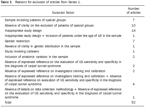 Considering that only one(16) among the 68 articles included in Series 1 detailed the calculation utilized for obtaining the sample size, the absence of such inclusion criterion was not considered as an excluding factor for the other articles. Apart from this latter criterion, ten articles have appropriately met all the other inclusion criteria. However six other articles were included as over inclusion by the third investigator, to avoid selection bias. The Series 2 resulting from the second selection comprised 16 articles. The third selection was aimed at assessing the methodological quality of the articles, following three criteria: randomization, masking of investigators and description of losses and exclusions. Of the 16 articles resulting from the second selection, four were included in Series 3. In the fourth selection, the quality of the four articles in Series 3 was analyzed by means of the CASP tool. All the articles selected in Series 3 were included in Series 4. It is important to comment on the reference standard utilized by the authors in the selected studies. As far as carpal tunnel syndrome is concerned, there is no reference standard unanimously accepted by the authors; some of them have utilized clinical data while others utilized electroneurography, and others an association of both reference standards. In the answers to the answering the questions of the CASP tool, all of these reference standards were considered. Among the four selected articles, three authors considered the association of clinical data and electroneurography as a reference standard(16-18) and one considered electroneurography alone(19). Table 2 summarizes the evidences in the articles selected in Series 4. 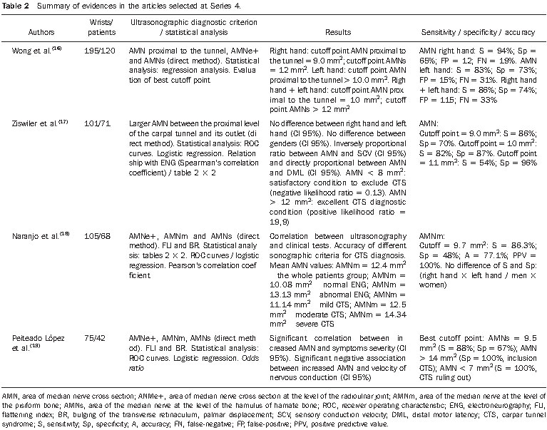 All the ultrasonography studies were performed with high resolution equipment (multifrequency transducers ranging from 5 to 13 MHz). In three studies, the observers were rheumatologists(1719) while in one(16), radiologists, all of them experienced in the method. In three studies(16,18,19) the reliability was evaluated among the readers, with a good correlation being observed among them, by obtaining the intraclass correlation coefficients(study 16: 0.710.90; study 18: 0.93; study 19: 0.912 0.987), highlighting the fact that the utilization of a standardized technique is sufficient to achieve a good reliability, even with observers with a baseline knowledge on ultrasonography, a relevant aspect in the generalization of the use of such method as a diagnostic evidence of carpal tunnel syndrome( 19). All examinations were performed with the patients sitting in front of the investigator, with flexed elbows and hands in the supine position, resting on a rigid surface. In all the studies, except the one developed by Wong et al.(16), ROC (receiver operating characteristics) curves were utilized in order to explore the relationship between sensitivity and specificity of the different described sonographic measurements and to define the best cutoff point for such measurements. Wong et al.(16) have utilized a linear regression software (Answer Tree, version 2.1; SPSS, Chicago, IL, USA) to evaluate such relationship. In the last phase, all the four articles selected in Series 4 were hierarchized according to the level of evidence and respective grade of recommendation of the scientific publication as proposed by Phillips et al.(15). Considering the fact that such Series included prospective, unicenter cohort studies with a good reference standard, the four articles obtained an evidence level 1b and grade of recommendation A, i.e., those studies present excellent evidence levels to routinely recommend the conduct proposed in the articles. DISCUSSION The diagnosis of carpal tunnel syndrome has always raised debate among clinical physicians, neurologists, surgeons, electroneurographers and radiologists, with no diagnostic criteria uniformity. What is the sonographic method accuracy in the diagnosis of carpal tunnel syndrome? The answer to this question would be highly beneficial for patients and would help to address the medical and legal problems arising from clinical, surgical legal requirements. In the current days, the best option for the decision making related to prevention, diagnosis and treatment of disorders is the practice of medicine based on evidences. The present study was developed in the search for the best scientific evidence related to the sonographic diagnosis of carpal tunnel syndrome. The utilization of the proposed method demonstrated how difficult the task was, requiring researchers availability, patience and skill for searching in databases, selecting and qualitatively analyzing the articles. The utilized model, in which two researchers worked independently in each phase of the process, with consensus meetings and, in the case of doubts, solution by a third more experienced researcher, demonstrated to be an indispensable requirement for the process completion. The search for articles in the electronic databases, with different forms of interaction with the user, has demonstrated to be the most important phase of the research, taking a greater length of time to be completed. The number of articles found was not significant to justify the utilization of a bibliographic management software (such as Endnote). Of the 68 articles selected in the first phase, 52 (76.4%) presented methodological shortcomings related to sampling, study design, diagnostic criteria and results presentation, causing the exclusion of such articles. After the application of the mentioned inclusion and exclusion criteria, the methodological quality of the articles selected in phase 2 (16 articles) was performed by means of a first objective filtering to measure the sample randomization, investigators masking, losses and exclusions. No specific scale was utilized. Instead, a tool developed by the authors of the present study was utilized as an adaptation of the criterion of patients masking because of the difficulty in achieving a double-blind masking in studies aimed at measuring the accuracy of a diagnostic method in which the index test (ultrasonography) and the test utilized one as a reference standard (electroneurography) are difficult to be disguised. This first filtering resulted in the selection of four articles. The main exclusion criterion was the omission regarding the sample randomization, with such criterion, isolatedly or associated with others, responsible for the exclusion of ten articles. The remaining exclusions occurred because of inappropriate investigators masking. Aiming at a more critical and deep analysis of the selected articles, the authors utilized a specific tool developed by CASP for evaluation of papers approaching diagnostic methods. As a result, the four articles selected in the first filtering (16, 17, 18 and 19) were included in the final series. It is necessary to draw a parallel between the selected articles, discussing and evaluating the methodological similarities and differences among them. All the studies were prospective, randomized and were developed with investigators masking. It is important to highlight that the studies developed by Wong et al.(16) and Ziswiler et al.(17) were the first prospective studies with the objective of measuring the diagnostic value of ultrasonography in carpal tunnel syndrome. In the four selected articles, 476 wrists were evaluated in a universe of 301 patients. The ratio between women and men in the studied samples ranged between 1.8(17) and 7.4(19). In the remaining studies, such ratio ranged between 4.4(16) and 4.7(18), practically the variation observed in the literature review data. The mean age in the samples ranged between 47 and 51 years, in agreement with data in the literature, which demonstrates that the incidence and prevalence of carpal tunnel syndrome is higher above the age of 40. It was observed that the cross sectional measurement of the median nerve (AMN) was the most relevant criterion for the sonographic diagnosis of carpal tunnel syndrome and that, in the selected studies, the most frequent cutoff value was between 9 and 10 mm2 (sensitivity = 8286.3%; specificity = 4887%). Another relevant information to be taken into consideration is the fact that the authors found cutoff values for the AMN at which the diagnosis of carpal tunnel syndrome can be ruled out (AMN < 8 mm2) or ratified (AMN > 13/ 14 mm2), with no need of complementary electroneurography. CONCLUSIONS The authors of all the studies concluded that the sonographic evaluation of patients with clinical suspicion of carpal tunnel syndrome can be performed as a first line test, with a good cost-benefit ratio, reducing the need of electroneurographic studies for such patients. Also, it is important to observe that ultrasonography can diagnose associated disorders and neural anatomic variations, in addition to the advantages of being a dynamic method easy to perform, at a relatively low cost as compared with electroneurography. The electroneurographic evaluation would be indicated in the case of symptomatic patients with negative diagnosis by ultrasonography. All the authors were unanimous in relation to the necessity of controlled and randomized prospective studies with larger samples for better understanding the actual value of ultrasonography in the diagnosis of carpal tunnel syndrome. According to the hierarchization of the evidence level of the selected studies (1b), the guidances contained in such articles can be considered as recommended evidences for application in the daily practice of decision making for professionals of different areas involved in the diagnosis of idiopathic or work-related carpal tunnel syndrome. Acknowledgements Special thanks to Conselho Nacional de Desenvolvimento Científico e Tecnológico (CNPq) (National Council for Scientific and Technological Development). REFERENCES 1. Atroshi I, Gummesson C, Johnsson R, et al. Prevalence of carpal tunnel syndrome in a general population. JAMA. 1999;282:1538. 2. Prick JJW, Blaauw G, Vredeveld JW, et al. Results of carpal tunnel release. Eur J Neurol. 2003;10: 7336. 3. Stoller DW. Magnetic resonance imaging in orthopaedics & sports medicine. 3rd ed. Baltimore, MD: Lippincott Williams & Wilkins; 2007. 4. Resnick DL, Kang HS, Pretterklieber ML. Internal derangements of joints. 2nd ed. Philadelphia, PA: WB Saunders; 2006. 5. Papaioannou T, Rushworth G, Atar D, et al. Carpal canal stenosis in men with idiopathic carpal tunnel syndrome. Clin Orthop. 1992;(285):2103. 6. Fernandes JL, Viana SL. Diagnóstico por imagem em reumatologia. Rio de Janeiro, RJ: Guanabara Koogan; 2007. 7. Aroori S, Spence RA. Carpal tunnel syndrome. Ulster Med J. 2008;77:617. 8. Koyuncuoglu HR, Kutluhan S, Yesildag A, et al. The value of ultrasonographic measurement in carpal tunnel syndrome in patients with negative electrodiagnostic tests. Eur J Radiol. 2005;56: 3659. 9. Machado DA, Martins WP. Síndrome do túnel do carpo. EURP. 2009;1:13640. 10. [No authors listed]. Practice parameter for carpal tunnel syndrome (summary statement). Report of the Quality Standards Subcommittee of the American Academy of Neurology. Neurology. 1993;43:24069. 11. Marinho VCC. Systematic reviews of controlled trials in general and oral health care. Braz J Oral Sci. 2003;2:21526. 12. Jadad AR, Moore RA, Carroll D, et al. Assessing the quality of reports of randomized clinical trials: is blinding necessary? Control Clin Trials. 1996;17:112. 13. Higgins JPT, Green S. Cochrane Handbook for Systematic Reviews of Interventions Version 5.0.2. [updated September 2009]. The Cochrane Collaboration, 2009. [cited 2010 Jan 17]. Available from: www.cochrane-handbook.org 14. Critical Appraisal Skills Programme (CASP). Diagnostic test studies. [cited 2010 Jan 18]. Available from: http://www.sph.nhs.uk/what-we-do/ public-health-workforce/resources/critical-appraisals- skills-programme 15. Phillips B, Ball C, Sackett D, et al. Oxford Centre for Evidence-based Medicine Levels of evidence. Grades of recommendation. [cited 2010 Feb 18]. Available from: http://www.cebm.net/ index.aspx?o=1025 16. Wong SM, Griffith JF, Hui ACF, et al. Carpal tunnel syndrome: diagnostic usefulness of sonography. Radiology. 2004;232:939. 17. Ziswiler HR, Reichenbach S, Vögelin E, et al. Diagnostic value of sonography in patients with suspected carpal tunnel syndrome: a prospective study. Arthritis Rheum. 2005;52:30411. 18. Naranjo A, Ojeda S, Mendoza D, et al. What is the diagnostic value of ultrasonography compared to physical evaluation in patients with idiopathic carpal tunnel syndrome? Clin Exp Rheumatol. 2007;25:8539. 19. Peiteado López D, Bohórquez Heras C, De Miguel Mendieta E, et al. Validez y utilidad de la ecografía en el síndrome del túnel carpiano. Reumatol Clin. 2008;4:1006. 1. Specialist in Ultrasonography by Colégio Brasileiro de Radiologia e Diagnóstico por Imagem (CBR), Fellow Masters Student of Forensic Expertise at Faculdade de Odontologia de Pernambuco Universidade de Pernambuco (UPE), Camaragibe, PE, Brazil. 2. PhD of Collective Health, Associate Professor at Faculdade de Odontologia de Pernambuco Universidade de Pernambuco (UPE), Camaragibe, PE, Brazil. 3. PhD of Health Sciences, Associate Professor at Faculdade de Odontologia de Pernambuco Universidade de Pernambuco (UPE), Camaragibe, PE, Brazil. 4. Specialist in Labor Medicine, Fellow Masters Student of Forensic Expertise at Faculdade de Odontologia de Pernambuco Universidade de Pernambuco (UPE), Camaragibe, PE, Brazil. 5. Graduate Student of Odontology, Fellow Masters Student of Forensic Expertise at Faculdade de Odontologia de Pernambuco Universidade de Pernambuco (UPE), Camaragibe, PE, Brazil. 6. Graduate Student of Odontology, Fellow Masters Student of Collective Health at Faculdade de Odontologia de Pernambuco Universidade de Pernambuco (UPE), Camaragibe, PE, Brazil. Mailing Address: Dra. Evelyne Pessoa Soriano Estrada de Aldeia km 13, nº 200, Condomínio Torquato Castro 1, Aldeia Camaragibe, PE, Brazil, 54783-010 Email: evelynesoriano@yahoo.com.br Received November 26, 2010. Accepted after revision January 27, 2011. Study developed during a Masters Fellowship in Forensic Expertise at Faculdade de Odontologia de Pernambuco Universidade de Pernambuco (UPE), Camaragibe, PE, Brazil. |
|
Av. Paulista, 37 - 7° andar - Conj. 71 - CEP 01311-902 - São Paulo - SP - Brazil - Phone: (11) 3372-4544 - Fax: (11) 3372-4554