Radiologia Brasileira - Publicação Científica Oficial do Colégio Brasileiro de Radiologia
AMB - Associação Médica Brasileira CNA - Comissão Nacional de Acreditação
 Vol. 43 nº 6 - Nov. / Dec. of 2010
|
CASE REPORT
|
|
Von Meyenburg complex: case report and literature review* |
|
|
Autho(rs): Luiz Fernando Vitule1; Flávia Mitsuse Simionato2; Milena Loureiro de Melo3; Rafael Yoshitake3 |
|
|
Keywords: Hamartoma; Intrahepatic bile ducts; Cholangiocarcinoma; Polycystic kidney; Imaging diagnosis; Ultrasonography. |
|
|
Abstract: INTRODUCTION
Biliary hamartomas, also known as von Meyenburg complex, are classically described as benign liver malformations(1). Von Meyenburg complex is rarely diagnosed and constitutes a typically asymptomatic finding(2,3). Imaging findings, generally incidental, lead to other diagnostic investigations and invasive approaches, particularly in oncologic patients, which result in considerable expenses and psychological distress for these patients(3,4). However, there has been debate in the literature as regards a possible malignant potential of von Meyenburg complex(5). The present article is aimed at reporting a case of von Meyenburg complex associated with renal cysts in a 27-year-old female patient, considering the scarcity of descriptions of similar cases, besides imaging findings that mimic metastases and the possible malignant potential currently discussed in the literature. CASE REPORT A white, female, 27-year-old patient born in São Paulo, SP, Brazil, was admitted to the emergency center of a general hospital in the city of São Paulo, in March/2010, with belt pain in the abdomen and vomiting for two days. The patient denied the previous occurrence of similar symptoms or any history of known disease. History of a sister with renal cysts was reported. Abdominal ultrasonography was requested and demonstrated multiple, diffuse, hyperechogenic nodules in the liver (Figure 1), numerous, bilateral renal cysts (Figure 2), hydropic bladder with thick bile, subtle biliary ducts dilatation and thickened pancreas. With the hypothesis of acute biliary pancreatitis, the patient received clinical treatment and was submitted to cholecystectomy after the acute picture. During the surgical procedure, fragments of hepatic nodules were collected for biopsy. Histological analysis demonstrated tortuosity and cystic dilations involved by granulomatous fibrotic tissue of intrahepatic biliary ducts suggestive of von Meyenburg complex (Figure 3). The patient is currently asymptomatic and is undergoing ambulatory follow-up. 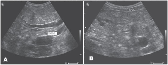 Figure 1. Liver ultrasonography demonstrating the presence of multiple hyperechogenic nodular images with irregular margins, some of them presenting acoustic shadowing and others with posterior wall enhancement in the periportal region (A) and diffusely distributed (B), with up to 1.2 cm in size. 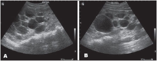 Figure 2. Sonographic images of right (A) and left (B) kidneys showing the presence of multiple cortical cysts of varied sizes, diffusely distributed throughout the parenchyma. 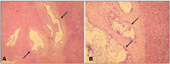 Figure 3. Histopathological analysis of surgical hepatic specimens. A: preserved hepatic tissue with irregular and dilated biliary ducts (arrows). B: Biliary duct cysts with granulomatous foreign body reaction with cholesterol crystals (arrows). DISCUSSION Von Meyenburg complex is considered as a benign liver malformation that histologically presents as cystic dilations of biliary ducts with 1 to 15 mm in diameter, involved by abundant fibrotic tissue(1,2). Due to the fact that typically this disease does not cause clinical complaints, its finding is frequently incidental on imaging studies. The prevalence of von Meyenburg complex is in the range of 0.6% in 2.000 biopsies(2). The sonographic findings of von Meyenburg complex are variable, including multiple, small, hyperechogenic images, with poorly delimited margins, with or without posterior reverberation, or even hypoechogenic images with a target pattern and well delimited margins(3,6). As regards the number of hamartomas at imaging studies, they may present as a uninodular lesion, but most frequently they are multinodular(1,3,6). Such characteristic leads to further investigation, particularly in the presence of some known tumor, considering its similarity with metastases, which represents an additional burden for the health system besides physical and psychological distress for the patient(3). Yet, cases of exploratory laparotomy have been described(4). So, in face of oncologic cases, sonographic and computed tomography findings may be inconclusive(7). However, magnetic resonance imaging presents as a highly accurate alternative for the diagnosis and follow-up of von Meyenburg complex(7,8). In the present case report, the presence of hepatic nodules observed at ultrasonography led to the suspicion of von Meyenburg complex, considering the patients age and the absence of a relation with her clinical condition. Considering the possibility of biopsy during the cholecystectomy, other imaging studies were not utilized. Studies with a great number of cases of von Meyenburg complex, evaluating association with malignancy were not found in the literature. However, there are several case reports and cases series describing association of von Meyenburg complex with cholangiocarcinoma, so the von Meyenburg complex benignity is currently questioned and considered as a possible risk factor for the development of cholangiocarcinoma(5). There is a correlation between adult polycystic disease and von Meyenburg complex; the greater the number of biliary hamartomas, the greater the association with adult polycystic disease(9). Some studies with autopsy report von Meyenburg complex as a non uncommon finding although this is a rare finding both in the clinical practice and in the radiological literature(3,7). This occurs because of the asymptomatic presentation of this condition, besides the small dimensions and non-recognition of the lesions(3,7). Therefore, because of the increasing number of cases with malignant transformation, statistical studies are necessary for a deeper investigation of the association between von Meyenburg complex and cholangiocarcinoma. In the clinical follow-up of renal cysts in cases of adult polycystic disease, the relevance of screening for von Meyenburg complex is emphasized, because of the association between both diseases. Additionally, in spite of the usual benign behavior of von Meyenburg complex, it is prudent to follow-up such cases, considering the possible malignant potential of this condition. REFERENCES 1. Lev-Toaff AS, Bach AM, Wechsler RJ, et al. The radiologic and pathologic spectrum of biliary hamartomas. AJR Am J Roentgenol. 1995;165:30913. 2. Thommesen N. Biliary hamartomas (von Meyenburg complexes) in liver needle biopsies. Acta Pathol Microbiol Scand A. 1978;86:939. 3. Machado MM, Rosa ACF, Barros N, et al. Aspectos ultra-sonográficos dos hamartomas dos ductos biliares (complexo de von Meyenburg): resultado de uma busca ativa de oito anos. Radiol Bras. 2003;36:1536. 4. Durán-Vega HC, Luna-Martínez J, González-Guzmán R, et al. Hamartoma of the bile ducts. Report of a case and review of the literature. Rev Gastroenterol Mex. 2000;65:1248. 5. Xu AM, Xian ZH, Zhang SH, et al. Intrahepatic cholangiocarcinoma arising in multiple bile duct hamartomas: report of two cases and review of the literature. Eur J Gastroenterol Hepatol. 2009;21:5804. 6. Machado MM, Rosa ACF, Barros N, et al. Múltiplos pequenos nódulos hepáticos hiperecogênicos sem reverberação sonora posterior: outra forma de apresentação dos hamartomas dos ductos biliares. Radiol Bras. 2005;38:38991. 7. Zheng RQ, Zhang B, Kudo M, et al. Imaging findings of biliary hamartomas. World J Gastroenterol. 2005;11:63549. 8. Nagano Y, Matsuo K, Gorai K, et al. Bile duct hamartomas (von Mayenburg complexes) mimicking liver metastases from bile duct cancer: MRC findings. World J Gastroenterol. 2006;12:13213. 9. Redston MS, Wanless IR. The hepatic von Meyenburg complex: prevalence and association with hepatic and renal cysts among 2843 autopsies [corrected]. Mod Pathol. 1996;9:2337. 1. PhD, Radiology Residency Coordinator at Hospital Geral de Pedreira (HGP), Pedreira, SP, Radiologist at Instituto de Radiologia do Hospital das Clínicas da Faculdade de Medicina da Universidade de São Paulo (InRad/HC-FMUSP) and Sistema Estadual de Diagnóstico por Imagem I (SEDI I), São Paulo, SP, Brazil. 2. MD, Radiologist at Hospital Geral de Pedreira (HGP), Pedreira, SP, Brazil. 3. MDs, Residents in Radiology and Imaging Diagnosis at Hospital Geral de Pedreira (HGP), Pedreira, SP, Brazil. Mailing Address: Dr. Luiz Fernando Vitule Rua Ouro Branco, 75, ap. 51B, Jardim Paulista São Paulo, SP, Brazil, 01425-080 E-mail: fernandovitule@terra.com.br Received August 30, 2010. Accepted after revision September 30, 2010. * Study developed at Hospital Geral de Pedreira (HGP), Pedreira, SP, and Sistema Estadual de Diagnóstico por Imagem I (SEDI I), São Paulo, SP, Brazil. |
|
Av. Paulista, 37 - 7° andar - Conj. 71 - CEP 01311-902 - São Paulo - SP - Brazil - Phone: (11) 3372-4544 - Fax: (11) 3372-4554