Radiologia Brasileira - Publicação Científica Oficial do Colégio Brasileiro de Radiologia
AMB - Associação Médica Brasileira CNA - Comissão Nacional de Acreditação
 Vol. 43 nº 5 - Sep. / Oct. of 2010
|
ORIGINAL ARTICLE
|
|
Freeware medical image viewers: can we rely only on them? |
|
|
Autho(rs): Filipe Ramos Barra1; Renato Ramos Barra2; Alaor Barra Sobrinho2 |
|
|
Keywords: DICOM; PACS; Medical images viewer; Teleradiology; Radiology information systems. |
|
|
Abstract: INTRODUCTION
With the technological progress, radiology services are increasingly resourcing to information technology and the different imaging modalities are being integrated. Computed tomography (CT), magnetic resonance imaging (MRI), positron-emission tomography (PET-CT) as well as ultrasonography, angiography, plain radiography and even modern fluoroscopy generate digital images to be analyzed on workstations. Such images are eventually utilized for cases recording, classes, conferences and, scientific meetings among other activities. A difficulty faced in these cases lies on the fact that such images are available in the DICOM (digital imaging and communications in medicine) format, a global standard for medical images, and cannot be visualized or manipulated in a Windows environment or through Power Point, demanding the use of specific applications. There are softwares that convert DICOM images into known formats, simple CD viewers and more complex viewers that run as clients in a network PACS (picture archiving and communication system). Considering that most modern imaging diagnosis centers have at least a small network connecting the CT or MRI equipment to a workstation, the use of viewers running as a PACS client is much more practical and fast. The utilization of proprietary PACS may not be feasible because of their high cost. Several freeware softwares for DICOM images visualization are available at internet, but only a few run as PACS clients. As freeware and open-source softwares are analyzed, one realizes that such softwares require a very frequent updating, to an extent that upon searching for softwares that were previously evaluated, one observes that several of those applications are no longer available, or are no longer being updated, and some are no longer freeware, with few alternatives remaining currently available(14). On the other hand, new and powerful softwares have been developed, incorporating more complex functions. The freeware considered as the gold standard among the medical viewers is the OsiriX that, among the currently available applications, presents the highest number of functions and tools. However, it is only available for the MacOS X(5) operating system. In Brazil, there are some research groups with good softwares and results; however some of them are proprietary. Among these applications, those developed by Universidade Federal de Santa Catarina (UFSC), Faculdade de Medicina de Ribeirão Preto of Universidade de São Paulo (FMRPUSP) and Pontifícia Universidade do Rio Grande do Sul (PUC-RS)(68) should be highlighted. The Cyclops Medical Station developed by UFSC is a powerful tool, however it is no longer available on the internet. The objective of this study is evaluating and testing several softwares, selecting those capable of running as PACS clients, besides evaluating the feasibility of using them in personal computers. The present study is not aimed at advertising and attributing value to any software in particular. MATERIALS AND METHODS A search was performed at Google by utilizing the keywords free dicom viewer and free dicom viewing. Additionally, a search was carried out at the specialized medical freeware Idoimaging, RTstudents and Medfloss websites(911). Some softwares that were known and used by the authors were also included. Considering that some softwares did not comprise detailed descriptions of their different functions, the authors have opted for testing those Windows-compatible capable of running as viewers. Approximately 70 softwares were found. Eleven of them were capable of searching and recovering DICOM images (DICOM Q/R), while running as a client in a PACS network. Five applications were excluded for several reasons (Table 1) and six were finally selected (ClearCanvas Workstation 2.0, KPACS 1.6.0, Onis 2.1, Synedra View Personal 1.0.12.3, Mito 1.0, Tudor DicomViewer 1.3). 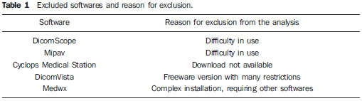 Considering that one of the proposals of the present study was evaluating the possibility of using medical viewers in personal computers, the authors have opted for testing the softwares in a computer with baseline configurations: Intel Celeron 1.6 GHz with 2 GB of RAM memory, 40 GB of storage capacity, and on-board graphics accelerator, running Windows XP SP2. Another computer (AMD Athlon 64 Dual Core 2.7 GHz with 4 GB RAM memory RAM, 1 TB of storage capacity and ATI Radeon HD 4350 graphics accelerator) was also utilized running Windows 7, in order to test compatibility of the softwares with this operating system. Some functions that were considered as the most essential ones in the radiologists daily practice were evaluated taking the services needs into consideration, as follows: multiplanar reconstruction (MPR); maximum intensity projection (MIP) reconstruction; volume rendering (VR) and image fusion capabilities; capacity of synchronizing different image series and showing reference lines with possibility of adding comments on the studies; capacity of showing images in a sequential mode (cine); working with multiple monitors; running as a PACS server; recording CDs with DICOM images; making the studies data anonymous; exporting JPG, BMP and DICOM images, and compatibility with Windows 7. RESULTS The results of the software analyses are shown on Tables 2 and 3. The following topics include comments on the softwares highlighting their strengths and weaknesses. 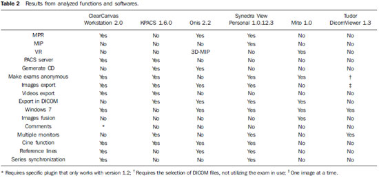 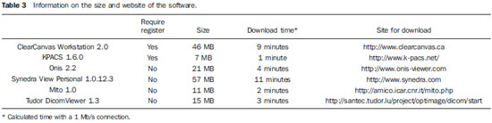 ClearCanvas Workstation It is a part of an open-source solutions package that also comprises an imaging server and a RIS (radiology information system). Among the applications evaluated in the present study, it is the only accredited software (Canada Department of Health) for use with diagnosis purposes. The current version (2.1) allows MPR, however it is not possible to change image display on the screen neither saving the reconstructed images. It synchronizes two images series, allowing the localization of a specific point on the different series (Figure 1). It does not export the DICOM images neither generate CDs, however it runs as a PACS server. As a server, it is quite heavy, using fixed 96 Mb of memory. It allows the utilization of usercreated plugins. One of them (study tagging), allows the addition of comments to the studies and later searches, which is very useful for filing cases, however it runs only on the version 1.2, which does not perform MPR. 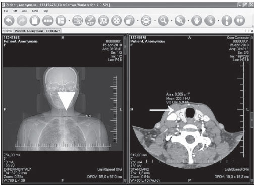 Figure 1. ClearCanvas software screen. Two series are displayed, with identification of the reference line and the spatial locator (arrowhead). There is also a region of interest (ROI) on the right lobe of the thyroid (arrow). KPACS Among the evaluated softwares, this is the oldest and is the only one that is non-Windows 7-compatible. It is a freeware version of a more complete commercial package. The function buttons are large and located at the bottom of the screen, with a different interface that lacks a nice appearance (Figure 2). It is quite simple, does not perform MIP, MPR or VR, however it runs as a PACS server, allowing the use of multiple monitors, besides generating CDs with a built-in viewer. As a PACS client it is a little slow, sometimes giving the impression that the system has locked up. 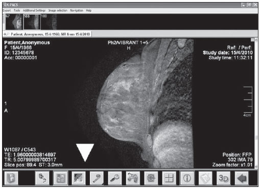 Figure 2. KPACS software screen. Notice that the function buttons are large and located at the bottom of the screen (arrowhead). Onis A freeware version of a commercial program presenting a simple and agreeable interface. In the current version (2.2) the databank capacity is limited to 15 patients. It is the only software among the tested ones that does not make the exams data anonymous. Another difference is the possibility of visualizing the images without having to download them from the server (on-demand), thus not requiring much storage capacity. It performs MPR, allowing the visualization of the desired plane as a single image on the screen (Figure 3). It generates 3D models with MIP techniques and allows the generation of CDs with a built-in, lightweight (9 Mb) and simple viewer (without the MPR function). This software is quite fast, in spite of all its capabilities. 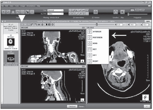 Figure 3. Onis software screen. The multiplanar reconstruction function (MPR) is being used. Notice that it is possible to select the desired plane (arrow), as well as the type of layout (arrowhead). The buttons are small and agreeable. Synedra View Personal A powerful PACS client. The interface is quite unique, with the buttons located at the bottom of the screen, which, initially, may make its use difficult. It is ISO 9001 and ISO 13485 certified, this latest one being a specific certification for medical devices. It allows the utilization of two monitors and performs both MPR and MIP, with the possibility of adjusting the slices thickness and also the way they are grouped, MIP, medium or mini-MIP (minimum intensity projection) (Figure 4). Its most important shortcoming is the fact that it lacks a data bank, i.e., it does not save the visualized exams, which can only be done manually for later visualization. It is also possible to save the performed reconstructions, an interesting feature. This is a rapid and friendly software. 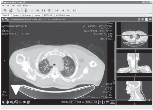 Figure 4. Synedra View Personal software screen. The multiplanar reconstruction function is being used, showing the three orthogonal planes. On the main picture, the maximum intensity projection function (MIP) is being used, showing the reconstruction of a 25 mm-thick section. The function buttons are located at the bottom of the screen (arrowhead). Mito Mito (medical imaging toolkit) is an open source software with some interesting features. Among the evaluated softwares, it is the only one capable of performing images fusion (Figure 5). It performs VR and MIP. It does not allow images to be saved in the JPG and BMP formats and cannot open more than one series at a time. As compared with others, it is a little bit slow. 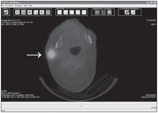 Figure 5. Mito software screen. A PET-CT image is shown. With the images fusion function one can observe a hyperfunctioning lymph node at right (arrow). Tudor DicomViewer 1.3 A simple software with few functions. It requires Java Runtime Environment® installation. It does not perform MPR, MIP or VR. It is capable of saving images in JPG or BMP, however, only one image at a time (Figure 6). In spite of its simplicity, it allows the use of three monitors. The exams can be opened with the ImageJ, a software that is included, and presents innumerable resources and plugins, MPR and VR, for example. 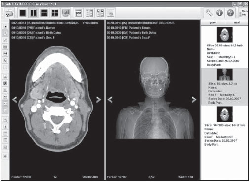 Figure 6. Tudor Dicom Viewer screen. Two series are shown. Notice the absence of reference lines or synchronization. DISCUSSION Over the past few years, notebooks and personal computers have become very popular and widely available, and are becoming increasingly powerful. A digital transformation also occurred in radiology services. With the availability of digital apparatuses (CT, MRI, PET-CT, ultrasonography) older X-ray and fluoroscopy apparatuses were converted, allowing the direct and indirect acquisition of digital images. Thanks to the use of RIS and PACS systems and report workstations, films and paper have been abandoned in the whole process, from the images acquisition to the final report issuing. For this purpose, it is necessary to utilize updated softwares compatible with such changes, which make the use of freeware more difficult, considering that they do not generate a return on the investment made by their developers, since their updating processes take a long time to be completed or are not performed at all. Several tools are utilized in the radiologists daily practice. Current tomography apparatuses are capable of acquiring volumes and, with the use of MPR, reconstructions can be made, sometimes isotropic, in the desired plane, being useful in the images interpretation. MIP is a powerful tool for the analysis of CT angiography, MR angiography, and MR cholangiography, but it also facilitates the identification of pulmonary nodules at chest CT. With the use of VR, more agreeable and easily visualizable both by the laymen and the assisting physician who have requested the study, coming closer to reality, thus enhancing the value of the imaging method. The PET/CT fusion image is useful in the accurate anatomic identification of PET uptake areas. The possibility of adding comments on the images facilitates later searches for findings or diseases, considering that with the greater storage capacity, personal computers have been increasingly utilized to file cases. In cardiac radiology, the cine tool is essential, as it provides dynamic images of the functioning heart. Synchronizing different imaging series and showing reference lines help in the identification of lesions or specific points, a very useful capability at MRI. The utilization of medical viewers for diagnostic purposes is somewhat controversial. The Agência Nacional de Vigilância Sanitária (Anvisa)(the Brazilian agency for health surveillance) does not provide specific regulations on this matter, therefore the authors resource to foreign agencies. Among the evaluated programs, ClearCanvas is certified by the Canadian Department of Health (Health Canada MDR) and both ClearCanvas and Synedra are certified under ISO 13485:2003 (specific international standard for medical equipment). CONCLUSION The internet is a vast source of information and softwares. Several DICOM viewers, many of them freeware, are currently available These are excellent work tools, representing good alternatives to commercial softwares. The selection of the appropriate software is many times difficult. The software with more functions is not always the best. Most of times, the best option is the one that best suits the users needs, whether be it MPR, image fusion, CD generation with a built-in viewer or a simple and rapid PACS client. In cases where the software will be permanently linked to the server network, visualizing a single image at a time, Synedra and Onis are indicated for being fast and not highly demanding in terms of computer memory; the difference between them is that the first one performs MIP and the second, VR. In cases where the exams are interpreted far from the network server or even in cases where there is the need to store them in files or for later analysis, ClearCanvas is indicated. At IMEB, for example, two softwares are currently in use, one of them more complete and fast for CT, MRI and ultrasonography that does not perform MIP (ClearCanvas), and another simpler one for PET-CT with fusion capability (Mito). Clear Canvas has been chosen because of its higher number of functions required in the IMEB daily routine and for its easier utilization. One should remind that it is important to analyze the personal needs and then select a software that best suits such needs, and if at all possible testing an additional alternative. REFERENCES 1. Liao W, Deserno TM, Spitzer K. Evaluation of free non-diagnostic DICOM software tools. vol. 6919. SPIE; 2008. [cited 2010 Apr 10]. Available from: http://spie.org/x648.html?product_id=770431 2. Zeman RK, Lyshkow H, Garra BS, et al. Viewing DICOM-compliant CT images on a desktop personal computer: use of an inexpensive DICOM receive agent and freeware image display applications. AJR Am J Roentgenol. 1999;172:3058. 3. Escott EJ, Rubinstein D. Free DICOM image viewing and processing software for your desktop computer: whats available and what it can do for you. Radiographics. 2003;23:134157. 4. Varma DR. Free DICOM browsers. Indian J Radiol Imaging. 2008;18:126. 5. Rosset A, Spadola L, Ratib O. OsiriX: an opensource software for navigating in multidimensional DICOM images. J Digit Imaging. 2004;17:20516. 6. The Cyclops Group. Research on Medical Imaging Software [Internet]. Florianópolis, Brasil: The Cyclops Group; c1992-2010; [cited 2010 Apr 10]. Available from: http://cyclops.telemedicina.ufsc.br/ 7. Caritá EC, Matos ALM, Azevedo-Marques PM. Ferramentas para visualização de imagens médicas em hospital universitário. Radiol Bras. 2004;37:43740. 8. Andrade MA, Costa MVS, Silva AMM. Javabased plugin for tomographic reconstruction for SPECT data. Med Phys. 2006;33:2015. 9. Crabb A. Free DICOM and medical image viewer / Converter software, open source DICOM conversion [Internet]. Baltimore, USA; c2002-2010; [revised 2009 Nov 19; cited 2010 Apr 10]. Available from: http://www.idoimaging.com/ 10. RTstudents.com. Radiology for students and professionals [Internet]. USA: c2004-2010; [revised 2009 Nov 19; cited 2010 Apr 10]. Available from: http://www.rtstudents.com/pacs/free-dicomviewers.htm 11. Schmuhl H. Medical free/libre and open source software [Internet]. Munich, Germany; [cited 2010 Apr 10]. Available from: http://www.medfloss.org/ 1. MD, Resident in Radiology and Imaging Diagnosis at IMEB Imagens Médicas de Brasília, Brasília, DF, Brazil. 2. Nuclear Physicians, Department of Nuclear Medicine, IMEB Imagens Médicas de Brasília, Brasília, DF, Brazil. Mailing address: Dr. Filipe Ramos Barra SHIN, QI 04, conjunto 05, casa 24, Lago Norte Brasília, DF, Brazil, 71510-250 E-mail: filipebarra@gmail.com Received May 9, 2010 Accepted after revision September 13, 2010 Study developed at IMEB Imagens Médicas de Brasília, Brasília, DF, Brazil |
|
Av. Paulista, 37 - 7° andar - Conj. 71 - CEP 01311-902 - São Paulo - SP - Brazil - Phone: (11) 3372-4544 - Fax: (11) 3372-4554