Radiologia Brasileira - Publicação Científica Oficial do Colégio Brasileiro de Radiologia
AMB - Associação Médica Brasileira CNA - Comissão Nacional de Acreditação
 Vol. 40 nº 6 - Nov. / Dec. of 2007
|
ORIGINAL ARTICLE
|
|
Radioprotection, doses and risks in the radiological assessment of paranasal sinuses in children, in hospitals of Belo Horizonte, MG |
|
|
Autho(rs): Marco Aurélio de Sousa Lacerda, Helen Jamil Khoury, Teógenes Augusto da Silva, Camila Maria de Sousa Lacerda, Alexandre Ferreira Carmo, Márcio Tadeu Pereira |
|
|
Keywords: Pediatric radiology, Patient dose, Organ dose |
|
|
Abstract:
IPhD of Nuclear Sciences, Assistant Researcher at Centro de Desenvolvimento da Tecnologia Nuclear/Comissão Nacional de Energia Nuclear (CDTN/CNEN), Belo Horizonte, MG, Brazil
INTRODUCTION Special attention should be paid to radiographic examinations performed in pediatric patients, considering they present cells with high radiosensitivity and longer life expectancy, significantly increasing the stochastic effects from radiological examinations as compared with examinations in adults(1). Furthermore, innumerable peculiarities inherent in pediatric radiology require specific procedures for the performance of radiographic examinations in children. In this context, the Commission of European Communities has published a document establishing quality criteria for diagnostic radiographic imaging of pediatric patients, defining the requirements for a normal, basic radiograph, specifying anatomical image criteria and significant image details, providing guidance as regards the choice of the radiographic technique, and proposing reference values for radiation dose per patient in the most frequent radiographic examinations(2). Cook et al.(3) have developed similar guidelines, in addition to reference criteria, patient preparation, practical instructions, exposure factors and doses related to four age ranges. Computed tomography is the gold standard for the diagnosis of sinusitis, one of the most frequent diseases in children(4,5). However, if acute sinusitis is suspected, conventional radiography is the investigative method of choice(6). The radiographic projections usually adopted for the diagnosis of acute sinusitis are(6,7): a) Waters or occipitomental view, for preferential evaluation of the maxillary sinuses; b) Caldwell or occipitofrontal view for evaluation of frontal and anterior ethmoidal sinuses; c) Hirtz or submental vertex view, where posterior ethmoidal and sphenoidal sinuses area evaluated; d) lateral view, allowing the evaluation of all the facial sinuses, particularly the frontal and sphenoidal ones, the maxillary sinuses posterior wall and floor, the nasal fossae and, also, the rhinopharynx. Some clinical studies have demonstrated that a single Waters view (occipitomental) presents a very high agreement with the four-view sinus series(8). With a single occipitomental view, 87% accuracy in the diagnosis of acute sinusitis in children can be achieved(9). In this context, the British Guidelines on best practice in the x-ray imaging of children(3) provides quality criteria only for the occipitomental and lateral views. The present study was aimed at evaluating the frequency of radiographic projections, as well as radioprotection conditions, radiographic techniques employed, entrance air kerma, and doses to the most exposed organs in pediatric patients of hospitals in the city of Belo Horizonte, MG, Brazil.
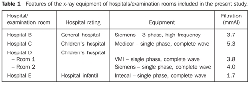
MATERIALS AND METHODS The authors have collected patients' data (sex, age, weight and height) and radiographic techniques parameters (kV, mA.s, focus-skin distance and exposure time) adopted for radiographic assessment of facial sinuses in children between 1 and 16 years of age in four Belo Horizonte hospitals. The main features of the x-ray equipment evaluated in the present study are shown on Table 1.
During the radiographic procedures in the hospitals, radioprotection conditions were evaluated, by means of observation of the following items: utilization of lead protective devices for the patient, utilization of antiscattering grids, collimation cylinder and added tube filtration. The x-ray tube output in the five examination rooms was determined for different kilovoltage values (kV) applied to the tube, with a fixed tube loading (Q). For measuring the air kerma rate, a previously calibrated Radcal/MDH 10X5-6 ionization chamber connected to an also calibrated Radcal/MDH 9015 electrometer was utilized. The ionization chamber was positioned on the examination table, on the center of the radiation field, at a 100 cm distance from the focus, and at 20 cm from the table. Based on the x-ray tube output and irradiation parameters utilized for each of the patients, it was possible to estimate the incident air kerma (INAK) and the entrance air kerma (ESAK), by means of the following equations:
where: Ri is the x-ray tube output for the radiographic technique employed in the examination, interpolated from the output curve as a function of the kilovoltage [Ri = a · (kV)b], a and b corresponding to the curve adjustment parameter(10); Q is the result of the tube current (I) by the exposure time (t), employed in the examination, in milliampere-second (mA.s); Dref. is the distance adopted for the output measurement (1 m); DFP is the focus-skin distance, in meters; BSF is the non-dimensional backscattering factor related to the field size, equipment filtration and the radiographic technique employed. In the present study a fixed value (1.30) was adopted for BSF(11). The software PCXMC was utilized for estimating the doses to the most exposed organs based on the incident air kerma, the patients' characteristics, and the radiographic techniques employed(12).
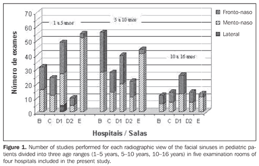
RESULTS Figure 1 shows the number of examinations for each radiographic projection on facial sinuses of pediatric patients divided into three age ranges: 1-5 years, 5-10 years, and 10-16 years, in five examination rooms of four hospitals.
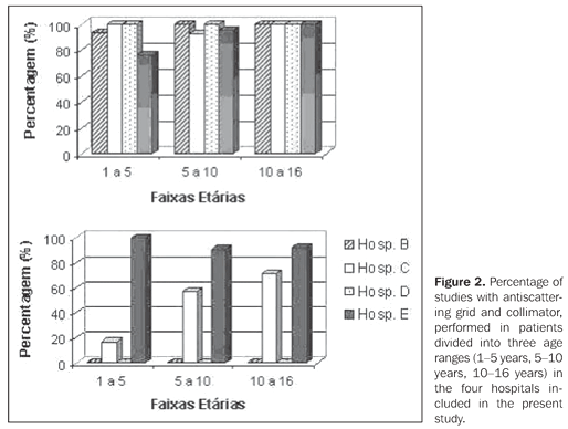
Figure 2 shows, respectively, for the same age ranges, the percentage of examinations utilizing antiscattering grid and collimator of the x-ray tube in the four hospitals included in the present study.
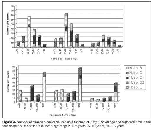
Figure 3 shows, respectively, the number of examinations of facial sinuses as a function of the x-ray tube voltage and exposure time in the four hospitals, for patients in the same age ranges.
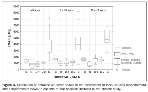
Figure 4 shows the distribution of estimated entrance air kerma rates for each radiographic projection for patients in the three age ranges, in each examination room of the hospitals included in the present study. This latest figure shows only the occipitofrontal and occipitomental views which are most frequently requested by assisting physicians.
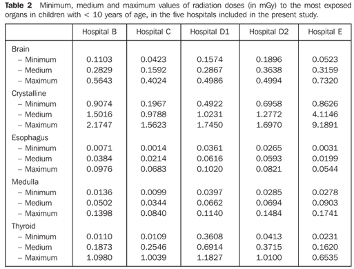
Table 2 shows minimum, medium and maximum values of doses to most exposed organs (brain, crystalline, esophagus, medulla, and thyroid) estimated for occipitomental and occipitofrontal views. Considering that the software PCXMC does not calculate the dose to the crystalline, this value was approximately estimated by the skin entrance dose (personally given information by Tapiovaara M).
DISCUSSION Figure 1 shows that the projections most frequently utilized for facial sinuses assessment are the occipitofrontal and occipitomental ones. Only in the hospital E, a single occipitomental view was frequently requested. In the other institutions, occipitofrontal and occipitomental views were requested in conjunction. Lateral sinus view was observed only in the hospital D, although in a small part of the examinations (< 10%). For this reason, the lateral view has not been included in the other analyses of the present study. As regards lead protective devices during the examinations, the authors observed that only the hospital D utilized thyroid shielding in about 25% of examinations. In the other hospitals, the utilization of this type shielding was not observed. On the other hand, by the analysis of Figure 2, It can be observed that in the hospitals B and D collimation cylinders were not utilized according to the recommendations of the British guidelines on best practice in the x-ray imaging(3) and one of the main manuals of radiological technique(7). The non-utilization of collimation cylinders in radiographic sinus evaluation may unnecessarily expose the thyroid (this is a highly radiosensitive organ) and esophageal regions, besides increasing the dose to other organs such as crystalline, brain and medulla. This fact may be observed on Table 2. For this reason, the utilization of a cylindrical collimator coupled with the x-ray equipment for studies of facial sinuses. An interesting fact observed in the hospital D is that a lead plate with a central orifice placed on the chassis, in a way that the appearance of the image obtained is similar to the one if a collimator cylinder were utilized. This corroborates the absence of a culture of radiological protection, considering that technicians try to meet the medical requirements (visualization of a round-shaped image) with no concern regarding the protection of the patient. Also, in Figure 2, it is possible to observe that in almost all examinations antiscattering grids were utilized, contrarily to the recommendations included in the British guidelines on best practice(3), about the non-utilization of antiscattering grids in the examination of facial sinuses of children with < 10 years of age. The utilization of antiscattering grids is important for reducing the scattered radiation. However, the intensity of the radiation beam is reduced, requiring higher tube loading (mA.s), and increasing the dose to the patient. Considering that the scattered radiation intensity depends, among other factors, on the patient thickness; and, considering the lower thickness of pediatric patients, antiscattering grids are deemed to play a non-significant role in the reduction of scattered radiation, so its utilization will only increase the dose to the patient. Figure 3 shows the number of facial sinuses studies (occipitofrontal and occipitomental in conjunction) as a function of the tube voltage and exposure time. Hospital D employs higher kilovoltage (kV); and hospitals C and E, lower kilovoltage and longer exposure time. Along the development of the present study, the authors could observe that only the hospital B x-ray equipment was able to provide exposure times shorter than 10 ms, and, therefore this is the only hospital in the present study meeting the minimum best practices requirements for radiographic examination in children(2,3). Notwithstanding, despite the equipment capability, the number of examinations with longer exposure time (more than 40 ms) is still considerably high in this hospital. As a result, the entrance air kerma rates found in this hospital are higher than those found in hospitals C and D, and lower than those found in the hospital E, as per Figure 4. This figure also demonstrates that hospital E presented a high variation in entrance air kerma rates. Mean entrance air kerma values for five examination rooms of four hospitals were, respectively: 1398 µGy, 829 µGy, 877 µGy, 1168 µGy and 3886 µGy for patients in the age range between one and five years; 1561 µGy, 1107 µGy, 1184 µGy, 1327 µGy and 4400 µGy for patients in the age range between five and 10 years; and 1613 µGy, 960 µGy, 1520 µGy, 1518 µGy and 5091 µGy for patients in the age range between 10 and 16 years. All of these values were above the reference levels proposed by the British guidelines(3). One can observe that mean values found at hospital E are three-fold higher than those found at the other hospitals. Considering that these values correspond to estimates of mean entrance air kerma/view, the two-view examinations (occipitofrontal and occipitomental) which are usual in the hospitals B, C, and D, expose the pediatric patients to lower risks than those resulting from the single-view examination (occipitomental) performed in the hospital E. Table 2 demonstrates that, despite the significantly highest entrance air kerma rates in the hospital E, the doses to thyroid and esophagus were lower. This can be explained by the extensive utilization, by this hospital, of a collimator cylinder coupled with the x-ray tube. The results found by the present study corroborate the relevance of the utilization of collimator cylinder in radiographic facial sinuses examinations. The estimated values of dose to the crystalline of patients in the hospital E, probably, are lower than those on Table 2 because they were approximate by the entrance air kerma rates. Considering the frequent utilization of collimator cylinder in this hospital, this approximation tends to overestimate the value for dose to the crystalline. Anyway, the high values for doses to this organ indicate the necessity of utilizing, if technically feasible, posteroanterior projections instead of the anteroposterior projections usually adopted by all of the hospital included in the present study for children with less than six years of age.
CONCLUSIONS Frequency, radioprotection, doses and risks in radiographic assessment of paranasal sinuses of pediatric patients in hospitals of Belo Horizonte, MG, Brazil were analyzed in the present study. The authors could observe that, in the majority of hospitals, occipitomental and occipitofrontal views are frequently requested in conjunction. This implies a significant increase in the radiation dose delivered to the patient, calling into question the justification for a two-view examination, considering clinical studies published in the literature(8,9). Risks for patients can be considerably reduced by means of an optimization of procedures, particularly regarding x-ray field collimation (cylinder), utilization of high x-ray tube voltage, low exposure times (and, consequently, lower tube loadings), and non-utilization of antiscattering grids. Furthermore, the utilization of posteroanterior projections instead of anteroposterior projections, if technically feasible, also would contribute to a significant reduction in the radiation doses to the crystalline. Acknowledgements The authors express their gratitude to the physicians, radiology technicians, nurses, directors and other health professionals in the institutions participating in the present study. The student Alexandre Ferreira Carmo thanks the Conselho Nacional de Desenvolvimento Científico e Tecnológico (CNPq), for the scholarship granted by the Probic - Programa de Bolsas de Iniciação Científica.
REFERENCES 1. International Commission on Radiological Protection. Recommendations of the International Commission on Radiological Protection. ICRP Publication 60. New York, NY: Pergamon Press, 1991. [ ] 2. European Commission. European guidelines on quality criteria for diagnostic radiographic images in paediatrics. EUR 16261. Luxembourg: Office for Official Publications of the European Communities, 1996. [ ] 3. Cook JV, Shah K, Pablot S, et al. Melhor prática em radiologia pediátrica: um manual para todos os serviços de radiologia. Rio de Janeiro, RJ: Editora Fiocruz, 2006. [ ] 4. Araújo Neto SA, Souza AS, Pereira IMR, Baracat ECE. Alterações incidentais dos seios da face na tomografia computadorizada do crânio e órbitas em crianças. Radiol Bras 2005;38:245–250. [ ] 5. Gebrim EMMS. Alterações incidentais dos seios da face na tomografia computadorizada em crianças. Radiol Bras 2005;38(4):III–IV. [ ] 6. Jacomelli M, Souza R, Pedreira Júnior WL. Abordagem diagnóstica da tosse crônica em pacientes não-tabagistas. J Bras Pneumol 2003;29:413–420. [ ] 7. Bontrager KL. Tratado de técnica radiológica e base anatômica. Rio de Janeiro, RJ: Guanabara Koogan, 1999. [ ] 8. Williams JW Jr, Roberts L Jr, Distell B, Simel DL. Diagnosing sinusitis by x-ray: is a single Waters view adequate? J Gen Intern Med 1992;7:481–485. [ ] 9. Ros SP, Herman BE, Azar-Kia B. Acute sinusitis in children: is the Water's view sufficient? Pediatr Radiol 1995;25:306–307. [ ] 10. American Association of Physicists in Medicine. Protocols for the radiation safety surveys of diagnostic radiological equipment. AAPM Report No. 25. New York, NY: American Institute of Physics, 1988. [ ] 11. Petoussi-Henss N, Zankl M, Drexler G, Panzer W, Regulla D. Calculation of backscatter factors for diagnostic radiology using Monte Carlo methods. Phys Med Biol 1998;43:2237–2250. [ ] 12. Tapiovaara M, Lakkisto M, Servomaa A. PCXMC: a PC-based Monte Carlo program for calculating patient doses in medical x-ray examinations. Report STUK-A139. Helsinki, Finland: Finnish Centre for Radiation and Nuclear Safety, 1997. [ ]
Received January 23, 2007. Accepted after revision February 23, 2007.
* Study developed at Centro de Desenvolvimento da Tecnologia Nuclear/Comissão Nacional de Energia Nuclear (CDTN/CNEN), Belo Horizonte, MG, Brazil. |
|


Av. Paulista, 37 - 7° andar - Conj. 71 - CEP 01311-902 - São Paulo - SP - Brazil - Phone: (11) 3372-4544 - Fax: (11) 3372-4554