Radiologia Brasileira - Publicação Científica Oficial do Colégio Brasileiro de Radiologia
AMB - Associação Médica Brasileira CNA - Comissão Nacional de Acreditação
 Vol. 40 nº 6 - Nov. / Dec. of 2007
|
ORIGINAL ARTICLE
|
|
The value of transvulvar ultrasonography in the assessment of major anatomical parameters in the management of female stress urinary incontinence |
|
|
Autho(rs): Frederico Teixeira Brandt, Carla Daisy Costa Albuquerque, Felipe Rinald Barbosa Lorenzato, Daniela Saraiva Guerra Lopes, Adriana Scavuzzi Carneiro da Cunha, Rosângela Falcão da Costa |
|
|
Keywords: Transvulvar ultrasound, Perineal ultrasound, Urethrovesical junction, Stress urinary incontinence, Tension-free vaginal tape, Sling procedure |
|
|
Abstract:
IPrivate Docent, Professor of Urology, Universidade Federal de Pernambuco (UFPE), Recife, PE, Brazil
INTRODUCTION Urinary incontinence affects million of women, with high worldwide prevalence, achieving 28% in some European countries, and up to 37% among North American women, according to a recent survey conducted in the United States of America(1). The magnitude of this problem can be measured by the amount of about 15 billion dollars annually spent in USA for diagnosis and treatment of this condition, besides the high impact on the women's quality of life, leading to low self-esteem, constraint and social isolation. Only 25% of patients affected by some degree of urinary incontinence and about half of those with significant incontinence seek medical assistance(1-6). Several surgical methods such as sling techniques have been adopted to achieve the four fundamental objectives of the treatment for stress urinary incontinence (SUI) with minimal complications. The theoretical principles are the following: positioning of the vesicourethral segment in its normal topography; adequately sustained continence under increased intra-abdominal pressure; absence of injury to the urethrovesical segment; and absence of urinary obstruction by urethral compression(7-10). In spite of having been developed in 1907, only in the last decades the sling technique started being more frequently utilized in the management of female SUI employing an autologous, heterologous or synthetic material placed in a retropubic position and through the vagina, involving the proximal urethra to support the posterior urethral wall. The rate of success of this technique ranges between 70% and 100%, with widespread acceptance as the first option for treating I-type and II-type SUI where, at strain, the urethrovesical junction (UVJ) descends lower than the pubis(7-10). Historically, there was a marked concern with the clinical results from the surgical treatment for SUI, without considering the objective analysis of both the pre- and postoperative UVJ and proximal urethra (PU) positioning(8,10). In the current decade, some studies started showing more concern about anatomical aspects of the UVJ and PU, particularly in relation their vertical positioning. So, the concept of primary measurement of parameters such as urethrovesical junction vertical distance (UVJVD), urethrovesical junction horizontal distance (UVJHD), pubourethral distance (PUD), and PU, was associated with the evaluation of the vertical UVJ mobility in order to identify more accurately the urethral topography in a system of orthogonal coordinates, comparing these pre- and postoperative sonographic measurements for correction of SUI by means of several surgical techniques(8,10). Although the sling surgical technique in the management SUI is globally widespread, producing satisfactory results, it is highly expensive as heterologous material, synthetic or not, are utilized, hindering or even preventing its application(11,12). The utilization of the homologous fascia lata has substantially reduced the cost of this surgery, so this technique is available for a higher number of patients, especially in less privileged regions(12). The present study is aimed at describing the anatomical changes resulting from fascia lata sling (FLS) and tension-free vaginal tape (TVT) procedures in the management of female SUI, by means of post-voiding transvaginal ultrasound. Topographic parameters such as UVJVD, UVJHD, PUD, and PU are evaluated by pre- and postoperative transvaginal ultrasound.
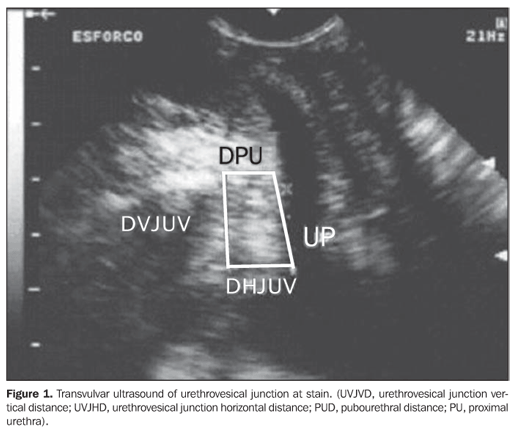
MATERIALS AND METHODS In the period between August 2004 and January 2006, the authors developed the present transverse and prospective study in the Unidade de Pesquisa em Incontinência Urinária (UPIU) (Division of Research in Urinary Incontinence) of Hospital das Clínicas da Universidade Federal de Pernambuco (HC-UFPE). The research protocol was reviewed and approved by the UFPE Committee for Ethics in Research previously to the enrollment of the first volunteer. After being given an explanation on aspects of the study, the patients who had accepted to participate were invited to sign a term of free and informed consent. Forty randomly selected women were included in the present study; 20 of them were submitted to FLS, and 20 to TVT. Inclusion criteria were: age range between 30 and 60 years, symptoms or signs of SUI, vertical UVJ displacement > 9 mm (determined by transvulvar ultrasound), absence of other diseases associated with urinary incontinence, absence of previous history of surgery involving the bladder, urethra or vagina. Exclusion criteria were: onset of acute disease hindering the procedure, patient's giving up on the surgery, diagnosis of pregnancy during the investigational period. All of the 40 patients answered a structured questionnaire and underwent a thorough urogynecological examination. Additionally, they were evaluated by a single sonographist with more-than-five-year experience in transvulvar ultrasonography, so minimizing inter- and intraobserver biases, for sonographic documentation in the preoperative period, and 30 days after the surgery by means of clinical follow-up. The measurement by transvulvar ultrasound was performed, with the patient in the dorsolithotomy position, with legs bent, immediately after voiding and having a barely empty bladder (< 50 ml of urine). The transducer previously covered by a condom and lubricated with water-soluble gel was placed touching the vulva, in a localization that allowed the sonographist to identify the urethra, the bladder, the vesical cervix and the pubic symphysis - structures presenting characteristic echotextures. The equipment used for the examinations was an Aloka SSD500, connected to a 7 MHz convex vaginal transducer and an electronic selector for real image measurement, equipped with computer and photographic camera with instantaneous resolution capability. The measurements were based on an orthogonal system of Cartesian coordinates originating from the inferior border of the pubic symphysis. The ordinates axis was perpendicularly oriented in relation to the pubis and the abscissas axis was tangent to the lower pubic region. The UVJ distance was estimated in millimeters, in relation to the ordinates axis, and the results were recorded in positive or negative numbers according to its position. Two measurements were performed as a routine on the monitor screen, the first one with the patients at rest, and the second during the Valsalva maneuver, aiming at determining the amplitude of the displacement of the anatomical structures of interest. The displacement resulting from the difference between the measurements was evaluated as follows: above the inferior border of the pubic symphysis, it was standardized with a positive (+) value; below the inferior border of the pubic symphysis, with a negative (-) value; above the inferior border of the pubic symphysis at rest, and below the inferior border of the pubic symphysis during Valsalva maneuver, the result from summation of these values. The following parameters were analyzed: - UVJVD: length of a longitudinal line between the inferior border of the pubic symphysis and a straight transverse line originating from the UVJ; - UVJHD: length of the straight transverse line originating from the UVJ to the longitudinal line originating from the pubic symphysis; - PUD: length of a horizontal line from the inferior border of the pubic symphysis to the urethra; - PU: distance between the UVJ and the pubourethral distance middle point. The criteria utilized for the diagnosis of SUI (UVJ hypermobility) have been suggested by Brandt et al., who describe the UVJ vertical displacement as the most accurate parameter for this purpose(8,13) (Figure 1).
The TVT surgery was performed according to the technique primarily described by Ulmsten(14). The fascia lata sling procedure involved the removal of an autologous segment of aponeurosis from the internal face of the thigh, measuring on average 2 cm × 6 cm. The insertion of this aponeurotic sling was performed by means of an abdominal Pfannenstiel incision. One month after the surgery, the patients were submitted to a new transvulvar ultrasound for a reevaluation of the UVJ and PU measurements. These data were compared with those obtained when the patients were enrolled in the present study. The data were analyzed with the Epi-Info 2002 software version 1.0. The chi-squared (c2) test was utilized for analysis of the contingency tables, the Fisher exact test for mean differences and variances ratios, with a statistical significance level (p value) = 5%.
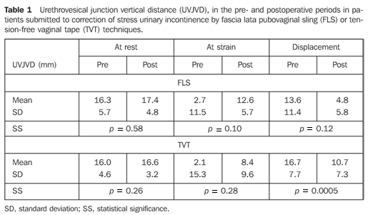
RESULTS The patients' age ranged between 30 and 60 years (mean age = 50.3 ± 9.6 years). Mean variation of pre- and postoperative UVJ vertical distances measurements in patients submitted to correction of stress urinary incontinence by fascia lata pubo vaginal sling or TVT techniques are described on Table 1.
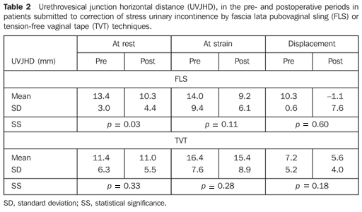
Mean variation of sonographic measurements of urethrovesical junction horizontal distance are described on Table 2.
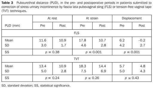
Mean ultrasound findings regarding measurements of PUD are described on Table 3.
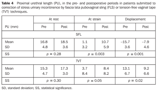
Among the findings of the authors in the present study, the most significant transvulvar sonographic measurements were those related to the PU measurements described on Table 4.
DISCUSSION The existence of more than a hundred surgical techniques for the management of SUI may mean that the physiopathology of this disease, and consequently, the logics of its surgical correction are still to be completely known(8,10,15). It seems that the main issues involved are related to the urethral resistance factor with urinary loss pressure, and the UVJ and PU positioning. On the one hand, the urodynamic testing is considered as a valuable tool, on the other hand, the utilization of pre- and postoperative ultrasound have increasingly been considered as a relevant investigative tool in the evaluation of the UVJ and PU(8,10,15). Among the different types of surgery for management of SUI, the former sling-type surgeries were updated with basis on the postulates defined on the TVT technique, with frequent reports on advantages in relation to the PU positioning as compared with other similar procedures. However, the majority of authors utilize the urodynamic testing as a supplementary investigational method, without taking relevant sonographic parameters into consideration(8,10). As regards the supplementary pre- and postoperative investigational method, although there is some divergence about the ideal diagnostic method for SUI, there is a reasonable agreement that the majority of these patients present isolated or associated anatomical alterations in the UVJ and PU. Additionally, it is important to note that approximately 95% of women complaining of SUI present UVJ and PU hypermobility(8,10,16-18). Independently from the urodynamics, there is a remarkable progress in the understanding of factors that may affect the results of a UVJ and PU sonographic study, such as the vesical volume and the necessary strain to cause UVJ and PU mobility, so increasing the ultrasound interest and reliability(13,19). These considerations have led the authors of the present study to adopt transvulvar ultrasound for a specific evaluation of the UVJ and PU functional topography. On the other hand, the urodynamic testing evaluates only some preoperative param eters, in an indirect attempt to observe the UVJ mobility, and fails to objectively record the UVJ and PU anatomical positioning both from the static and dynamic point of view(8,10,18,20). The fascia of the rectum-abdominal muscle is quite utilized, but the superiority of the fascia lata sling surgery is recognized, particularly for the lower tissue injury as a result of the possibility of obtention of larger tissue segments, the lower risk for complications on the donor site, shorter hospital stay, and, as a result, an earlier return of the patients to their daily activities(12). It was important to observe by means of transvulvar ultrasound, that the UVJVD to the inferior border of the pubic symphysis considered as one of the main anatomical parameters related to the urinary incontinence, has significantly changed only after the TVT surgery, specifically in relation to the UVJ displacement, as shown on Table 1. That is to say, ultrasound can demonstrate that TVT technique significantly hinders the UVJ downwards displacement, allowing a certain degree of voiding obstruction. Likewise, the FLS method also corrected the SUI in most of patients submitted to this surgery and even so has resulted in a not-so-remarkable suspension of the UVJ. On the other hand, from the dynamic, physiopathological point o view, the UVJHD was not affected by any of these techniques. It is important to note that the FLS technique resulted in a more significant change in the resting angle of the UVJ, but this static anatomical change did not remained significant under the dynamic abdominal pressure (Table 2). The results of the present study also demonstrated that the FLS surgery for the management of SUI resulted in reduced PUD at strain, and decreased movement of the PU, differently from the TVT technique results (Table 3). Transvulvar ultrasonography has allowed the authors to observe that maybe among their findings the most important one was the elongation of the proximal urethra, both at strain and displacement after correction of SUI by either of the techniques described in the present study (Table 4). So, this is the importance of restoring the proximal urethral pressure by means of an increase in area resulting from the PU elongation to achieve an effective urinary continence. In summary, transvulvar ultrasound, taking the inferior border of the pubic symphysis, demonstrate that the pubovaginal sling surgery could simultaneously reduce the UVJHD at rest, and extend the PU length at strain, allowing these structures to return to a more anatomical position, and restoring the urinary continence by reducing the bladder neck mobility. Thirty days after the surgeries, the authors could observe that both the FLS and the TVT methods were effective in 85% of the patients, These results are compliant with data reported in the literature(9). It is important to highlight that a cure rate cannot be estimated in a short term, neither is this hypothesis supported by the present study, considering the consensus that a mean ten-year follow-up is necessary to confirm a cure. In fact, the present study is aimed at reinforcing the thesis that SUI is directly related to a passive urinary continence established principally by the anatomical positioning of the UVJ and PU. Therefore, the surgical correction should be focused on the functional recovery of these structures to restore the passive urinary continence. The relevant role played by the postoperative ultrasound should be highlighted as an objective follow-up parameter, where a 5 mm cut-off for UVJVD is the mean measurement for young, continent patients(20) suggesting a satisfactory surgical correction(21), that is to say, leading to an effective urination control and possibly playing a role as a predictive factor for SUI recurrence, a matter already approached by other authors(22), and that is the objective of a new ongoing investigation by the authors of the present study. Therefore it can be assumed that both the lateral vastus fascia lata sling and the TVT techniques are equivalent in relation to the immediate clinical results and elongation of the proximal urethra. Besides the few postoperative anatomical differences above described requiring further studies with larger samples, it is necessary to consider that the fascia lata sling technique implies lower operative costs as compared with those of the synthetic materials utilized in TVT, and shorter hospital stay in relation to the techniques which utilized abdominal fasciae.
CONCLUSION Transvulvar ultrasound plays a significant role in the pre- and postoperative assessment of women with SUI. This method can objectively record the measurements of the essential parameters in the surgical restoration of the normal voiding, besides allowing the comparison among several results from different surgical techniques. With the aid of transvulvar ultrasonography, the authors could conclude that the TVT surgery has elongated the PU at strain and displacement, as well as reduced the UVJVD; and the FLS procedure reduced the distance between the UVJ and the pubis both by the decrease in the UVJHD at rest and in the PUD at strain and displacement. Furthermore, the FLS technique resulted in a higher extension of the PU length at strain and displacement.
REFERENCES 1. Waetjen LE, Subak LL Shen H, et al. Stress urinary incontinence surgery in the United States. Obstet Gynecol 2003;101:671–676. [ ] 2. Hampel C, Artibani W, Espuna Pons M, et al. Understanding the burden of stress urinary incontinence in Europe: a qualitative review of the literature. Eur Urol 2004;46:15–27. [ ] 3. Contreras O. Stress urinary incontinence in the gynecological practice. Int J Gynaecol Obstet 2004;86:6–16. [ ] 4. Minassian VA, Drutz HP, Al-Badr A. Urinary incontinence as a worldwide problem. Int J Gynaecol Obstet 2003;82:327–338. [ ] 5. Birnbaum HG, Leong SA, Oster EF, Kinchen KS, Sun P. Cost of stress urinary incontinence: a claims data analysis. Pharmacoeconomics 2004; 22:95–105. [ ] 6. Kinchen KS, Orsini S, Crown W, Swindle R. A retrospective claims analysis of the direct costs of stress urinary incontinence. Int Urogynecol J Pelvic Floor Dysfunct 2003;14:403–411. [ ] 7. Bullock TL, Ghoniem G, Klutke CG, Staskin DR. Advances in female stress urinary incontinence: mid-urethral slings. BJU Int 2006;98 (Suppl 1): 32–40. [ ] 8. Brandt FT, Santos Junior MW, Albuquerque CDC, Lorenzato FRB, Viana LA, Cunha ASC. Modificações da junção uretrovesical e uretra proximal após a cirurgia de alça sem tensão em mulheres de 45 a 72 anos Rev Bras Saúde Mater Infant 2005;5:185–191. [ ] 9. Schraffordt Koops SE, Bisseling TM, Heintz AP, Vervest HA. The effectiveness of tension-free vaginal tape (TVT) and quality of life measured in women with previous urogynecologic surgery: analysis from The Netherlands TVT database. Am J Obstet Gynecol 2006;195:439–444. [ ] 10. Ribeiro CBL, Brandt FTB, Albuquerque CDC, Arraes F, Pinho Neto JS, Ávila M. Modificações da uretra proximal e da junção uretrovesical decorrentes da cirurgia do tipo Kelly-Kennedy. Acta Cir Bras 2001;17:21–23. [ ] 11. Wilson L, Brown JS, Shin GP, Luc KO, Subak LL. Annual direct cost of urinary incontinence. Obstet Gynecol 2001;98:398–406. [ ] 12. Norton P, Brubaker L. Urinary incontinence in women. Lancet 2006;367:57–67. [ ] 13. Brandt FT, Albuquerque CDC, Arraes AF, Albuquerque GF, Barbosa CD, Araújo CM. Influência do volume vesical na avaliação ultra-sonográfica da junção uretrovesical e uretra proximal. Radiol Bras 2005;38:33–36. [ ] 14. Ulmsten U. An introduction to tension-free vaginal tape (TVT) – a new surgical procedure for treatment of female urinary incontinence. Int Urogynecol J Pelvic Floor Dysfunct 2001;12 (Suppl 2):S3–4. [ ] 15. Howden NS, Zyczynski HM, Moalli PA, Sagan ER, Meyn LA, Weber AM. Comparison of autologous rectus fascia and cadaveric fascia in pubovaginal sling continence outcomes. Am J Obstet Gynecol 2006;194:1444–1449. [ ] 16. McGuire EJ. Urinary incontinence: a diverse condition. J Urol 2005;173:1453–1454. [ ] 17. Dietz HP. Why pelvic floor surgeons should utilize ultrasound imaging. Ultrasound Obstet Gynecol 2006;28:629–634. [ ] 18. Dalpiaz O, Curti P. Role of perineal ultrasound in the evaluation of urinary stress incontinence and pelvic organ prolapse: a systematic review. Neurourol Urodyn 2006;25:301–306. [ ] 19. Brandt FT, Nóbrega LV, Albuquerque CDC, et al. Aferição simultânea da pressão abdominal na avaliação ultra-sonográfica de mulheres com incontinência urinária de esforço. Radiol Bras 2006;39:91–95. [ ] 20. Brandt FT, Albuquerque CDC, Lorenzato FR, Amaral FJ. Perineal assessment of urethrovesical junction mobility in young continent females. Int Urogynecol J 2000;11:18–22. [ ] 21. Vierhout ME, Hol M. Vaginal ultrasound studies before and after successful colposuspension and in continent controls. Acta Obstet Gynecol Scand 1998;77:101–104. [ ] 22. Viereck V, Pauer HU, Hesse O, et al. Urethral hypermobility after anti-incontinence surgery – a prognostic indicator? Int Urogynecol J Pelvic Floor Dysfunct 2006;17:586–592. [ ]
Received November 26, 2006. Accepted after revision May 9, 2007.
* Study developed at Universidade Federal de Pernambuco (UFPE), Recife, PE, Brazil. |
|

Av. Paulista, 37 - 7° andar - Conj. 71 - CEP 01311-902 - São Paulo - SP - Brazil - Phone: (11) 3372-4544 - Fax: (11) 3372-4554