Radiologia Brasileira - Publicação Científica Oficial do Colégio Brasileiro de Radiologia
AMB - Associação Médica Brasileira CNA - Comissão Nacional de Acreditação
 Vol. 40 nº 5 - Sep. / Oct. of 2007
|
ORIGINAL ARTICLE
|
|
Can changes associated with aging hinder the identification of individuals submitted to lumbar spine radiography? A potential contribution of radiology to the forensic activity |
|
|
Autho(rs): Sílvia Falcão de Oliveira, Glória Maria Martins Gomes, Lucio Ronaldo Cardoso, Hilton Augusto Koch, Edson Marchiori, Bianca Gutfilen |
|
|
Keywords: Radiographic comparison, Identification, Forensic anthropology |
|
|
Abstract:
IMasters in Radiology by Universidade Federal do Rio de Janeiro (UFRJ), Experts Medical Examiners at Instituto Médico-Legal Afrânio Peixoto (IMLAP), Rio de Janeiro, RJ, Brazil
INTRODUCTION The pioneering utilization of radiological resources to elucidate problems related to forensic identification dates back to 1927. Culbert & Law wrote an interesting article where they report a case of identification based on the comparison between nasal accessory sinuses and mastoid processes on preoperative radiographs and on radiographs of a deceased individual, where they could enumerate 20 coincidental items(1). From that time on, the comparative analysis of ante- and post-mortem radiographs in association with studies of fingerprints and dental arcades, and currently with DNA study, started constituting the routine protocol in forensic identification studies(2). A study performed in 2002 by American scientists identified the poor quality of images on ante- and post-mortem radiographs as the major limitation for the use of this method(3), but nevertheless such limitations usually do not use to affect the performance of experienced professionals, provided protocols are observed. Therefore, a methodology should be adopted in the process of forensic identification in order to facilitate the procedures: radiologists should classify the bone morphological features into categories such as typical morphological differences, anatomical variants, aging degenerative processes, traumatisms and congenital malformations(4). The present study is aimed at evaluating the possibility of a radiological study of the lumbar spine determining the correct identification of an individual despite changes associated with aging, to demonstrate the potential role of lumbar spine radiography in the forensic activity.
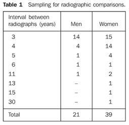
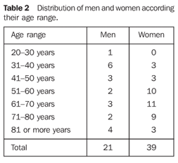
MATERIALS AND METHODS A survey aiming at finding adult patients with at least two radiographs of their lumbar spine was performed in the Radiological Archives of the Service of Radiodiagnosis at Hospital Universitário Clementino Fraga Filho. The inclusion criterion was patients with two radiographs of the lumbar spine acquired at three-year minimum interval, independently from the degree of degeneration observed or the presence of other osteoarticular diseases. Sixty pairs of studies performed at intervals ranging between three and thirty years (mean 4.8 years and median 4.0 years) were selected. The youngest patient was 20 years old, and the oldest, 88 years old (mean age 60.82 years and standard deviation, 16.14) (Tables 1 and 2).
Once the pairs of films had been selected, they had their register numbers hidden and were mixed up before being analyzed by two experienced radiologists. The comparative study included the analysis of morphological variations of vertebral bodies, spinous processes, transverse processes, pedicles and intervertebral spaces, degenerative aging processes, and also any particular findings that could be of help in the process of images pairing. The pairing criterion adopted was a single finding of an anatomical variant or specific detail, or the finding of two or more similarities among anatomical details, with no point of divergence Besides being evaluated at the negatoscope, the radiographic images were photographed and submitted to an images processing software (Adobe Photoshop), to enhance or reduce their contrast and/or brightness, or for segmentation aiming at optimizing the comparison process.
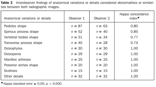
RESULTS Both observers could pair off all of the 60 studies. The process of putting the image pairs back together was developed through a careful observation of anatomical details. The first pairs of images were based on findings of anatomical variants or specific details - on the very spinal column or not - capable, by themselves, to allow the identification. The reading of the analyses performed by both observers demonstrated 20 coincidence points, with highlights on the following findings: "partial vertebra collapse", "spondylolysis", "bizarre transverse process", "very short transverse process", "elongated left transverse process", "upward transverse process", "transverse mega-apophysis" and "gross calcifications in the pelvic excavation - calcified myoma?". The other anatomical details were considered as a whole for establishing a positive identification. Correct pairing of radiographs of the whole sample was achieved by both observers, who presented countless coincidence points in their analyses, and the statistical analysis demonstrated a good-to-perfect interobserver agreement (Table 3).
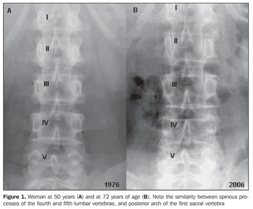
Signs of traumatisms and/or bone surgeries or congenital malformations, considered as a comparison criterion, have not been found among the radiographic images included in the sample. Figure 1 shows a pair of images corresponding to the case with the longest interval between the two radiographs (30 years).
DISCUSSION The 60 pairs of radiographic images were correctly identified by both observers, with a concordance index specifically calculated for each anatomical detail or pathological alteration analyzed. These indices ranged between 0.74 and 1.00, with the statistical analysis demonstrating a good-to-perfect interobserver agreement. The observers' confidence to define the images pairing was due to the finding of similarly shaped anatomical details - investigated and evaluated separately or in conjunction - and the absence of discrepancies amongst images. Findings of either pathological alteration on the very spinal column, such as osteophytes and other evidences of arthrosis, or occasional abnormal findings, such as "gross calcification in the pelvic excavation - calcified myoma?", or atypical anatomical details, such as "upward transverse process", have allowed some pairings. Essentially, the perfect pairing was based on morphological parameters. The large number of degenerative features found on the majority of radiographic images is attributed to the high age range of the patients included in the present study; however, osteopenia and osteophytes were pointed out as contributory factors in the pairing process. Manifest arthrosis does represent a relevant factor, as well as do alterations capable of considerably affecting anatomical patterns. Some anthropologists have been insisting that the interval between ante- and post-mortem radiographies should be no longer than few years, in cases where such radiographic images are suggested as a support to human remains identification(5). However, the results of the present study go against this idea: among the 60 cases included in the present study, six corresponded to individuals with intervals of more than tem years between studies. It is important to mention a case described in the literature where the ante-mortem radiograph had been acquired 13 years before the disappearance of an individual whose cadaver could be identified(6). The trabecular pattern can be utilized in comparative studies(7), but only in cases where the images are concerned with studies performed in not very distant times, considering that the trabecular structure is susceptible to reabsorption, even in early stages of osteoporosis, particularly in women, and may lead to erroneous interpretation(4). Reference to forensic authors demonstrates that anatomical details should be meticulously observed despite the absence of a mandatory minimum number of items to be compared for determining the identification of human remains. Usually, one to four coincidental characteristics and no discordance are considered as sufficient parameters(8). Detailed anatomical structures with natural variations, especially the skull, lumbarsacral column and chondrosternal joints, constitute the best elements for human remains identification(9). However, not only these structures can be of help in comparative studies. The literature suggests the mastoid apophysis(10), sella turcica of the sphenoid bone(10), hand and wrist(11), and clavicle(12). At global level, comparative analysis of ante- and post-mortem radiographs has represented a potential resource both in necroscopic studies of bone specimens and analyses of carbonized or putrefied cadavers, or even recent cadavers whose physiognomic lines individualization is still feasible(13). It is evident that the major contribution of this method is associated with recent cadavers, considering that, nearly always in such cases, relatives show up to claim the cadaver The procedure could be routinely adopted in forensic identification processes, and it is a responsibility of coroners providing the family relatives with guidance in the search of ante mortem radiographs of the deceased. Considering that the comparison between radiographic images of lumbar spine can determine a correct identification of individuals, despite changes associated with aging, it may be concluded that radiography represents a potential tool to be utilized in forensic identification studies, comparable in utility to fingerprints and dental evidences. Acknowledgement Professor Mário Newton Leitão Azevedo, Head for the Service of Rheumatology, Faculty of Medicine at Universidade Federal do Rio de Janeiro, for his collaboration in the survey for the sampling of the present study.
REFERENCES 1.Culbert WL, Law FM. Identification by comparison of roentgenograms of nasal accessory sinuses and mastoid process. JAMA J Am Med Assoc 1927;88:1634–1636. [ ] 2.Iscan MY. Rise of forensic anthropology. Yrbk Phys Anthropol 1988;31:203–230. [ ] 3.Kuehn CM, Taylor KM, Mann FA, Wilson AJ, Harruff RC. Validation of chest X-ray comparisons for unknown decedent identification. J Forensic Sci 2002;47:725–729. [ ] 4.Kahana T, Goldin L, Hiss J. Personal identification based on radiographic vertebral features. Am J Forensic Med Pathol 2002;23:36–41. [ ] 5.Angyal M, Dérczy K. Personal identification on the basis of antemortem and postmortem radiographs. J Forensic Sci 1998;43:1089–1093. [ ] 6.Valenzuela A. Radiographic comparison of the lumbar spine for positive identification of human remains. Am J Forensic Med Pathol 1997;18: 215–217. [ ] 7.Mann RW. Use of bone trabeculae to establish positive identification. Forensic Sci Int 1998;98: 91–99. [ ] 8.Kahana T, Hiss J. Forensic radiology. Br J Radiol 1999;72:129–133. [ ] 9.Quatrehomme G, Fronty P, Sapanet M, Grévin G, Bailet P, Ollier A. Identification by frontal sinus pattern in forensic anthropology. Forensic Sci Int 1996;83:147–153. [ ] 10.Voluter G. The V-test. Radiol Clin 1959;Supl 28: 5–7. [ ] 11.Greulich WW. Skeletal feature: visible on the roentgenogram of hand and wrist which can be used for establishing individual identification. Am J Roentgenol Radium Ther Nucl Med 1960; 83:756–764. [ ] 12.Sanders I, Woesner ME, Ferguson RA, Noguchi TT. A new application of forensic radiology: identification of deceased from a single clavicle. Am J Roentgenol Radium Ther Nucl Med 1972;115: 619–622. [ ] 13.Murphy WA, Spruill FG, Gantner GE. Radiologic identification of unknown human remains. J Forensic Sci 1980;25:725–735. [ ]
Received November 28, 2006. Accepted after revision March 8, 2007.
* Study developed in the Department of Radiology at Faculdade de Medicina da Universidade Federal do Rio de Janeiro (UFRJ), Rio de Janeiro, RJ, Brazil. |
|

Av. Paulista, 37 - 7° andar - Conj. 71 - CEP 01311-902 - São Paulo - SP - Brazil - Phone: (11) 3372-4544 - Fax: (11) 3372-4554